대한두경부종양학회지, 제33권 제1호, 2017. pp.21-29 Korean Journal of Head & Neck Oncology, Vol.33, No.1
http://dx.doi.org/10.21593/kjhno/2017.33.1.21 ISSN 1229-5183(Print)
전장유전체수준 메틸레이션 분석을 통한 두경부암 특이 메틸레이션 바이오마커의 발굴
장재원1⋅박기완1⋅홍소혜1⋅정승남1⋅류려화1⋅김진만2⋅오태정3⋅구본석1+
충남대학교 의과대학 이비인후-두경부외과학교실1, 병리학교실2, 지노믹트리3
Genome-wide Methylation Analysis and Validation of Cancer Specific Biomarker of Head and Neck Cancer
Jae Won Chang, MD, PhD1, Ki Wan Park, MD1, So-Hye Hong, MS1, Seung-Nam Jung, PhD1, Lihua Liu1, Jin Man Kim, MD, PhD2, Taejeong Oh, PhD3 and Bon Seok Koo, MD, PhD1+
Department of 1Otorhinolaryngology-Head and Neck Surgery,
2Pathology, School of Medicine, Chungnam National University, Daejeon, Republic of Korea
3Research and Development Center, GenomicTree, Inc., Daejeon Bioventure Town, Daejeon Republic of Korea
= Abstract =
Methylation of CpG islands in the promoter region of genes acts as a significant mechanism of epigenetic gene silencing in head and neck squamous cell carcinoma (HNSCC). DNA methylation markers are particularly advantageous because DNA methylation is an early event in tumorigenesis, and the epigenetic modification, 5-meth- ylcytosine, is a stable mark. In the present study, we assessed the genome-wide preliminary screening and were to identify novel methylation biomarker candidate in HNSCC. Genome-wide methylation analysis was performed on 10 HNSCC tumors using the Methylated DNA Isolation Assay (MeDIA) CpG island microarray. Validation was done using immunohistochemistry using tissue microarray of 135 independent HNSCC tumors. In addition, in vitro proliferation, migration/invasion assays, RT-PCR and immunoblotting were performed to elucidate molec- ular regulating mechanisms. Our preliminary validation using CpG microarray data set, immunohisto-chemistry for HNSCC tumor tissues and in vitro functional assays revealed that methylation of the Homeobox B5 (HOXB5) and H6 Family Homeobox 2 (HMX2) could be possible novel methylation biomarkers in HNSCC.
Key W ords:Genome-wide methylation analysis, Methylation biomarker, Homeobox B5 (HOXB5), H6 Family Homeobox 2 (HMX2), Head and neck squamous cell carcinoma (HNSCC)
R eceived R e v i s e d A ccepted
: May 4, 2017 : May 12, 2017 : May 16, 2017
+Corresponding author: Bon Seok Koo, MD, PhD Professor
Department of Otorhinolaryngology-Head and Neck Surgery, School of Medicine, Chungnam National University, 282 Munhwa-ro, Jung-gu, Daejeon, 35015, Republic of Korea Tel: +82-42-280-7695 Fax: +82-42-253-4059
E-mail: [email protected]
서 론
모든 암은 유전자의 변화에 의해서 발생하며, 이는 종
양유전자의 활성화와 종양억제유전자의 비활성화로 요 약된다.1,2) 종양유전자의 활성화에는 유전자 증폭(gene amplification), 점돌연변이(point mutation), 염색체 전좌 (chromatin translocation)등이 관여하며, 종양억제 유전자 의 불활성화에는 유전자 변이, 유전자 상실(gene dele- tion), 그리고 프로모터CpG island의 과메틸화가 관여한 다. 여기서 유전자 증폭, 점돌연변이, 염색체 전좌, 유전 자상실 등은 DNA 염기서열의 변동을 수반하는 유전적 변화(genetic change)인 반면, CpG island과메틸화는DNA 염기서열의 변동없이 유전자의활성을 억제할 수 있다는 점에서 유전적 변화와 구별하여 후성유전적 변화(epige-
netic change)라고 부른다.2)후성유전적 변화에는 CpG is- land 과메틸화 외에도 히스톤 변경, 크로마틴 구조의 변 경, microRNA 등이 포함된다.1)
DNA 메틸화는 DNA methyl transferase에 의해서 cytosine pyrimidine 링의 5번째 탄소에 메틸기(CH3)가 공유결합으 로 첨가되는 현상으로 유전체에 생기는 가장 대표적인 후 성유전적 변화이다.2) DNA 메틸화는 정상적인 개체의 발생 에서도 genomic imprinting, X-chromosome inactivation 등 다양한 생명현상에서 중요한 역할을 하는 것으로 알려져 있지만 다양한 암종의 발생에서 중요한 역할을 한다는 연구결과가 축적되고 있다.3)암 조직에서는 정상 세포와 는 다른 두 종류의 DNA 메틸화 현상이 나타나는데, 유전 체 전반에 걸친 저메틸화 (hypomethylation) 현상과 유전 자 발현 조절부위 (promoter)에 위치한 CpG island (CpG 디뉴클레오티드의 밀도가 높은 시퀀스)의 과메틸화 (hy- permethylation) 현상이 그것이다. 저메틸화 현상은 주로 유전자와 유전자 사이 지역(intergenic region)에서 나타 나며, 이는 염색체를 불안정하게 만들어 세포분열 과정 에서 염색체의 재조합(recombination), 전이(translocation), 절단(deletion), 재배열(rearrangement) 등을 일으키는 것으 로 생각되며, 암 억제유전자(tumor suppressor), 세포주기조 절(cell cycle) 유전자, DNA 수리(repair) 관련 유전자, 세포 접착(adhesion) 관련 유전자가 조절부위의 CpG island의 과 메틸화에 의해 발현이 억제되면, 세포는 비정상적으로 증 식하며, 유전적 안정성을 유지하지 못하게 되어 추가적인 돌연변이를 유발하여 암이 유발된다고 알려져 있다.4,5)
두경부암에서도 특정부위의 methylation 혹은 유전체 의 메틸화 profile과 예후의 관계에 대하여 일부 보고는 되어 있고, promoter 과메틸화 여부와 임상 병리학적 병 기와의 연관관계, 면역염색을 통한 특정 유전자의 발현 감소 및 소실을 관찰하려는 시도가 이루어지고 있으나, 전유전체적인 분석을 통해 메틸화 바이오마커를 탐색한 연구는 아직까지 부족한 실정이다.5-7) 또한 후성유전적 변화는 인종이나 환경과 같은 요인에 영향을 받기 때문 에 우리나라 환자들을 대상으로 전유전체 수준의 메틸화 profile에 대한 확인을 통해 기존에 발견되지 않았던 바이 오마커를 발굴하는 것이 필요하기에 본 연구를 통하여 한국인 두경부암에 특이적인 신규 메틸화 바이오마커를 발굴하고자 하였다.8,9)
대 상 및 방 법
메틸화 DNA 분리 및 분석 (Methylated DNA isolation assay, MeDIA)
CpG microarray analysis 는 전체유전자를 대상으로 메 틸화된 DNA를 탐색하여 이를 특이적으로 선택 분리할 수 있는 MethylCaptureTM 방식을 이용하였다.10) 절차를 간단히 기술하면 다음과 같다. 10명의 두경부암 환자에 서 genomic DNA를 종양과 주변의 종양이 아닌 조직에서 추출한 뒤, 음파를 이용하여 DNA를 단편화 시켰다. 단편 화된 genomic DNA 0.5μg를 2μg의 유전공학적으로 조 작된 메틸화 DNA에 특이적으로 결합하는MBD2b 단백 질의 기능기만 축소하여 만든 MBD2bt 단백질과 함께 4℃에서 4시간동안 반응시킨 다음 whole-genome amplifi- cation kit (Sigma-Aldrich)를 이용하여 genome을 증폭한 이후 Cy5 dye를 이용하여 labelling 한다. 종양이 아닌 조 직에서 역시 DNA를 추출하여 Cy3으로 labelling 한다.
DNA를 PCR purification kit (Qiagen)을 이용하여 정제한 이후, 총 27,800개의 알려진 CpG island를 표지할 수 있는 237,000개의 oligonucleotide의 probe와 혼성화 반응시켜 메틸화 DNA를 확인한다. 이때 quality control을 위하여 이 미 알고 있는 사람세포주의 특정 메틸화 유전자를 MeDIA 후 gene-specific PCR 방법으로 확인하였다.10)
두 경 부 암 세 포 주 와 조 직 에 서 메 틸 화 확 인 MeDIA를 이용한 전유전체의 메틸화 분석을 통해 얻은 후보유전자를 두경부암 세포주와 조직을 이용하여 검증 하였다. 세포주를 이용한 검증에서 대조군으로 human fibroblast (hFB), 두경부암 세포주로는 FaDu, SNU1041, SNU1076, SCC15, SCC25, YD8을 이용하였다. 두경부암 조직은 2015~2016년 충남대학교 병원 이비인후과에 내 원하여 두경부암으로 진단받은 환자 20명과 양성종양 (non-tumor)로 수술받은 환자 12명의 조직을 이용하여 DNA 염기서열의 정성적, 정량적 분석을 간편하고 정확 하게 수행할 수 있는 next-generation sequencing 기반의 잘 알려진 실험기법인 pyrosequencing 을 이용하여 분석 하였다.11)
후 향 적 두 경 부 암 조 직 을 이 용 한 임 상 검 증 2005년에서 2012년까지 충남대학교 병원 이비인후과 에 내원하여 두경부암으로 진단받고 수술하여 인체자원 은행에 paired 된 비암성조직 (non-tumor tissue)와 함께 조직이 확보되어 있는 환자 135명의 의무기록을 후향적 으로 분석하였으며, 동일한 환자로 확보된 Tissue micro-
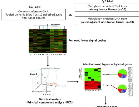
Fig. 1. Stepwise filtering processes for candidate gene selection. The methylated DNA was separately enriched from 10 primary head and neck tumors and paired, adjacent nontumor tissues by a MeDIA technique. The methylated DNAs (Cy5) were in- dividually compared with amplified common reference DNA (Cy3) without methylation enrichment. After removed lower singnal probe from unsupervised hierarchical clustering based on Pearson’s correlation, group A reliable probes were selected using principal component analysis. The heatmap shows the methylation status of significant 334 hypermethylated and 507 hypomethy- lated genes. Seven novel candidate genes hypermethylated in primary head and neck tumor were finally selected.
array를 면역조직화학염색을 통하여 발굴된 메틸화 바이 오마커의 발현양상을 분석하였다.
세 포 주 를 이 용 한 기 전 확 인
두경부암 세포주인 SCC15, YD8 세포주를 이용하여 메틸화 바이오마커의 발현조절에 따른 단일세포수준의 cell proliferation 을 WST-1 cell proliferation assay(Roche Diagnostics Corporation, Indianapolis, IN, USA) 를 이용하여 실험하였으며, migration 및 invasion 의 변화를 Transwell chambers (24-well, Costar, Cambridge, MA, USA)와 Matrigel (Coring, MA, USA)을 이용하여 확인하였다.12)
또한 Western blotting을 이용하여 발암 및 전이과정에 중요한 상피-간질세포 이행 (Epithelial-to-mesenchymal transition, EMT)과 관련된 표지자: anti-vimentin, an- ti-Slug, anti-β-actin (1:1,000; Cell Signaling Technology Inc.), anti-E-cadherin (1:1,000; Santa Cruz Biotechnology, Santa Cruz, CA, USA)를 확인하였다. HOXB5와 HMX2의 확인을 위한 항체로는 anti-HOXB5 (1:1000; abcam; ab26079), anti-HMX2 (1:1000; GeneTex; GTX81571)를 사용하였다.
결 과
두 경 부 암 의 전 장 유 전 체 수 준 메 틸 화 분 석 을 통 한 신 규 두 경 부 암 바 이 오 마 커 발 굴
두경부암에서 비정상적으로 메틸화된 유전자의 CpG island를 포괄적으로 확인하기 위해 10명의 두경부암 환 자에서 암조직과 주위의 비암조직에서 채취된 genomic DNA의 MeDIA CpG microarray 방식을 이용하여 비교한 결과, 정상과 비교하여 암조직에서 특이적으로 메틸화 양상에 차이가 있는 reliable 한 메틸화 표적이 841개(저메 틸화 표적 507개, 과메틸화 표적 334개) 확인되었다 (Fig.
1). 그 중에서 1) positive call in multiple probe, 2) promoter 혹은 regulatory regions, 3) novelty의 기준으로 선별하여 과메틸화 표적 334개 중, 총 7개의 기존에 연구되지 않은 메틸화 바이오마커 후보 유전자 (SLC30A3, LHX8, GATA4, HOXB5, ZNF274, HMX2, HOXA7)를 확인하였다.
두 경부 암 세 포 주에 서 발 굴 한 마 커 의 유용 성 검 증 MeDIA microarray와 통계적방법을 이용하여 발굴한 7개 의 바이오마커 후보 유전자의 두경부암 세포주에서의 발 현을 확인하기 위하여 두경부암 점막의 정상세포를 대변
Fig. 2. The mRNA expression of candidate methylation biomarker in various head and neck cancer cell lines. (A) RT-PCR showed that HOXB5 and HMX2 showed relatively lower mRNA expressions than normal hFB. hFB, human fibroblast. (B) Demethylation by DAC treatment rescued HOXB5 and HMX2 expression in both head and neck cancer cell lines.
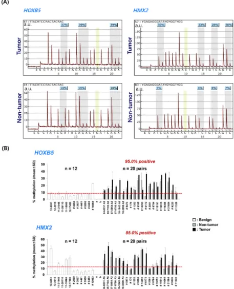
Fig. 3. Quantitative analysis of HOXB5 and HMX2 methylation by pyrosequencing. (A) Pyrogram from quantification of methylation at the HOXB5 and HMX2 promoter in both tumor and non-tumor tissues. (B) Methylation index of tumor (n = 20), paired non-tumor tissue (n = 20), and benign mass (n = 12).
하는 섬유세포 (hFB)와 두경부암 세포주 (FaDu, SNU1041, SNU1076, SCC15)에서 각 후보유전자의 mRNA발현을 살
펴본 결과, ZNF274와 HOXA7은 실험한 모든 두경부암 세 포주에서 정상세포와 유사하게 발현되고 있어 메틸화 마
Fig. 4. Kaplan-Meier plots for (A) overall survival (OS) and (B) disease-free survival (DFS) based on HOXB5 or HMX2 expression level as determined by immunoblotting in tumor tissues.
커의 유용성이 적은 것으로 확인되었으며, SLC30A3, LHX8, GATA4는 정상세포주에서 발현이 거의 되지 않고 있어 역 시, 메틸화 마커의 유용성을 확인하기 어려워, 최종적으 로 Homeobox B5 (HOXB5)와 H6 Family Homeobox 2 (HMX2) 가 메틸화 마커로서 가능성이 있음을 확인하였다 (Fig. 2A).
발굴한 후보 바이오마커의 메틸화 의존성을 확인하기 위하여 DNA methyltransferase의 억제제인 5-Aza-2'-deox- ycytidine (decitabine; DAC)를 이용하여 두경부암 세포주 (SCC25, YD8)에서 HOXB5와 HMX2를 탈메틸화 시켰을 때, HOXB5의 경우 SCC25에서 발현이 증가하였으며, HMX2의 경우 두 세포주 모두에서 발현이 증가하는 것을 확인하여 HOXB5와 HMX2의 메틸화 의존성을 확인하였 다 (Fig. 2B).
두 경 부 암 조 직 에 서 발 굴 한 마 커 의 유 용 성 검 증 세포수준에서 메틸화 바이오마커의 가능성을 확인한 HOXB5와 HMX2의 메틸화 정도를 조직 수준에서 정량적 으로 분석하기 위해 bisulfite pyrosequencing을 시행하여 methylation index(%)를 종양과 비종양에서 비교하였다
(Fig. 3A). Fig. 3B의 결과에서 확인할 수 있듯이, HOXB5와 HMX2 모두 paired 된 비종양 부위에 비해서 종양 부위에 서 methylation index가 유의하게 높은 것을 확인할 수 있 다 (HOXB5의 경우 95%의 시료, HMX2의 경우 85%의 시 료). 뿐만 아니라, 12명의 양성종양 환자의 조직과 비교하 였을 때도, 종양조직에서 methylation index의 평균이 유의 하게 높은 것을 확인하여, HOXB5와 HMX2의 메틸화 바이 오마커의 가능성을 다양한 조직 수준에서 확인하였다.
후 향 적 두 경 부 암 조 직 을 이 용 한 임 상 검 증 앞에서 확인한 두경부암 메틸화 바이오마커인 HOXB5 와 HMX2의 임상적 유용성을 확인하기 위하여, 후향적 의무기록 분석을 통한 임상정보가 연결된 135명의 환자 의 조직에서 HOXB5와 HMX2±의 발현을 확인하였다. 환 자의 평균추적관찰기간은 45.9 34.4개월 (2-180개월) 이었 다. 단변량분석을 시행하였을 때, HOXB5와 HMX2 모두에 서 나이, 성별, 림프절 전이, 조직학적인 분화도에 따른 유의한 발현차이는 없었으나, T stage 와 AJCC stage에서 는 HOXB5와 HMX2 모두 stage III+IV에서 통계적으로 유
Factor β p Exp(β) 95% CI
Advanced T stage -1.667 0.001 0.189 (0.071, 0.501)
Advanced AJCC stage 0.342 0.500 1.407 (0.522, 3.796)
AJCC, American Joint Committee on Cancer; CI, confidence interval Variables No. of
patients
HOXB5 expression HMX2 expression
Low High p Low High p
Age, year
< 65
≥ 65 66
69
31(47.0) 35(53.0)
35(50.7) 34(49.3)
0.663
33(45.2) 40(54.8)
33(53.2) 29(46.8)
0.353
Gender Male Female
118 17
57(86.4) 9(13.6)
61(88.4) 8(11.6)
0.721
66(90.4) 7(9.6)
52(83.9) 10(16.1)
0.254
T stage I+II III+IV
73 62
25(37.9) 41(62.1)
48(69.6) 21(30.4)
0.000*
28(38.4) 45(61.6)
45(72.6) 17(27.4)
0.000*
LN metastasis No Yes
77 58
38(57.6) 28(42.4)
39(56.5) 30(43.5)
0.902
38(52.1) 35(47.9)
39(62.9) 23(37.1)
0.204
AJCC stage I+II
III+IV
46 89
16(24.2) 50(75.8)
30(43.5) 39(56.5)
0.018*
19(26.0) 54(74.0)
27(43.5) 35(56.5)
0.032*
H ist o lo g ic a l grade Well Moderate Poor
39 63 33
22(33.3) 28(42.4) 16(24.2)
17(24.6) 35(50.7) 17(24.6)
0.501
27(37.0) 29(39.7) 17(23.3)
12(19.4) 34(54.8) 16(25.8)
0.069
Table 1. Associations between HOXB5/HMX2 expression and clinicopathological features in patients with head and neck squ- amous cell carcinoma.
Factor β p Exp(β) 95% CI
Advanced T stage -1.362 0.005 0.256 (0.098, 0.667)
Advanced AJCC stage 0.065 0.900 1.067 (0.391, 2.912)
AJCC, American Joint Committee on Cancer; CI, confidence interval
Table2. Binary logistic regression for the associations of HOXB5 expression with pT stage, and AJCC stage
Table 3. Binary logistic regression for the associations of HMX2 expression with pT stage, and AJCC stage
의하게 낮은 발현을 보였다 (Table 1). T stage와 AJCC stage 를 이용하여 다변량 분석 (Binary logistic regression analysis)을 시행하였을 때, 진행된 T stage (Advanced T stage) 에서 통계적으로 유의하게 HOXB5 (Table 2)와 HMX2 (Table 3) 모두 낮은 발현을 확인하였다.
또한, 두 메틸화 바이오마커의 발현에 따른 전체생존률 (Overall survival, OS)과 무병생존률 (Disease free surviv- al, DFS)을 Kaplan-Meier curve를 이용하여 분석하였다.
두 메틸화 바이오마커 모두에서 발현이 높을수록 전체생 존률과 무병생존률이 높았으며, 발현이 낮을수록 전체 생존률과 무병생존률이 낮은 경향성을 확인할 수 있었으나, HOXB5의 경우 통계적으로도 유의하였던 반면 (OS, 50.7%
vs. 76.1%, p = 0.001; DFS, 52.1% vs. 74%, p = 0.001), HMX2 의 경우 그 차이가 경미하여 통계적인 유의성이 확인되 지는 않았다.
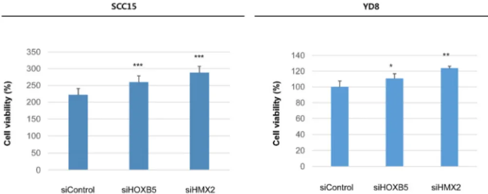
두 경 부 암 세 포 주 에 서 HOXB5와 HMX2의 기 능 분 석 두경부암에서 HOXB5와 HMX2의 암 억제유전자로서 의 기능을 확인하기 위해 두 유전자의 발현이 내인적으 로 올라가 있는 두경부암 세포주인 SCC15와 YD8에서 이를 siRNA를 이용하여 발현을 억제하였을 때의 기능상 변화를 확인하는 실험을 진행하였다.
먼저 WST-1 assay를 이용하여 cell proliferation 을 확인 하였을 때, 두 세포주 모두에서 HOXB5와 HMX2를 억제 하였을 때, proliferation 이 통계적으로 유의하게 증가하는 것이 확인되어, 암 억제유전자 (Tumor suppressor gene)의 가능성이 확인되었다 (Fig. 5).
다음으로 Transwell chamber를 이용하여 migration assay 를 시행하였으며, Matrigel을 추가하여 invasion assay를 시행하였을 때, 역시 두 세포주 모두에서 HOXB5와 HMX2 를 억제하였을 때, migration과 invasion 모두 증가하여,
Fig. 5. HOXB5 and HMX2 suppression promote HNSCC proliferation. To evaluate the effect of HOXB5 and HMX2 suppression on cell proliferation, a WST-1 assay was performed after transient transfection of small interfering RNA (siRNA). Each figure is representative of three independent experiments. *P<0.05; **P<0.01; ***P<0.001.
Fig. 6. HOXB5 and HMX2 suppression promote HNSCC migration and invasion. To elucidate the effect of HOXB5 and HMX2 suppression on cell migration and invasive phenotype, a Transwell chamber assay without/with Matrigel was performed. Each cell line was seeded onto a filter (pore size, 8 μm) (coated with Matrigel for invasion assay) in the upper chamber. After 48 h of transfection with each siRNA, the cells attached to the lower chamber were stained with crystal violet. Each figure is representative of three independent experiments. Scale bar = 50 μm.
Fig. 7. HOXB5 and HMX2 regulates epithelial-to-mesenchymal transition (EMT). Each cell line was transiently transfected with siRNA. Expression of HOXB5 or HMX2, E-cadherin (epithelial marker), EMT markers (vimentin), and the level of EMT-inducing tran- scription factors (Slug) were measured by Western blot analysis. Each band is representative of three independent experiments.
일관성 있게 두 유전자가 암 억제유전자임을 증명하였다 (Fig. 6).
분자적 수준에서 암세포의 proliferation, migration 및 invasion과 관련 되어있는 EMT hall-marker을 Western blotting을 통하여 단백질 수준에서 분석하였을 때, 두 세 포주 모두에서 HOXB5와 HMX2를 억제하였을 때, E-cad- herin 발현이 증가하고, Vimentin과 Slug의 발현은 감소하
는 것을 확인하였는데, 이는 위에 시행한 phenotype 실험 과 일관되는 결과였다 (Fig. 7).
고 찰
정밀의료 (Precision medicine)로 암치료의 패러다임이 이동하면서 암유전체에 대한 연구가 활발하게 이루어지
고 있으며,13)최근에는 Single-Cell Sequencing에 의하여 전장유전체 및 전사체 데이터를 생산하고, 이를 통하여 암이질성(tumor heteorogeneity) 및 암진화 (tumor evolu- tion)의 이해를 위한 연구가 활발하게 진행 중에 있다.13,14) 이러한 암이질성과 암진화에 후성유전학적인 변화 또한 관여한다는 것이 밝혀지면서, 후성유전학 또한 급속도 로 발전하고 있으며,15) promoter의 과메틸화 여부와 임상 병리학적 병기와의 상관관계, 면역조직화학염색을 통한 특정 유전자의 발현 감소 및 소실을 관찰하는 시도가 두경부암에서도 이루어지고 있다.5)그 결과로 세포부착 능에 관여하는 막당단백질인 E-cadherin,16,17)세포사멸과 관련된 효소인 Death associated protein kinase(DAPK),6) 세포주기 조절유전자인 p16, p15, p1418,19)외에도 Deleted in colorectal cancer (DCC)19)와 Methylated in tumor (MINT) family CpG islands4)등의 과메틸화가 두경부암에서 보고 되어 있으나, 이미 잘 알려진 암 억제유전자들을 대상으 로만 연구가 이루어져 암화 과정에서 메틸화에 영향을 받는 다른 유전자에 대한 연구는 미흡한 실정이다. 또한 최근의 연구동향이 후성유전적 변화로 확장되는 추세임 을 차치하여도, 두경부암의 경우 흡연, 음주, 바이러스 등 환경적인 요인이 발암과정에 깊이 관여하는 것으로 받아들여 지고 있기 때문에,6,20)후성유전적인 유전체의 변화가 발암과정에 중요한 것은 자명하다. 따라서 본 연 구에서는 기존에 후성유전적으로 연구가 활발히 진행되 지 않은 두경부암에서 전유전체적인 분석을 통해서 기존에 알려지지 않은 메틸화 바이오마커를 찾고자 하였다.21)
HOXB5는 17번 염색체에 존재하며, DNA 결합 도메인 의 homeobox에 결합하는 전사인자를 만들어내는 유전 자로 배아의 발달 과정 중 기관형성과 구획의 정체성을 조절하고, 세포수준에서 proliferation, migration을 조절 한다는 것이 알려져 있다.22)HMX2 는 10번 염색체에 존 재하며, 역시 발생과정에서 기관형성을 조절하는 전사 인자를 만들어내는 유전자로, 세포분열 및 분화를 촉진 하는 것으로 알려져 있으며, 특히 내이기형 (inner ear malformation), 전정기능부전 (vestibular dysfunction), 청 력소실 (hearing loss)과 관계된다고 연구되어 있으나 암 과의 관련성은 많이 알려져 있지 않다.23) 본 연구에서 전유전체 수준의 MeDIA CpG microarray와 임상조직 대 상 bisulfite pyrosequencing 을 통하여 두경부암에서 과메 틸화되어 있으며, 암 억제유전자로서의 가능성을 최초 로 확인하였고 나아가 진행된 병기와 관련되어 있으며, 유의한 예후인자가 될 수 있다는 것을 확인하였다는 점 에서 의미가 있다.
임상적인 유용성 뿐만 아니라, 본 연구에서는 HOXB5와
HMX2가 두경부암에서 발암과정에 관련되는 분자적 기전 을 세포수준에서도 확인하고자 하였다. HOXB5와 HMX2 가 과발현되어 있는 두경부암세포에서 RNA interference 를 이용하여 발현을 억제하였을 때 proliferation, migration 및 invasion 이 증가하는 것으로 미루어 보아, 위의 두 유전 자가, 암 억제유전자일 가능성을 확인하였으며, 분자적 수준에서는 HOXB5와 HMX2가 EMT를 억제하는 것을 확 인하였다. 이는 기존에 세포 내에서 HOXB5와 HMX2가 발생과정에서 기관형성에 관여한다고 알려진 것과도 연 관될 수 있는 결과이다.
본 연구결과를 통해 HOXB5와 HMX2가 두경부암 특이 메틸화 바이오마커로 이용될 수 있으며, 후성유전적 바 이오마커가 유전적 바이오마커와 비교하여 가지는 장점 으로 인하여, 조기진단, 예후판정에 임상적으로 적용할 수 있는 가능성이 더 크다고 할 수 있다. 예컨대, DNA의 메틸화는 질환의 초기에 일어나는 것으로 알려져 있고, 높은 민감도와 특이도를 가지며 최소의 증상에서도 보여 질 수 있을 만큼 충분한 양으로 존재하기 때문에 암의 조기진단에 유전적 마커에 비하여 유리하다.20)뿐만 아 니라, 일반적으로 후성유전적 변화는 유전자의 발현과 같은 유전학적 변화의 다양성에 비해 그 정도가 심하지 않게 때문에 다수의 마커가 필요한 유전자 발현 프로파 일링에 비해서 단 하나의 마커로도 암을 초기에 비침습 적으로 진단 할 수 있다.20)더욱이, 기존에 많이 사용되고 있는 단백질 마커나 RNA 보다 훨씬 안정적으로 존재하 며, 서로 다른 환자에서 한 유전자 내에서의 여러 위치를 분석해야 하는 다른 DNA 변이와는 달리 메틸화는 한 유전자의 같은 위치에서 일어나므로 분석방법 상의 장점 도 있어 매우 유망하고 효율적인 진단도구로 사용할 수 있다.20,24)
본 연구결과는 향후 대단위의 public한 유전체 database 를 이용하여 검증이 필요하며, 위 유전자의 메틸화를 유 발하는 인자, 메틸화로 인한 발암에 관계된 분자적 메커 니즘의 변화, 자손에 대한 유전적 영향 등에 대한 보다 많은 연구가 필요할 것을 생각한다. 그러나, 먹고, 마시 고, 숨쉬는 기능과 같이 생명유지에 필수적인 기능과 직 결되어 그 기능의 보존을 위해 두경부암의 조기진단 및 치료가 절실하지만 아직 확립된 조기진단 바이오마커가 없는 현실과, 두경부암의 발암과정에 환경적인 요인이 중요하다는 점을 고려할 때, 두경부암 특이 후성유전적 메틸화 바이오마커를 발굴하는 연구는 두경부암의 조기 진단 뿐만 아니라, 기존의 항암치료에 대한 저항을 극복 할 수 있는 새로운 병합항암치료법의 개발에도 도움이 될 수 있을 것으로 생각한다.20)
Acknowledgement
이 논문은 2016년도 정부(교육부)의 재원으로 한국연 구재단의 지원을 받아 수행된 기초연구사업임 (No. 2016 R1D1A1B04932112).
중심 단어:전유전체메틸레이선분석, 메틸레이션 분자표지자, 두경부암
References
1) Verma M. The Role of Epigenomics in the Study of Cancer Biomarkers and in the Development of Diagnostic Tools. Adv Exp Med Biol. 2015;867:59-80.
2) Esteller M. Cancer epigenomics: DNA methylomes and his- tone-modification maps. Nat Rev Genet. 2007;8:286-298.
3) Esteller M. The necessity of a human epigenome project.
Carcinogenesis. 2006;27:1121-1125.
4) Ogi K, Toyota M, Ohe-Toyota M, Tanaka N, Noguchi M, Sonoda T, et al. Aberrant methylation of multiple genes and clinicopatho- logical features in oral squamous cell carcinoma. Clinical Cancer Research. 2002;8:3164-3171.
5) Park IS. Epigenetics in Head and Neck Cancer. Korean J Otorhinolaryngol-Head Neck Surg. 2009;52:943-948.
6) Sanchez-Cespedes M, Esteller M, Wu L, Nawroz-Danish H, Yoo GH, Koch WM, et al. Gene promoter hypermethylation in tumors and serum of head and neck cancer patients. Cancer research.
2000;60:892-895.
7) Lechner M, Fenton TR. The Genomics, Epigenomics, and Transcriptomics of HPV-Associated Oropharyngeal Cancer-- Understanding the Basis of a Rapidly Evolving Disease. Adv Genet. 2016;93:1-56.
8) Zhang G, Wang K, Schultz E, Khoo SK, Zhang X, Annamalay A, et al. Western environment/lifestyle is associated with increased genome methylation and decreased gene expression in Chinese immigrants living in Australia. Environ Mol Mutagen.
2016;57:65-73.
9) Baglietto L, Ponzi E, Haycock P, Hodge A, Bianca Assumma M, Jung CH, et al. DNA methylation changes measured in pre-diag- nostic peripheral blood samples are associated with smoking and lung cancer risk. Int J Cancer. 2017;140:50-61.
10) Oh T, Kim N, Moon Y, Kim MS, Hoehn BD, Park CH, et al.
Genome-Wide Identification and Validation of a Novel Methylation Biomarker, SDC2, for Blood-Based Detection of Colorectal Cancer. The Journal of Molecular Diagnostics.
2013;15:498-507.
11) Oh T, Kim N, Moon Y, Kim MS, Hoehn BD, Park CH, et al.
Genome-Wide Identification and Validation of a Novel Methylation Biomarker, <em>SDC2</em>, for Blood-Based
Detection of Colorectal Cancer. The Journal of Molecular Diagnostics.15:498-507.
12) Chang JW, Jung S-N, Kim J-H, Shim G-A, Park HS, Liu L, et al.
Carboxyl-Terminal Modulator Protein Positively Acts as an Oncogenic Driver in Head and Neck Squamous Cell Carcinoma via Regulating Akt phosphorylation. Scientific Reports.
2016;6:28503.
13) Giefing M, Wierzbicka M, Szyfter K, Brenner JC, Braakhuis BJ, Brakenhoff RH, et al. Moving towards personalised therapy in head and neck squamous cell carcinoma through analysis of next generation sequencing data. Eur J Cancer. 2016;55:147-157.
14) Brocks D, Assenov Y, Minner S, Bogatyrova O, Simon R, Koop C, et al. Intratumor DNA methylation heterogeneity reflects clo- nal evolution in aggressive prostate cancer. Cell Rep.
2014;8:798-806.
15) Lin DC, Mayakonda A, Dinh HQ, Huang P, Lin L, Liu X, et al.
Genomic and Epigenomic Heterogeneity of Hepatocellular Carcinoma. Cancer Res. 2017;77:2255-2265.
16) Nakayama S, Sasaki A, Mese H, Alcalde RE, Tsuji T, Matsumura T. The E‐cadherin gene is silenced by CpG methylation in human oral squamous cell carcinomas. International journal of cancer.
2001;93:667-673.
17) Chang HW, Chow V, Lam KY, Wei WI, Wing Yuen AP. Loss of E‐cadherin expression resulting from promoter hypermethylation in oral tongue carcinoma and its prognostic significance. Cancer.
2002;94:386-392.
18) Huang M-J, Yeh K-T, Shih H-C, Wang Y-F, Lin T-H, Chang J-Y, et al. The correlation between CpG methylation and protein expression of P16 in oral squamous cell carcinomas. International journal of molecular medicine. 2002;10:551-554.
19) Shaw RJ, Brown JS, Woolgar JA, Lowe D, Rogers SN, Vaughan ED. The influence of the pattern of mandibular invasion on re- currence and survival in oral squamous cell carcinoma. Head &
neck. 2004;26:861-869.
20) Issa J-P. DNA Methylation As a Clinical Marker in Oncology.
Journal of Clinical Oncology. 2012;30:2566-2568.
21) Hoesli RC, Ludwig ML, Michmerhuizen NL, Rosko AJ, Spector ME, Brenner JC, et al. Genomic sequencing and precision medi- cine in head and neck cancers. Eur J Surg Oncol. 2017;43:884-892.
22) Hong CS, Jeong O, Piao Z, Guo C, Jung MR, Choi C, et al.
HOXB5 induces invasion and migration through direct tran- scriptional up-regulation of beta-catenin in human gastric carcinoma. Biochem J. 2015;472:393-403.
23) Miller ND, Nance MA, Wohler ES, Hoover-Fong JE, Lisi E, Thomas GH, et al. Molecular (SNP) analyses of overlapping hemizygous deletions of 10q25.3 to 10qter in four patients: evi- dence for HMX2 and HMX3 as candidate genes in hearing and vestibular function. Am J Med Genet A. 2009;149a:669-680.
24) Feinberg AP. The epigenetics of cancer etiology. Semin Cancer Biol. 2004;14:427-432.