INTRODUCTION
The genes of cancer/testis antigens (CTA) such as melanoma anti- gen gene (MAGE) and synovial sarcoma on X chromosome (SSX) are thought to be silent in normal adult tissues except testis. How- ever, these genes are expressed at a high frequency in a large vari- ety of cancer cells. Therefore, the corresponding transcripts rep- resent attractive targets for cancer immunotherapy and cancer diagnosis (1-4).
The MAGE A family consists of several subtypes, including MAGE-1 to MAGE-12. During the past several years, many re- searchers have studied the expression of individual MAGE A genes for cancer diagnosis. However, the use of MAGE genes in the
detection of a small number of cancer cells by reverse transcrip- tion-polymerase chain reaction (RT-PCR) has been limited by the low expression frequency of individual MAGE genes in var- ious cancer tissues. It has been reported that the probability of a cancer cell expressing at least one MAGE gene is very high (5- 7). In order to improve the detection rate of MAGEgenes, we have recently designed common primers that can bind simulta- neously to the cDNA of MAGE-1, -2, -3, -4a, -4b, -5a, -5b and -6 (MAGE 1-6). We also developed a MAGE 1-6 assay that can simultaneously detect the transcripts of MAGE 1-6 (8).
The SSX gene family, which was originally identified as fusion partners to the SYT gene in synovial sarcomas, consists of 9 sub- type genes (SSX 1-9). The known SSX family members share high homology at the protein and DNA level. Also, naturally occur- ring serologic responses mounted by cancer patients against one SSX family member cross-react with other members of the fam- ily (9-11). The high level of homology between the subtypes at the DNA and protein levels suggests that it may be possible to design a common primer for the SSX family. In this study, we
97
Objectives. The melanoma antigen gene (MAGE) and synovial sarcoma on X chromosome (SSX) gene families are silent in most normal adult tissues, but are expressed in a variety of malignant lesions. Therefore, detection of MAGE and SSX transcription may be useful for the diagnosis of head and neck cancers. The aim of this study is to detect MAGE and SSX gene transcripts of head and neck cancers using the MAGE 1-6 assay and the SSX 1-9 assay.
Methods. The transcripts of MAGE 1-6 and SSX 1-9 genes were detected by the MAGE 1-6 assay and the SSX 1-9 assay respectively, in cancer cell lines, cancer tissue, and induced sputum specimens from head and neck cancer patients.
Results. The transcripts of MAGE 1-6 and SSX 1-9 genes were detected in 82.8% and 75.9% of head and neck cancer tis- sues (N=29) respectively, and 96.6% of cancer tissues expressed at least one of MAGE 1-6 or SSX 1-9 genes. In the induced sputum of head and neck cancer patients (N=18), the transcripts of MAGE 1-6 and SSX 1-9 genes were detected in 72.2% and 77.8%, respectively, and 94.4% of the sputum specimens were positive for either the MAGE 1-6 or the SSX 1-9 assay.
Conclusion. These results suggest that the combination of MAGE 1-6 and SSX 1-9 assays may be useful in the diagnosis of head and neck cancer.
Key Words. MAGE and SSX gene, Head and neck cancer, RT-PCR
Detection of MAGE and SSX Gene Expressions by RT-nested PCR Using Common Primers in Head and
Neck Cancer
Dal Won Song, MD, PhD Seung Jin Shin, MD Dong Eun Kim, MD Seung Gon Jung, MD Jong Wook Park, MD, PhD
1Kang Dae Lee, MD, PhD
2Departments of Otolaryngology and
1Immunology, School of Medicine, Keimyung University, Daegu;
2Department of Otolaryngology, College of Medicine, Kosin University, Busan, Korea
�Received April 8, 2008
Accepted after revision April 25, 2008
�Corresponding author : Dal Won Song, MD
Department of Otolaryngology, School of Medicine, Keimyung University, 194 Dongsan-dong, Jung-gu, Daegu 700-712, Korea Tel : +82-53-250-7715, Fax : +82-53-256-0325
E-mail : [email protected] Original Article
designed a common SSX primer, and developed an SSX 1-9 assay that can detect cancer cells expressing at least one of the 9 SSX genes by RT-nested PCR using the common SSX primers.
MATERIALS AND METHODS
Cell culture
Thirty five cancer cell lines derived from stomach cancer (SNU 484, SNU 620, SNU 638, SNU 668), colon cancer (SNU C1, SNU C4, SNU C5, HT29, HCT 116), head and neck cancer (AMC- HN3, AMC-HN4, AMC-HN7), leukemia (U937, HL 60) cervi- cal cancer (Caski, C4-II, ME-180, Hela, CUNC-6, SiHa), lung cancer (NCI-H292, NCI-H522, NCI-H1703, A-549), prostate cancer (PC 3, DU 145), hepatocellular carcinoma (HEPG2, SNU 182, SNU 354, SNU 387, SNU 398, SNU 423), kidney cancer (HEK 293), breast cancer (MDA 231), and osteosarcoma (SAOS 2) were studied for the expression of SSX 1-9 genes. These can- cer cell lines were grown in DMEM or RPMI1640 supplemented with 10% FBS in a 5% CO
2incubator at 37℃. Then total RNA was extracted with the trizol chloroform method.
Cancer tissue and sputum specimens
The cancer tissue samples were obtained from 29 head and neck cancer patients who were surgically treated at the Department of Otolaryngology, School of Medicine, Kosin University, Busan, Korea. The tissue samples were immediately frozen and kept at -70℃. Then total RNA was extracted with the trizol chloroform method.
Induced sputum samples were obtained from 18 patients with head and neck cancer and 22 patients with benign pulmonary diseases such as pneumonia, bronchitis, and tuberculosis. Induced sputum was collected after inhalation of 100 g of Ventolin (Gl- axoSmithKline, London, UK) and nebulization with 3% hyper- tonic saline solution for 10 min. The induced sputum was mixed with 10 mL of specimen protector (iC&G Co., Daegu, Korea) and stored at -20℃ until RNA isolation. The messenger RNA in sputum was extracted using a sputum mRNA extraction kit (iC&G Co.) according to the manufacturer’s instructions. Briefly, after thawing the sputum that had been digested with the speci- men protector, the undigested debris was removed by centrifu- gation, and capture beads were then added to the liquefied super- natants. After mixing the liquefied sputum and beads for 30 min at room temperature, the beads were separated in a magnet rack and washed 3 times with washing buffer. Messenger RNAs attached to the beads were eluted with an elution buffer and stored at -70℃ until the reverse transcription reaction.
MAGE 1-6 assay
Detection of transcripts of MAGE 1-6 gene was performed using the Cancer Hunter kit (iC&G Co.). Briefly, reverse transcription reactions were carried out in a 20 L reaction mixture contain-
ing 7 L of RT master mixture, 0.5 L of RNase inhibitor, 1 L of RTase and 11.5 L of eluted RNA solutions. The reaction mixture was incubated at 42℃ for 60 min, 95℃ for 5 min and then stored at -20℃ until the polymerase chain reaction (PCR).
The first PCR was carried out in a 20 L reaction mixture con- taining 8 L of PCR master mixture, 0.5 L of outer primers, 2 L of the RT reaction products and 9.5 L of distilled water. The cycling parameters were as follows: denaturation was initiated at 94℃ for 2 min, followed by 30 cycles of 94℃ for 30 sec, 60℃
for 45 sec and 65℃ for 60 sec. The final extension incubation was performed at 65℃ for 5 min. After the first PCR, 20 L of nest- ed PCR mixture containing 8 L PCR master mixture, 0.5 L inner primers and 11.5 L DW was added to the first PCR tube. Nested PCR was carried out under the same conditions as the first PCR except for the annealing temperature, which was set at 62℃. The nested PCR products were separated on 1% agarose gels impreg- nated with ethidium bromide (0.5 g/ mL). The DNA sequences of MAGE primers were as follows: (MMRP1, 5 ′ -CTGAAGGA- GAAGATCTGCC-3 ′ ; MMRP2, 5 ′ -CTCCAGGTAGTTTTCCT- GCAC-3 ′ ; MMRP3, 5'-CTGAAGGAGAAGATCTGCCWGT- G-3 ′ , W is A or T; MMRP4, 5 ′ -CCAGCATTTCTGCCTTTGT- GA-3 ′ ).
Designing common primers for SSX 1-9 assay
The sequences of SSX 1-9 gene were obtained from National Center for Biotechnology Information (Besthesda, MD, USA), and the DNA homology of each SSX gene was analyzed using the DNAsis program (Hitachi, Tokyo, Japan). The DNA sequences that are commonly present in SSX 1-9 subtype genes that were used for designing the common primers were as follows: (outer sense [OS] primer, 5 ′ -GTGCCATGAACGGAGAYGA-3 ′ , Y is C or T; outer antisense [OAS] primer, 5 ′ -TCTGTGGGTCCAG- GCATGT-3 ′ ; inner antisense [IAS] primer 5'-TGTYTCCCCC- TTTTGGGTCC-3 ′ , Y is C or T).
SSX 1-9 assay
Detection of transcripts of SSX 1-9 genes was performed using a Cancer Hunter kit (iC&G Co.) with some modifications. In the first and nested PCR, the common primers for SSX 1-9 gene were used in place of common primers for MAGE 1-6. All other procedures and temperature conditions for PCR were identical to the MAGE 1-6 assay.
RESULTS
Primer design and development of SSX 1-9 assay In order to design common primers that bind to 9 SSX cDNAs together, DNA sequences of the SSX subtypes were compared.
The SSX-1 sequence showed more than 90% DNA homology
with all other SSX subtype sequences (data not shown). Using
highly homologous DNA sequences, common primers that can
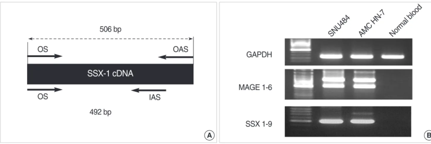
bind simultaneously to SSX 1-9 cDNAs were designed, and used for the SSX 1-9 assay. The binding site of common primers on SSX-1 gene and the products amplified by the SSX1-9 assay and the MAGE 1-6 assay are summarized in Fig. 1. All primers (OS, OAS, and IAS) had 100% homology with the sequences of the 9 SSX cDNAs, and were used for the SSX 1-9 assay. The tran- scripts of MAGE 1-6 genes and SSX 1-9 genes were detected in SNU484 and AMC HN-7 cell lines, but not in normal blood cells.
The expected DNA sizes amplified by the SSX 1-9 assay and the MAGE 1-6 assay were 490-492 bp and 448-472 bp, respectively.
Detection of the transcripts of SSX 1-9 genes in cancer cell lines
In order to evaluate the sensitivity of the SSX 1-9 assay, mRNA was isolated from 35 cancer cell lines described previously in this
paper, and transcripts of SSX 1-9 genes were detected by the SSX 1-9 assay. Thirty one (88.6%) of the 35 cancer cell lines were positive (Fig. 2). All 3 head and neck cancer cell lines were pos- itive in both gene assay (data not shown).
Detection of the transcripts of SSX 1-9 and MAGE 1-6 in cancer tissues
The transcripts of MAGE 1-6 and SSX 1-9 genes were detected in 24 (82.8%) and 22 (75.9%) of 29 fresh cancer tissues from head and neck cancer patients. Eighty six percent (18/21) and 72.7%
(16/21) of squamous cell carcinoma cells expressed MAGE 1-6 genes and SSX 1-9 genes, respectively. Sixty three percent (5/8) of other pathologic types of cancers expressed both MAGE 1-6 genes and SSX 1-9 genes (Fig. 3, Table 1).
Detection of the transcripts of SSX 1-9 and MAGE 1-6 in sputum samples
In order to investigate the abilityof MAGE 1-6 and SSX 1-9 assays to detect small numbers of cancer cells mixed in sputum, SNU484 (MAGE- and SSX-positive cell lines) were added to normal sputum. After digesting the sputum with specimen pro- tector, mRNA was isolated, and MAGE 1-6 and SSX 1-9 assays were performed. Both assays detected transcripts in samples Fig. 1. The binding sites of common primers on SSX-1 cDNA (A) and PCR products amplified by MAGE 1-6 and SSX 1-9 assay (B).
GAPDH OS
OS
OAS
IAS 506 bp
492 bp
SNU484 AMC HN-7 Normal blood
MAGE 1-6
SSX 1-9
B A
SSX-1 cDNA
Fig. 2. Detection of SSX 1-9 transcripts of cancer cell lines by SSX 1- 9 assay. M: Marker; 1: SNU 484; 2: SNU 620; 3: SNU 638; 4: SNU 668;
5: SNU C1; 6: SNU C4; 7: SNU C5; 8: HT29; 9: HCT 116; 10: AMC- HN3; 11: AMC-HN4; 12: AMC-HN7; 13: U937; 14: HL 60; 15: Caski;
16: C4-II; 17: ME-180; 18: Hela; 19: CUNC-6; 20: SiHa; 21: NCI-H292;
22; NCI-H522; 23: NCI-H1703; 24: A-549; 25: PC 3; 26: DU 145; 27:
HEPG2; 28: SNU 182; 29: SNU 354; 30: SNU 387; 31: SNU 398; 32:
SNU 423; 33: HEK 293; 34: MDA 231; 35: SAOS 2.
M 1 2 3 4 5 6 7 8 9 10 11 12
Stomach
M 13 14 15 16 17 18 19 20 21 22 23 24 Leukemia
M 25 26 27 28 29 30 31 32 33 34 35
Prostate Liver Kidney Breast Bone
Cervix Lung
Colon Head & Neck
Fig. 3. Detection of MAGE 1-6 and SSX 1-9 gene expressions in can- cer tissue of the patients with head and neck cancer by MAGE 1-6 assay and SSX 1-9 assay.
SSX 1-9
SSX 1-9 MAGE 1-6
MAGE 1-6
M 1 2 3 4 5 6 7 8 9 10 11 12 13 14 15
M 16 17 18 19 20 21 22 23 24 25 26 27 28 29
containing more than 20 SNU484 cells (Fig. 4).
Eighteen samples of induced sputum from head and neck can- cer patients and 22 samples of induced sputum from patients with benign lung disease were collected. After digestion of sputum with specimen protector and isolation of mRNA in liquefied sputum, MAGE 1-6 and SSX 1-9 assays were performed. The transcripts of MAGE 1-6 and SSX 1-9 genes were detected in 72.2% (13/18) and 77.8% (14/18) of fresh induced sputum samples from head and neck patients (Fig. 5, Table 2). The MAGE 1-6 assay detect- ed transcripts in 33.3% of sputum samples from TNM stage T1 tumors, 88.9% of T2 tumors, 75% of T3 tumors and 100% of T4 tumors. The SSX 1-9 assay detected transcripts in 100% of sputum samples from T1 tumors, 77.8% of T2 tumors, 75% of T3 tumors and 100% of T4 tumors (Table 3). In order to evalu- ate the specificity of both assays, transcripts of the MAGE 1-6 and SSX 1-9 assays in induced sputum samples from patients with benign lung disease were detected. The MAGE 1-6 assay was positive in 4.5% (1/22) and the SSX 1-9 assay was positive in 13.6% (3/22) of the samples (Fig. 6). These results represents MAGE 1-6 assay is more specific than SSX 1-9 asaay.
Combination of MAGE 1-6 and SSX 1-9 assay in the cancer tissue and induced sputa of head and neck cancer patients
In head and neck cancer patients, the MAGE 1-6 assay was posi- Fig. 4. Detection of the transcripts of MAGE 1-6 and SSX 1-9 genes in
sputum containing SNU484 by MAGE 1-6 assay and SSX 1-9 assay.
GAPDH
No. of cancer cell in normal sputum
MAGE 1-6
SSX 1-9
0 1 5 20 100
Fig. 5. Detection of MAGE 1-6 and SSX 1-9 gene expressions in sputum of the patients with head and neck cancer by MAGE 1-6 assay and SSX 1-9 assay.
MAGE 1-6
MAGE 1-6 SSX 1-9
SSX 1-9
M 1 2 3 4 5 6 7 8 9 10
M 11 12 13 14 15 16 17 18
Fig. 6. Representative results of MAGE 1-6 and SSX 1-9 expression in sputum of the patients with benign lung disease (n=22) by MAGE 1-6 or SSX 1-9 assay.
MAGE 1-6
SSX 1-9 GAPDH
1 2 3 4 5 6 7 8 9 10 11 12
MAGE: 4.5%, SSX: 13.6%
Pathology Primary site
No. of positive reaction (%)
MAGE SSX
Table 1. Analysis of the profile of MAGE and SSX gene expression in cancer tissue according to the pathology of head and neck cancer
Squamous cell carcinoma Larynx (9)* 9 (100) 7 (77.8) Nasopharynx (2) 0 (0) 2 (100) Oropharynx (4) 3 (75) 1 (25) Hypopharynx (2) 2 (100) 2 (100) Oral cavity (4) 4 (100) 4 (100) Verrucous carcinoma Larynx (1) 1 (100) 1 (100) Undifferentiated carcinoma Nasopharynx (1) 0 (0) 1 (100)
Lymphoma Lymphatics (2) 1 (50) 1 (50)
Thyroid ca Thyroid (3) 3 (100) 2 (66.7)
Adenoid cystic carcinoma Submandibular 1 (100) 1 (100) gland (1)
Total (n=29) 24 (82.8) 22 (75.9)
*No. of total cases tested. MAGE: melanoma antigen gene; SSX: synovial sarcoma on X chromosome.
Pathology Primary site
No. of positive reaction (%)
MAGE SSX
Table 2. Analysis of the profile of MAGE and SSX gene expression in sputum according to the pathology of head and neck cancer
*No. of total cases tested. MAGE: melanoma antigen gene; SSX: synovial sarcoma on X chromosome.
Squamous cell carcinoma Larynx (12)* 8 (66.7) 9 (75) Hypopharynx (1) 0 (0) 0 (0) Nasopharynx (1) 1 (100) 1 (100) Oropharynx (1) 1 (100) 1 (100) Adenocarcinoma Hypopharynx (1) 1 (100) 1 (100) Adenoid cystic carcinoma Oropharynx (1) 1 (100) 1 (100) Basaloid carcinoma Oropharynx (1) 1 (100) 1 (100) Total (n=18) 13 (72.2) 14 (77.8)
tive in 82.8% of cancer tissues and 72.2% of induced sputa. The SSX 1-9 assay was positive in 75.9% of cancer tissues and 77.8%
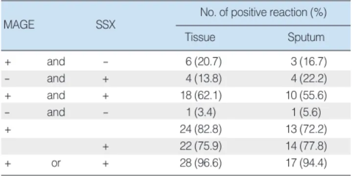
of induced sputa. When both MAGE 1-6 assay and SSX 1-9 assay were performed on the same specimen, 96.6% of cancer tissues and 94.4% of induced sputa were positive in either one of the two assays (Table 4).
DISCUSSION
Cancer antigens that have a high frequency of expression exclu- sively in cancer cells can be excellent markers for cancer diag- nosis. The expression of MAGE and SSX genes are highly spe- cific to cancer cells, but their use in the detection of a small num- ber of cancer cells by RT-PCR has been limited by the low expres- sion frequency of individual genes. If the transcripts of several cancer antigen genescould be detected simultaneously by RT-PCR, cancer detection rates could be increased. Two major methods have been described one is multiplex PCR, which combines sev- eral specific primers in the same reaction (12), and the other is PCR that uses common primers that simultaneously bind to mul- tiple targets. Whereas it is difficult to amplify more than 5 targets using multiplex PCR, multiple targets can be detected with few limitations by performing PCR using common primers. In a pre- vious study, we showed that RT-nested PCR using common pri- mers was a very useful method for detecting MAGE-expressing cancer cells (8).
Expression analysis has demonstrated that SSX is a member of the recently described cancer/testis antigen class. It is expressed in a variety of different human neoplasms, but not in normal tis- sues, with the exception of testis and a weak expression in the thyroid. The SSX family of genes consists of several subtypes Tureci et al. (13) studied the expression of SSX genes in 325 specimens of human neoplasms, and found that SSX-1, -2, and -4 subtypes are expressed in 8%, 15% and 15% of the cancers, respectively, while the expression of the SSX-5 or SSX-3 subtypes were rare or absent. Expression of at least one of the SSX family members was most frequently observed in head and neck cancer (75%), followed by ovarian cancer (50%), malignant melanoma (43%), lymphoma (36%), colorectal cancer (27%) and breast cancer (23%). These results suggest that simultaneous detection of tran-
scripts of multiple SSX genes by RT-nested PCR will be valuable in the diagnosis of SSX-expressing cancer. The known SSX fami- ly members share more than 90% homology at the DNA level.
In the present study, we designed new common primers and developed an SSX 1-9 assay that can detect cancer cells express- ing at least one of the 9 SSX genes. Our SSX 1-9 assay success- fully detected the transcripts of all SSX 1-9 genes in 31 out of 35 cancer cell lines.
In order to compare the sensitivity of MAGE 1-6 and SSX 1- 9 assays, and to evaluate the value of combining both assays in diagnosis, we detected the transcripts of MAGE and SSX genes in 29 cancer tissue samples of head and neck cancer patients.
The MAGE 1-6 and SSX 1-9 assays were positive in 82.8% and 75.9% of cancer tissues, and 96.6% of cancer tissues were posi- tive for at least one of the two assays. The SSX-positive rate in head and neck cancer tissues observed in the present study is higher than the rate reported by Tureci et al. (13). The results from the present study show that the SSX genes, as well as the MAGE genes, are expressed in head and neck cancer tissue, and that combination of the MAGE 1-6 assay and the SSX 1-9 assay can detect more than 94% of head and neck cancers.
RT-PCR-based amplification of transcripts that are expressed in cancer cells, but not in normal non-neoplastic cells, is increas- ingly being used as a sensitive diagnostic tool for detecting rare disseminated or exfoliated cancer cells to improve the accuracy of cancer staging and to aid in developing early detection pro- tocols. In particular, detection of transcripts of cancer markers in sputum may be very useful for cancer screening or early diagno- sis of cancer of the respiratory tracts. In this study, we evaluat- ed the sensitivites of MAGE 1-6 and SSX 1-9 assays in detecting cancer cells in induced sputum of head and neck cancer patients.
It has been reported that head and neck cancer can be diagnosed by sputum cytology (14, 15). The reports suggested that cancer cells from head and neck malignancies can be expelled from can- cer tissue and end up in sputum. Matsuda et al. (14) detected 29.4% of T1 lesions and 63.3% of T2 lesions of head and neck malignancies by sputum cytology. In the present study, 33.3%
of T1 lesions (N=3) and 88.9% of T2 lesions (N=9) were detect-
Cancer stage (N0)No. of positive reaction (%)
MAGE SSX
Table 3. Analysis of the profile of MAGE and SSX gene expression in sputum according to the cancer stage of head and neck cancer
MAGE: melanoma antigen gene; SSX: synovial sarcoma on X chromo- some.
T1 (3) 1 (33.3) 3 (100)
T2 (9) 8 (88.9) 7 (77.8)
T3 (4) 9 (75) 3 (75)
T4 (1) 1 (100) 1 (100)
MAGE SSX
No. of positive reaction (%)
Tissue Sputum
Table 4. Detection of MAGE and SSX gene expression by MAGE 1-6 or SSX 1-9 assay in patients with head and neck cancer
+ and - 6 (20.7) 3 (16.7)
- and + 4 (13.8) 4 (22.2)
+ and + 18 (62.1) 10 (55.6)
- and - 1 (3.4) 1 (5.6)
+ 24 (82.8) 13 (72.2)
+ 22 (75.9) 14 (77.8)
+ or + 28 (96.6) 17 (94.4)
MAGE: melanoma antigen gene; SSX: synovial sarcoma on X chromo- some.
ed by MAGE 1-6 assay, and 100% of T1 lesions (N=3) and 77.8
% of T2 lesions (N=9) were detected by the SSX 1-9 assay. All T1 and T2 lesions were detected by either one of the two assays, and there was no relationship between MAGE or SSX gene expres- sion and the tumor stage. These results suggest that it may be possible to detect small numbers of cancer cells that express at least one of MAGE 1-6 or SSX 1-9 genes in sputum or in malig- nant tissue by performing the MAGE 1-6 and the SSX 1-9 assays together. Thus the combination of these assays may become very useful for screening or early diagnosis of primary or recurrent head and neck cancers.
The MAGE 1-6 and SSX 1-9 gene transcripts were detected in 4.5% and 13.6% of sputum of patients with benign lung disease.
These false positive results may be due to the detection of dys- plastic cells in sputum. One possible explanation for the false positive amplification of MAGE or SSX transcripts is the activa- tion of MAGE genes by dysplastic cells during early tumorigen- esis (16). Another possible explanation is the low level of tran- scription of MAGE or SSX genes in normal cells (17, 18). We have been following up these patients at 3-6 month intervals some of patients were negative for both MAGE and SSX assays during the follow-up period, while others consistently tested positive. If the results show continuous positive on follow up test, it is need to do the screening work-up to rule out occult head and neck cancer.
CONCLUSION
We developed an SSX 1-9 assay to detect the transcripts and of 9 SSX genes simultaneously, and investigated the usefulness of SSX 1-9 MAGE 1-6 assays in the detection of cancer cells in tis- sue and sputum samples of head and neck cancer patients. The results of the present investigation suggest that the combination of MAGE 1-6 and SSX 1-9 assays may be a sensitive diagnostic tool in detecting small numbers of malignant cells in the cancer tissue and induced sputum of head and neck cancer patients.
REFERENCES
1. Bodey B. Cancer-testis antigens: promising targets for antigen directed antineoplastic immunotherapy. Expert Opin Biol Ther. 2002 Aug;2(6):
577-84.
2. Mashino K, Sadanaga N, Tanaka F, Yamaguchi H, Nagashima H, Inoue
H, et al. Expression of multiple cancer-testis antigen genes in gastroin- testinal and breast carcinomas. Br J Cancer. 2001 Sep 1;85(5):713-20.
3. Scanlan MJ, Gure AO, Jungbluth AA, Old LJ, Chen YT. Cancer/testis antigens: an expanding family of targets for cancer immunotherapy. Im- munol Rev. 2002 Oct;188:22-32.
4. Tajima K, Obata Y, Tamaki H, Yoshida M, Chen YT, Scanlan MJ, et al. Expression of cancer/testis (CT) antigens in lung cancer. Lung Cancer.
2003 Oct;42(1):23-33.
5. Chen CH, Huang GT, Lee HS, Yang PM, Yan MD, Chen DS, et al.
High frequency of expression of MAGE genes in human hepatocellu- lar carcinoma. Liver. 1999 Apr;19(2):110-4.
6. Eura M, Ogi K, Chikamatsu K, Lee KD, Nakano K, Masuyama K, et al.
Expression of the MAGE gene family in human head-and-neck squa- mous-cell carcinomas. Int J Cancer. 1995 Oct 20;64(5):304-8.
7. Otte M, Zafrakas M, Riethdorf L, Pichlmeier U, Loning T, Janicke F, et al. MAGE-A gene expression pattern in primary breast cancer. Cancer Res. 2001 Sep 15;61(18):6682-7.
8. Park JW, Kwon TK, Kim IH, Sohn SS, Kim YS, Kim CI, et al. A new strategy for the diagnosis of MAGE-expressing cancers. J Immunol Me- thods. 2002 Aug 1;266(1-2):79-86.
9. Clark J, Rocques PJ, Crew AJ, Gill S, Shipley J, ChanAM, et al. Iden- tification of novel genes, SYT and SSX, involved in the t(X;18)(p11.2;
q11.2) translocation found in human synovial sarcoma. Nat Genet. 1994 Aug;7(4):502-8.
10. Gure AO, Tureci O, Sahin U, Tsang S, Scanlan MJ, Jager E, et al. SSX:
a multigene family with several members transcribed in normal testis and human cancer. Int J Cancer. 1997 Sep 17;72(6):965-71.
11. Gure AO, Wei IJ, Old LJ, Chen YT. The SSX gene family: character- ization of 9 complete genes. Int J Cancer. 2002 Oct 10;101(5):448-53.
12. Morot-Bizot SC, Talon R, Leroy S. Development of a multiplex PCR for the identification of Staphylococcus genus and four staphylococcal species isolated from food. J Appl Microbiol. 2004;97(5):1087-94.
13. Tureci O, Chen YT, Sahin U, Gure AO, Zwick C, Villena C, et al. Ex- pression of SSX genes in human tumors. Int J Cancer. 1998 Jul 3;77 (1):19-23.
14. Matsuda M, Nagumo S, Horai T, Yoshino K. Cytologic diagnosis of laryngeal and hypopharyngeal squamous cell carcinoma in sputum. Acta Cytol. 1988 Sep-Oct;32(5):655-7.
15. Saito H, Ono I, Ebihara S, Yoshizumi T, Ikeda S, Ono R. [Cases of head and neck carcinoma detected by sputum cytology-lung cancer screen- ing and head and neck cancer]. Gan No Rinsho. 1985 Oct;31(13):1665- 8. Japanese.
16. Jang SJ, Soria JC, Wang L, Hassan KA, Morice RC, Walsh GL, et al.
Activation of melanoma antigen tumor antigens occurs early in lung carcinogenesis. Cancer Res. 2001 Nov 1;61(21):7959-63.
17. Bialkowska-Hobrzanska H, Bowles L, Bukala B, Joseph MG, Fletcher R, Razvi H. Comparison of human telomerase reverse transcriptase messenger RNA and telomerase activity as urine markers for diagno- sis of bladder carcinoma. Mol Diagn. 2000 Dec;5(4):267-77.
18. Piva MG, Navaglia F, Basso D, Fogar P, Roveroni G, Gallo N, et al.
CEA mRNA identification in peripheral blood is feasible for colorectal, but not for gastric or pancreatic cancer staging. Oncology. 2000 Nov;
59(4):323-8.
. .
. .
. .
. . . .
. . . .