40(2) : 150 154 (2009)
150
왜모자반 (Sargassum yezoense)에서 분리한 화합물의 α-glucosidase 및 산화스트레스 억제효과
이은하·함정엽·안홍열·김민철·김철영·판철호·엄병헌·정상훈∗
한국과학기술연구원 강릉분원
Inhibitory Effects of the Compounds Isolated from Sargassum yezoense on α-Glucosidase and Oxidative Stress
Eun Ha Lee, Jungyeob Ham, Hong Ryul Ahn, Min Chul Kim, Chul Young Kim, Cheol-Ho Pan, Byung Hun Um and Sang Hoon Jung
*Natural Products Research Center, Korea Institute of Science and Technology (KIST) Gangneung Institute, Gangneung 210-340, Korea
Abstract −
We examined ethanol extracts prepared from the Korean marine algae belonging to the Sargassaceae family for their inhibitory effects on
α-glucosidase activity and free radicals in vitro . Among five marine algae, the extracts of Sargassum yezoense were found to possess strongly
α-glucosidase inhibition and free radicals scavenging activities. Two compounds were isolated via bioactivity guided isolation and tested for their effects on
α-glucosidase, DPPH, ABTS
+and Photochem® analysis.
Their chemical structures were elucidated by spectral analysis and direct comparison with authentic compounds; their structures were identified as sargaquinoic acid (
1) and sargahydroquinoic acid (
2). The inhibitory effects of compound
1and
2(IC
50value:
14.2 and 12.8
µM, respectively) on
α-glucosidase were more potent that of deoxynojirimycin as a positive control (IC
50value:
18.0
µM). All compounds displayed antioxidative activity which was measured by DPPH, ABTS
+and Photoche
®apparatus.
Key words −
Sargassum yezoense , sargaquinoic acid, sargahydroquinoic acid,
α-glucosidase, antioxidative activities
당뇨병치료제를개발하기위하여표적으로삼고있는작 용기전은당질화합물의소화및흡수억제
,
췌장에작용하여인슐린분비촉진
,
간장에작용하여포도당대사조절,
인슐린감응성증강또는인슐린저항성개선
,
중추신경계에작용등으로연구가이루어지고있다
. 1, 2)
당질화합물의소화에관여하는효소인 α
-glucosidase
의억제제로는대표적으로
acarbose
와같은화합물이있으며,
당질화합물이섭취될때
,
소장의 α-glucosidase
를억제하여,
고분자당화합물의분해를감소시켜
,
당질화합물섭취후급격히 증가하는 혈당을억제하게 된다
. 3) Guar gum
이나pectin
과같은가용성섬유를이용하여소장의당흡수를방해하여완만한혈당강하를하기도한다
.
왜모자반은모자반과에속하는해조류중갈조류의일종 으로서
,
이런갈조류들에서독특한plastoquinones, merodi- terpenoids
과phlorotannins
등의물질들이보고된바있다. 4-7)
또한 왜모자반에서 분리된
plastoquinones
화합물인sargaquinoic acid
와sargahydroquinoic acid
는3T3-L1
세포에서
PPAR
α/
γ를활성화시킴으로써, adipocyte
의분화를촉진한다는연구결과도보고된바있다
. 8)
연구자등은최근에당뇨병및합병증의저해에유효한 해조류를탐색하였으며
,
그성분을규명하는연구를진행해왔다
.
이일환으로왜모자반의에탄올추출물에대해α-
glucosidase
억제효과및산화스트레스저해효과를측정하고이를보고하고자한다
.
또한생리활성물질추적의결과로분리된
2
종의화합물의구조에대해서도보고하고자한다. 재료 및 방법
실험재료 − 본 실험에 사용한 갈조류인
Sargassum thunbergii , S. horneri , S. confusum , S. yezoense
그리고S.
miyabei
는동해안에서직접채취하였으며, 1
주일동안건조후
95%
에탄올로추출하여사용하였다.
*교신저자(E-mail):[email protected]
(Tel):82-33-650-7203
시약 및 기기 −
NMR spectra
는Varian (500 MHz)
의program
을사용하였으며,
내부표준물질로는tetramethylsilane (TMS)
를 사용하였고, chemical shift value
는part per million (ppm)
단위로사용하였다. Thin layer chromatography
용
plate
는precoated silica gel 60 F 254 (Merck)
를사용하였다
.
분취용컬럼크로마토그래피는순상실리카겔분취-
중압액체크로마토그래피
(normal phase silica gel preparative middle pressure liquid chromatography, MPLC, CombiFlash
Companion)
를사용하였으며,
분취용컬럼크로마토그래피의
packing material
로는Silica gel 60 (
입자크기35-70
µM)
을사용하였다
.
α-glucosidase, 2,2-diphenyl-1-picrylhydrazyl (DPPH) radical, p-nitrophenyl
α-D-glucopyranoside, 2,2’- aziono-bis(3-ethylbenzthiazoline-6-sulfonic acid) radical (ABTS + ),
그리고deoxynojirimycin
은Sigma (St Louis, MO,
미국)
에서구입하여사용하였다.
그밖의시약은특급및
HPLC
용등급을사용하였다.
크로마토그래피용용매는J.T. Baker
사의HPLC
등급용용매를사용하였고,
증류수는3
차증류수를여과하여사용하였으며,
그외에시료추출을위한용매는특급시약을사용하였다
.
추출 및분리 − 모든시료는건조하여
95%
에탄올로추출후
,
감압건조하여활성측정에사용하였다.
물질분리를위하여
,
왜모자반건조중량187 g
을분쇄하고,
아세톤에냉침한후
,
감압건조하여23 g
의추출물을얻었다.
추출물1 g
을증류수에현탁하여, ethyl acetate (EtOAc)
로분획하고
,
감압 농축하여622 mg
의 분획물을얻었다.
이를n - hexane
과EtOAc
의혼합액을사용하여,
극성에따른실리카겔컬럼크로마토그래피를실시하여
10
개의소분획을얻었고
,
박층크로마토그래피를통하여비슷한양상을보이는분획인분획
2
와분획3
을모아, preparative
박층크로마토그래피를
n-hexane/EtOAc (15:1)
을3
회실시하여 화합물1(24.9 mg)
과화합물 2(114.5 mg)
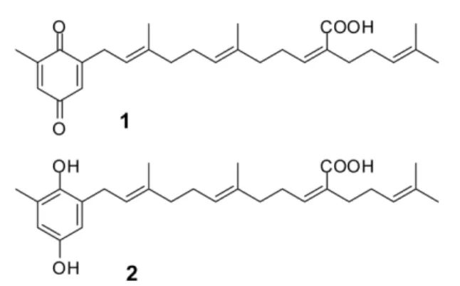
을분리하였다(Fig. 1).
화합물 1 (Sargaquinoic acid):
yellowish oil; 1 H NMR (500 MHz, CDCl 3 )
δ: 6.55 (1H, m), 6.46 (1H, dd, J = 2.0, 0.5 Hz), 6.00 (1H, t, J = 7.0 Hz), 5.17-5.08 (3H, m), 3.13 (2H, d, J = 7.5 Hz), 2.59 (2H, q, J = 7.5 Hz), 2.27 (2H, t, J = 7.0 Hz), 2.15-2.04 (8H, m), 2.06 (3H, s), 1.68 (3H, s), 1.63 (3H, s), 1.61 (3H, s), 1.59 (3H, s); 13 C NMR (125 MHz, CDCl 3 )
δ: 188.3, 188.2, 172.5, 148.7, 146.1, 145.5, 140.0, 134.8, 133.4, 132.5, 132.4, 130.7, 124.7, 124.0, 123.6, 118.2, 39.8, 39.2, 34.8, 28.4, 28.1, 27.7, 26.5, 25.9, 17.9, 16.4, 16.3, 16.2.; FT-IR (neat) v max 3436, 2924, 1683, 1654, 1441, 1292 cm -1 ; HRESIMS m / z 424.2611 (calcd for C 27 H 36 O 4 424.2614).
화합물 2 (Sargahydroquinoic acid):
yellowish oil; 1 H NMR (500 MHz, CDCl 3 )
δ: 6.49 (1H, d, J = 2.5 Hz), 6.46 (1H, d, J = 2.5 Hz), 5.99 (1H, t, J = 7.0 Hz), 5.27
(1H, t, J = 7.0 Hz), 5.12-5.07 (2H, m), 3.28 (2H, d, J = 7.0 Hz), 2.58 (2H, q, J = 7.5 Hz), 2.26 (2H, t, J = 7.0 Hz), 2.18 (3H, s), 2.14-2.05 (8H, m), 1.75 (3H, m), 1.67 (3H, s), 1.59 (3H, m), 1.58 (3H, s); 13 C NMR (125 MHz, CDCl 3 )
δ: 172.9, 148.9, 146.6, 145.8, 138.5, 134.9, 132.5, 130.7, 127.8, 125.7, 124.5, 123.6, 121.9, 115.6, 114.2, 39.7, 39.2, 34.7, 30.2, 28.5, 28.1, 26.3, 25.9, 17.9, 16.4, 16.3, 16.2.; FT-IR (neat) v max 3434, 2921, 1682, 1471, 1194 cm -1 ; HRESIMS m / z 426.2768 (calcd for C 27 H 38 O 4
426.2770).
α
-Glucosidase
저해활성 − α-Glucosidase
저해활성은기존에보고된방법을일부수정하여사용하였다
. 9)
추출물및화합물
50
µl
를0.3U/ml
α-glucosidase
효소액50
µl, 200 mM phosphate buffer (pH 7.0) 50
µl
와혼합하여37 o C
에서
15
분간 예비 배양 한 후3 mM p -nitrophenyl
α-D- glucopyranoside 100
µl
를가하여37 o C
에서10
분간반응시켰다
. 0.1 M Na 2 CO 3 750
µl
로 반응을정지시키고, 405 nm
에서흡광도를측정하였으며,
대응하는 α-glucosidase
의IC 50
값을계산하였다.
DPPH
자유라디칼소거활성 −DPPH
자유라디칼소거활성은기존에보고된방법을일부수정하여사용하였다
. 10)
추출물및화합물의검체를적당한농도로추출용매에희석 한용액
10
µl
와100
µM DPPH
용액190
µl
를균일하게혼합한다음
,
실온에서30
분간방치한후, spectrophotometer (Bio-TEk instruments, VT, USA)
로517 nm
에서흡광도를측정하였다
.
항산화효과는대조군에대한50%
흡광도의감소를나타내는검체의농도
(IC 50 )
로표시하였다.
각시료를
3
회반복실시하여평균하였다.
ABTS+
자유라디칼소거활성 −ABTS +
자유라디칼소거활성은기존의방법을일부수정하여사용하였다
. 11) 7 mM
Fig. 1.
Chemical structures of isolated compounds from S.
yezoense.
All compounds identified by NMR, IR and Mass spectrometry,
and direct comparison with authentic compounds. Their
chemical structures were sargaquinoic acid (
1) and sargahy-
droquinoic acid (
2).
ABTS + 5 ml
와140 mM potassium persulfate 88
µl
를섞은후
,
상온에서16
시간동안빛을차단한상태에서보관하였다
.
이용액을에탄올에1:44
의v/v
비율로묽힌후,
이묽힌용액
190
µl
와해조류추출물및화합물10
µl
와섞은후상온에서
6
분간반응시키고734nm
에서흡광도를측정하였다
. ABTS +
의 억제효과는Inhibition (%) = (Control O.D.-Sample O.D.)/Control O.D. × 100
으로측정하였으며,
대조군에대한
50%
흡광도의감소를나타내는검체의농도
(IC 50 )
로표시하였다.
각시료를3
회반복실시하여평균하였다
Trolox equivalent assay (Photochem
®analysis)
−항산화 활성을
Trolox
와 정량적으로 비교하기 위하여photochemiluminescence
방법으로항산화활성을측정하였다
. 12) ACL (Analitik Jena AG)
이luminol
로써photosensitizer
의기능과라디칼을검출하는두가지의기능을가지고있으 며
,
실험은manufacturer’s instruction
에기술된방법에따라수행하였다
.
항산화활성은Trolox equivalents
로나타내었다. 결과 및 고찰
본연구는동해안에서채집한
5
종의Sargassaceae family
의해조류로부터 α
-glucosidase
억제활성및항산화소재를찾고궁극적으로당뇨병을억제할수있는소재를찾는데 있다
.
당뇨병환자의소장내 α
-glucosidase
는정상인에비해활성이높아져있기때문에
,
음식물의열량을생산하는가장주된영양소인탄수화물의섭취시식후혈당이큰폭으 로상승하게마련이다
.
이런α-glucosidase
억제제는소장의brush border
에존재하는이당류분해효소를가역적으로억제하여장에서탄수화물흡수를지연시켜
,
식후혈당을감소시키고
,
인슐린비의존성당뇨병의고혈당으로인한인슐린분비지연의개선에효과적이다
. 2)
임상에서acarbose,
voglibose
등이사용되고있다.
이런α
-glucosidase
억제제는인슐린분비를통하지않고,
소장에서의탄수화물소화및흡수를저해함으로써기존약 물들이갖고있는저혈당현상
,
간독성유발,
베타세포기능저하등의부작용을최소화할수있는장점을가지고 있다
.
본연구에서는
5
종의Sargassaceae family
해조류를동해안에서채집하였고
,
그에탄올추출물에대해 α-glucosidase
억제활성을 측정하였다
. 5
종의 해조류 중 왜모자반( S.
yezoense )
이IC 50
값3.5
µg/ml
로가장우수한효능을나타내었다
(Table I).
고혈당과관련된많은생화학적경로들
(
포도당의자가산화
, polyol
경로,
단백질당화등)
에의해자유라디칼의생성이증가됨이알려졌고
,
그밖에도당뇨병에서는여러인자들에의해산화스트레스및조직의산화적손상이증가 될수있다
. 13)
당뇨병에서혈장,
적혈구막,
저밀도지단백등의지질과산화가증가됨이밝혀졌다
. 14)
이런산화스트레스를막을수있는기전은자유라디칼의직접적인소거
,
지질과산화의억제혹은생체내항산화효소계
(superoxide dismutase, catalase, glutathione peroxidase)
를활성화하는Table I.
Scientific name, family, and the inhibitory effects of marine algae on
α-glucosidase
Scientific name Family Inhibition (%) IC
50(
µg/ml) S. thunbergii Sargassaceae <5
a)>100 S. horneri Sargassaceae <5
a)>100 S. confusum Sargassaceae 75.9 19.0 S. yezoense Sargassaceae 84.5 13.5
S. miyabei Sargassaceae 84.1 13.3
a)
The inhibitory effect of marine algae on α -glucosidase was measured at 100 µ g/ml.
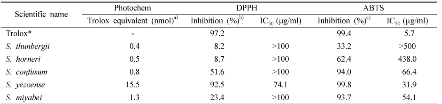
Table II.
Antioxidative effects of marine algae extracts on DPPH and ABTS
Scientific name Photochem DPPH ABTS
Trolox equivalent (nmol)
a)Inhibition (%)
b)IC
50(
µg/ml) Inhibition (%)
c)IC
50(
µg/ml)
Trolox* - 97.2 99.4 15.7
S. thunbergii 10.4 18.2 >100 33.2 >500
S. horneri 10.5 18.7 >100 62.4 438.0
S. confusum 10.8 51.6 >100 94.0 166.4
S. yezoense 15.5 92.5 74.1 99.8 131.9
S. miyabei 11.3 23.4 >100 93.7 154.1
*Trolox was used as positive control.
a)
The antioxidative capacity of marine algae was measured at 0.4 µ g/ml.
b)
The radical scavenging activities of marine algae on DPPH were measured at 100
µg/ml.
c)
The radical scavenging activities of marine algae on ABTS were measured at 500
µg/ml.
방법으로나눌수있다
.
본연구에서는항산화활성을측정하기위하여
, DPPH
및ABTS +
라디칼소거능활성을측정하였으며
, trolox
와의항산화도를비교하기위하여Photochem
®(Analitik Jena AG)
를사용하였다. 5
가지의해조류추출물중에
,
왜모자반이가장높은자유라디칼소거효과를나타내었으며
, DPPH
라디칼과ABTS
라디칼의IC 50
값은각각74.1
과
31.9
µg/ml
이었다. Trolox
와의동등성을Photochem
®으로 측정한결과 왜모자반 추출물
0.4
µg/ml
이Trolox
의15.5 nmol
과동등한것으로나타났다(Table II).
왜모자반이 α
-glucosidase
억제활성및산화스트레스저해작용이가장우수하여
,
활성물질분리를시도하였다.
추출물을실리카겔컬럼크로마토그래피및
preparative
박층크로마토그래피를 통하여
2
개의화합물을분리하였으며, NMR, IR
및Mass spectrometry
를 사용하여 화합물 1을sargaquinoic acid,
화합물 2를sargahydroquinoic acid
로구조동정하였다
.
두가지화합물에대하여상기의활성을측정하였다
.
화합물 1과화합물 2의α
-glucosidase
억제활성IC 50
은각 각14.2
와12.8
µM
로대조군으로사용한deoxynojirimycin
의
IC 50
값인18.0
보다높은저해활성을나타내었다(Table III).
화합물 1과화합물 2의자유라디칼소거효과를측정하 여본결과
, DPPH
라디칼의IC 50
값은각각51.9
과45.5
µM
로대조군의
IC 50
값인27.2
µM
보다약2
배정도의낮은활성이었다
. ABTS
라디칼의IC 50
값은각각78.4
와44.7
µM
로대조군의
IC 50
값인40.7
µM
보다약2
배정도의낮은활성이거나
,
화합물2의경우비슷한활성을나타내었다(Table IV).
Trolox
와의동등성을Photochem
으로측정한결과화합물1과 2의
0.5
µM
농도에서Trolox
의각각7.5
와6.1 nmol
와동등한것으로나타났다
(Table IV).
결 론
본연구를통해서동해안의
5
종의Sargassaceae family
해조류에대해 α
-glucosidase
억제활성및항산화활성을측정하였으며
, Sargassum yezoense
가가장우수한효과를 나 타내는 것을 확인하였다.
활성물질 분리를 시도하여sargaquinoic acid (
1)
와sargahydroquinoic acid (
2)
를분리하였고
,
그물질들에대하여 α-glucosidase
억제활성및항산화활성을측정하여본결과
,
두화합물모두강력한α- glucosidase
억제활성을나타내었고,
항산화활성은대조군인
trolox
에는미치지못하지만,
우수한항산화효과를갖는것으로확인하였다
.
따라서Sargassum yezoense
에서분리한 두가지화합물은향후당뇨병관련연구에유효한화합물 로사료되며,
앞으로그활성에대한작용기전과,
당뇨병관련다른인자에대한억제활성연구가지속적으로필요할 것이라사료된다
.
사 사
본연구는지식경제부지방기술혁신사업
(RTI05-01-02)
지원으로수행되었음
.
인용문헌
1. Moller, D. E. (2001) New drug targets for type 2 diabetes and the metabolic syndrome. Nature
414: 821-827.
2.
임중인. (2003)
당뇨병치료제의최근연구동향.
보건산업기3. Lebovitz, H. E. (1992) Oral antidiabetic agents. The emer-
술동향gence of
α-glucosidase inhibitors. Drugs
44 Suppl 3: 21-28.
4. Nakai, M., Kageyama, N., Nakahara, K. and Miki, W. (2006) Phlorotannins as radical scavengers from the extract of Sar- gassum ringgoldianum . Mar. Biotechnol. (NY)
8: 409-414.
5. Reddy, P. and Urban, S. (2009) Meroditerpenoids from the southern Australian marine brown alga Sargassum fallax . Phytochemistry
70: 250-255.
Table III
.
α-Glucosidase inhibitory effects of the compounds isolated from S. yezoense
Sample Inhibition (%)
a)IC
50(
µM)
1 79.1 14.2
2 84.6 12.8
Deoxynojirimycin* 78.8 18.0
*Deoxynojirimycin was used as positive control.
a)
Tested compounds from S. yezoense was measured at 25 µ M.
Table IV.
Antioxidative effects of marine algae extracts on DPPH and ABTS
Scientific name
Photochem DPPH ABTS
Trolox equivalent
(nmol)
a)Inhibition (%)
b)IC
50(
µM) Inhibition (%)
c)IC
50(
µM)
Trolox* - 93.8 27.2 198.2 40.7
1 7.5 83.9 51.9 76.72 78.4
2 6.1 87.5 45.5 89.68 44.7
*Trolox was used as positive control.
a)
The antioxidative capacity of marine algae was measured at 0.5 µ M.
b)
The radical scavenging activities of marine algae on DPPH were measured at 250 µ M.
c)
The radical scavenging activities of marine algae on ABTS
were measured at 500 µ M.
6. Jung, M., Jang, K. H., Kim, B., Lee, B. H., Choi, B. W., Oh, K. B. and Shin, J. (2008) Meroditerpenoids from the brown alga Sargassum siliquastrum . J. Nat. Prod.
71: 1714-1719.
7. Seo, Y., Park, K. E., Kim, Y. A., Lee, H. J., Yoo, J. S., Ahn, J. W. and Lee, B. J. (2006) Isolation of tetraprenyltoluquinols from the brown alga Sargassum thunbergii . Chem. Pharm.
Bull. (Tokyo)
54: 1730-1733.
8. Kim, S. N., Choi, H. Y., Lee, W., Park, G. M., Shin, W. S. and Kim, Y. K. (2008) Sargaquinoic acid and sargahydroquinoic acid from Sargassum yezoense stimulate adipocyte differ- entiation through PPARalpha/gamma activation in 3T3-L1 cells. FEBS Lett.
582: 3465-3472.
9. Lam, S. H., Chen, J. M., Kang, C. J., Chen, C. H. and Lee, S. S. (2008) alpha-Glucosidase inhibitors from the seeds of Syagrus romanzoffiana . Phytochemistry
69: 1173-1178.
10. Cao, G., Sofic, E. and Prior, R. L. (1997) Antioxidant and prooxidant behavior of flavonoids: structure-activity rela-
tionships. Free Radic. Biol. Med.
22: 749-760.
11. Re, R., Pellegrini, N., Proteggente, A., Pannala, A., Yang, M.
and Rice-Evans, C. (1999) Antioxidant activity applying an improved ABTS radical cation decolorization assay. Free Radic. Biol. Med.
26: 1231-1237.
12. Lee, E. H., Song, D. G., Lee, J. Y., Pan, C. H., Um, B. H. and Jung, S. H. (2008) Inhibitory effect of the compounds iso- lated from Rhus verniciflua on aldose reductase and advanced glycation endproducts. Biol. Pharm. Bull.
31: 1626-1630.
13. Baynes, J. W. and Thorpe, S. R. (1999) Role of oxidative stress in diabetic complications: a new perspective on an old paradigm. Diabetes
48: 1-9.
14. Sato, Y., Hotta, N., Sakamoto, N., Matsuoka, S., Ohishi, N.
and Yagi, K. (1979) Lipid peroxide level in plasma of dia- betic patients. Biochem. Med.
21: 104-107.
(2009년 6월 11일 접수)