Antioxidant Activity and Alpha-Glucosidase Inhibitory Activity of Stings of Gleditsia sinensis Extracts
Jeung-Min Lee
1, Jae-Hee Park
2, Won Mi Chu
2, Yi-Mook Yoon
1, Eunju Park
2and Hae-Ryong Park
1*
1
Department of Food Science and Biotechnology, Kyungnam University, Changwon 631-701, Korea
2
Department of Food and Nutrition, Kyungnam University, Changwon 631-701, Korea
Received September 20, 2010 /Accepted October 18, 2010This study was performed to investigate the physiological activities of stings of Gleditsia sinensis extracts. Antioxidant activity was evaluated by measuring total phenolic contents (TPC), comet assay, and 2.2-diphenyl-1-picrylhydrazyl (DPPH) radical scavenging activity (RSA). Anti-diabetic activity was measured by inhibition activities on α-glucosidase. Stings of Gleditsia sinensis extracts were prepared by extracting them with methanol and ethanol. The methanolic extracts showed the highest phenol content (1.12 g/100 g gallic acid equivalents). The α-glucosidase inhibitory activity of methanol ex- tracts were 17.9% higher, and that of ethanol extracts were 10.3% higher at a concentration of 1 mg/ml. These results indicate that stings of Gleditsia sinensis might be potential candidates as anti- oxidant and anti-diabetic agents.
Key words : Stings of Gleditsia sinensis, antioxidant, α-glucosidase, anti-diabetes
*Corresponding author
*Tel:+82-55-249-2689, Fax:+82-55-249-2995
*E-mail : [email protected]
서 론
경제가 발전함에 따라 생활수준의 향상으로 인하여 평균수 명이 증가함으로써 각종 퇴행성 질환에 대한 연구가 활발하게 진행되고 있다[4]. 여러 종류의 퇴행성 질환 중에서 노화는 아 직 확실한 치료제가 존재하지 않지만, 노화를 지연시켜주는 물질을 찾고자 하는 연구가 진행되고 있으며, 여러 가지 관련 원인을 찾고 그것을 해결하고자 하였다[17]. 그 중 활성산소는 세포 내에서 대사하는 과정에서 생성이 되는 산소로 세포막을 파괴하거나, 유전정보를 가진 DNA를 변형시켜 돌연변이를 일으키거나, 단백질을 변성시켜 신호전달 체계를 무너뜨리는 등 각종 질환의 원인이라고 할 수 있다[10]. 이런 활성산소에 대한 우리 몸의 방어 기전으로는 glutathione S-transferase, glutathione peroxidase, superoxide dismutase 등 여러 가지 효소가 작용을 하고 있지만[1], 발생되는 활성산소의 양을 전 부 제거하기에는 부족하여 여러 가지 질병의 원인이 되고 있 다[16]. 여러 질병 중 하나인 당뇨병은 요즘 급격하게 증가하는 추세이고 여러 종류의 합병증을 유발시키며, 그 발병시기가 점점 빨라지고 있는 추세이다[25]. 당뇨병은 혈관에 존재하는 당의 수준을 췌장에서 분비되는 인슐린으로 인해 조절이 되어 야 하지만, 췌장의 인슐린 분비의 이상으로 당이 급격하게 증 가되어 여러 종류의 합병증을 일으키는 질병으로 주로 인슐린 비의존성 당뇨병인 제2형 당뇨병이 대부분을 차지하고 있다 [12]. 당뇨병의 원인으로는 유전적인 원인이 강하며, 스트레스,
과로, 비만, 잦은 출산 등이 있으며[2], 치료제로는 여러 종류의 target을 이용하여 치료제가 개발되어 있지만[7], 각 종 부작용 으로 인해 치료가 더 위험해질 수 있다는 단점을 가지고 있다 [21].
본 연구에서는 당뇨병의 여러 가지 기전 중 소장의 소화효 소를 억제하여 분해되는 단당류의 양을 감소 시켜 소장에서 흡수되는 당이 감소함으로써 혈관 내 급격한 당의 증가를 막 아줄 수 있는 α-glucosidase의 활성을 억제하는 물질을 찾고자 하였다[11]. 현재 알려져 있는 α-glucosidase 억제제로는 acar- bose, voglibose, miglitol이 있으며, 그 외로는 천연물 유래의 추출물이나 미생물이 생산하는 대사산물을 이용하여 건강기 능성 식품형식으로 주로 생산되고 있다. 이러한 α-glucosidase 억제제의 부작용으로는 효소의 활성을 저해함으로 분해되지 못한 이당류가 대장에 이동되어 대장 속에 존재하는 박테리아 로 인해 분해되어 가스, 설사, 변비 등 배설에 관련된 질환을 야기할 수 있다[18]. 또한 효소 억제율이 높을 경우 흡수되는 단당류의 양의 감소로 저혈당으로 인한 쇼크가 나타날 수 있 으므로 당을 조절할 수 있는 억제율에 대한 정보는 현재 치료 제로 쓰이는 약물을 비교 대상으로 이용하여 연구가 진행되고 있으며[6], 이러한 점을 보완하기 위해 다른 천연물을 병용 처 리하는 방법으로 부작용을 감소시키고자 연구가 활발히 진행 되고 있다[19].
본 연구팀은 국내, 외에서 자생하고 있는 약용 식물의 추출
물을 대상으로 항산화 활성을 가지고 있으며 당뇨병 치료제로
써 가능성이 있는 물질을 찾고자 하였으며, 그 결과 조각자
추출물로부터 항산화 활성과 항당뇨 효과가 있다는 것을 확인
하였다. 조각자는 조각자 나무(Gleditsia sinensis LAM.)의 가시
부분으로써 콩과에 속하는 낙엽교목이며, 곳곳에 편평한 가시 가 존재하며, 잎은 어긋나고 1~2회 깃꼴 겹이다. 꽃은 잡성 암수한그루로 6월에 지름 6 mm 정도의 연한 노란색으로 피 며, 꽃받침 잎과 꽃잎은 각각 5개, 수술은 9~10개이다. 꼬투리 는 꼬이며, 길이는 5~15 cm로 기부의 지름이 8~12 mm이고 끝은 뾰족하다. 갈라진 가시의 길이는 15~70 mm 정도로 때 로는 갈라져 작은 가시로 되어있다. 바깥 면은 적자색~적갈 색이며 가시 끝부분은 적갈색으로 광택이 있으며 가는 주름이 있다. 전국 산기슭, 개울가에서 자라며, 봄철에 어린잎을 뜯어 서 햇볕에 말리며, 열매를 조협이라고 하여 가을에 여문열매 를 따서 건조 시킨다[26]. 특히, 가시는 조각자라고 하여 시기 에 상관없이 채취가 가능하며, 외용약으로 쓸 때는 가루를 내 어서 뿌리거나 기초제에 개어 바른다. 약리 실험결과 평활근 진경작용, 혈압강하작용, 호흡중추에 대한 흥분작용이 밝혀졌 다. 조각자에 대한 활성으로는 항암효과, 항균효과, 미백효과, 항HIV효과 등이 보고되어 있지만[14,15], 항당뇨 효과에 대한 활성은 보고되어 있지 않다.
따라서, 본 연구팀은 조각자 추출물로부터 항산화 활성과 항당뇨 효과에 대해 밝히고자 하였다.
재료 및 방법
시약 및 실험재료
본 실험에서 사용한 조각자(stings of Gleditsia sinensis)는 2010년 3월 경남 마산시 (주)금강제약으로부터 제공을 받아 실험에 사용하였으며, 항산화 활성 실험을 하기 위해 사용한 시약으로 Folin-Ciocalteau`s phenol, 2.2-diphenyl-1-picrylhy- drazyl, hydrogen peroxide, Histopaque 1077, low melting point agaroses, normal melting point agarose Triton X-100, disodium salt ethylenediaminetetraacetic acid, Tris-buffer, sodium chloride, sodium hydroxide, ethidium bromide, po- tassium chloride, potassium phosphate, sodium hydrogen phosphate, sodium, Na
2CO
3등은 Sigma Chemical Co. (St Louis, MO, USA)에서 구입 하였다. 항당뇨 효과를 확인하기 위해 α-glucosidase 효소로 사용한 rat intestinal acetone pow- der 시약은 Sigma Chemical Co. (St Louis, MO, USA)에서 구입하였고, glucose kit는 Bioclinical system에서 구입하였다.
그 외 연구에 사용된 여러 종류의 용매 및 시약은 모두 일급 이상의 등급을 사용하였다.
시료의 추출
조각자 5 g을 100 ml의 methanol 또는 ethanol 용매에 첨가 하여 상온에서 3일간 정치시켜 추출한 다음 여과지(Advantec, Tokyo, Japan)를 이용하여 여과를 한 후, 회전감압농축기 (EYELA, Tokyo, Japan)를 이용하여 40
oC에서 감압 농축을 하 였다.
Methanol 추출물은 GSM 그리고 ethanol 추출물은 GSE 이라 명명을 하였다. 각각의 추출물은 항산화 활성 및 항당 뇨 효과 실험을 위하여 DMSO와 methanol에 녹여 사용하 였다.
총 페놀 함량
총 페놀의 함량은 Singleton과 Rossi의 방법[22]을 실험에 맞게 변형하여 조각자 추출물인 GSM과 GSE 200 μl에 1 N-Folin-Ciocalteau reagent 200 μl를 첨가하여 3분 동안 실온 에 방치한 후 10% Na
2CO
3200 μl를 첨가 하여 암실에 1시간 방치한 후 13,400× g에서 5분 동안 원심분리 후 반응액을 690 nm에서 흡광도를 측정하였다. 표준검정곡선은 gallic acid를 사용하여 나타냈으며, 총 페놀의 함량은 시료 100 g을 추출 용매에 녹여 나오는 페놀의 함량을 구하였다.
DPPH 라디칼 소거능 측정
조각자 추출물인 GSM과 GSE의 DPPH 라디칼 소거능 (radical scavenging activity, RSA)을 알아보기 위해 Thitilertdecha등의 방법[23]을 실험에 맞게 변형하여 측정을 하였다. 추출한 시료 20 μl에 0.2 mM DPPH 용액 80 μl를 첨가 하여 10초 동안 섞은 후 실온에서 10분간 방치 후 492 nm에서 흡광도를 측정하였다.
DNA 손상 측정(Comet assay)
신선한 전혈 5 ml을 Histopaque 1077를 이용해 백혈구만 을 분리해 낸 후 본 실험에 사용하였다. 2종류의 조각자 추출 물 GSM과 GSE를 각각 1, 5, 10, 50 μg/ml의 농도로 백혈구 에 처리한 후 37
oC에서 30분간 반응시킨 후 백혈구에 산화적 손상을 주기 위해 200 μM의 hydrogen peroxide (H
2O
2)를 4
oC에서 5분간 처리 후 PBS로 세척하였다. Negative control 은 백혈구에 PBS만을 처리하였으며, positive control은 추출 물 대신 PBS를 사용하여 반응 시킨 후 200 μM H
2O
2를 처리 하였다. Comet assay를 위해 반응을 끝낸 백혈구를 75 μl의 0.7% low melting agarose gel (LMA)과 섞은 후, 1.0% nor- mal melting agarose (NMA)가 미리 코팅된 슬라이드위로 세포현탁액과 LMA의 현탁액이 골고루 분산되게 한 후 커버 글래스로 덮어 4
oC 냉장고에 보관하였다. 젤이 굳으면 커버 글래스를 벗기고 그 위에 다시 0.7% LMA 용액 75 μl로 한겹 더 덮었다. 미리 준비해 둔 차가운 alkali lysis buffer (2.5 M NaCl, 100 mM Na
2EDTA, 10 mM tris)에 사용 직전에 1%
Triton X-100을 섞은 후 슬라이드를 담가 저온, 암실에서 1시 간 동안 침지시켜 DNA의 double strand를 풀어주었다.
Lysis가 끝난 후, 슬라이드를 전기영동 수조에 배열하고 4
oC
의 차가운 전기영동 buffer (300 mM NaOH, 10 mM
Na
2EDTA, pH>13)를 채워 40분 동안 unwinding 시켜 DNA
의 alkali labile sites가 드러나게 한 후 25 V/300±3 mA의 전
압을 걸어 20분간 전기영동을 실시하였다. 빛에 의해 DNA 가 부가적으로 손상되는 것을 방지하기 위해 위의 과정은 전 기영동 수조를 어두운 천으로 덮은 채 실시하였다. 전기영동 이 끝난 후 0.4 M Tris buffer (pH 7.4)에 5분씩 담가 세척하 는 과정을 3회 반복하여 슬라이드를 건조시켰다. 20 μg/ml 농도의 ethidium bromide로 핵을 염색하여 커버글래스로 덮 은 뒤 형광현미경(Leica, Wetzlar, Germany) 상에서 관찰하 였다. CCD camera (Nikon, Tokyo, Japan)를 통해 보내진 각 각의 세포핵 이미지는 comet image analyzing system (Komet 5.0, Kinetic Imaging, Liverpool, UK)이 설치된 컴퓨 터상에서 분석하였다. 백혈구의 H
2O
2에 의한 DNA 손상 및 각 시료에 의한 손상억제 정도는 핵으로부터 이동해서 꼬리 부분으로 떨어져 나간 꼬리 부분 내 DNA % 함량(tail in- tensity)을 측정하여 나타내었다. 각각의 처리군에서 2개의 슬라이드를 만들어 각각 100개 세포의 DNA 손상 정도를 측 정하고 각 처리군은 최소 3회 반복 실험하였다.
α -glucosidase 저해 활성
α -glucosidase로 사용하기 위해 rat intestinal acetone pow- der (1.86 unit/mg) 5 mg에 0.1 M maleate buffer (pH 6.9) 1 ml를 첨가하여 충분히 섞은 후 30분간 차가운 상태를 유지시 켜 주면서 초음파 분쇄를 시켜준 후 다시 11,000 rpm, 4
oC에서 30분간 원심분리를 시켜 중간층을 사용하였다. 50 μl enzyme solution, 50 μl extract solution과 100 μl 0.1 M maleate buffer 를 첨가한 후 충분히 섞어 10분간 37
oC에 incubation 하였다.
여기에 이당류인 4% maltose substrate solution을 첨가하여 다시 37
oC 30분간 incubation 시켜준 후 꺼내어 10분 정도 차 가운 상태를 유지시켜준다. Glucose kit 1 ml에 10 μl를 첨가시 켜 37
oC에서 5분 incubation한 후 흡광도 405 nm에서 측정하 여 그 수치를 이용하였다. 또한 효소 처리 시간에 따른 변화를 알아보기 위하여 위의 방법과 동일하게 하되 enzyme solution 과 extract solution을 처리하는 시간을 10, 30 그리고 60분을 처리하여 실험을 실시하였다.
통계처리
실험을 통하여 얻어진 결과는 SPSS/Windows 11.0 (Statistical Package for the Social Sciences, SPSS Inc., Chicago, IL, USA) program을 이용하여 평균±표준편차로 나 타내었고, 실험군 간의 평균값의 통계적 유의성은 추출용매에 따른 총 페놀함량의 차이는 Student t-test에 의해 p<0.05 수준 에서 유의성을 검증하였다. 추출 용매별 또는 각 농도별 DPPH 라디칼 소거능의 차이와 조각자 추출물의 DNA 손상 억제 정도의 비교는 one way ANOVA법으로 분산분석을 하 였으며, 유의성 검정은 p<0.05 수준에서 Duncan's multiple range test로 실시하였다.
결과 및 고찰
조각자 추출물의 항산화 효과
총 페놀함량의 측정은 항산화 활성을 가지는 물질의 대부분 이 페놀을 함유하고 있으며, 추출물 상태에서 페놀의 양의 알 아봄으로써 페놀이 가지고 있는 활성인 항산화 활성 등의 생 리활성 기능을 알 수 있다[8]. 본 논문에서는 표준품으로 gallic acid를 선택하여 추출물에 들어있는 polyphenol성 물질의 함 량을 측정하였다. 조각자 추출물인 GSM과 GSE를 이용하여 총 페놀 함량을 알아본 결과 100 g을 기준으로 보면 GSM에서 는 1.12 g이 나왔으며, GSE에서는 0.6 g을 함유하고 있었다.
이상의 결과로 GSM에서 polyphenol 함량이 가장 많다는 것 을 확인하였다(Table 1).
이 결과는 Lee 등 외의 연구[13]에서 밀순 추출물의 항산화 효과 측정과 비교하여 보았을 경우, g당 환산할 경우 페놀의 함량이 GSM의 경우 11.2 mg이며, GSE의 경우 6 mg으로 밀순 추출물과 비교하면 충분히 항산화 효과를 가진다고 확인할 수 있다.
또한 항산화 활성을 측정하는 실험 중 DPPH 라디칼 소거 능은 실제적으로 항산화 활성과 연관성이 높으며, 짙은 자색 의 시약이 추출물을 통해 노란색으로 탈색되는 정도를 확인하 여 항산화 물질의 수소공여능 정도를 알아보는 실험이다[3].
주로 항산화 물질로 알려져 있는 tocopherol, ascorbic acid, polyhydroxy 등 방향족 화합물에 의해 환원이 되며, 조각자 추출물을 250, 500, 그리고 1,000 μg/ml의 농도로 측정한 결과 GSM은 24.2%, 44.7%, 68.8%이며, GSE는 32.0%, 50.3%, 70.4%
로 결과를 확인하여 GSE에서 DPPH 라디칼 소거능 활성이 좋은 것으로 확인되었다(Table 2). 이것은 총 페놀함량의 결과 와 비교해 보았을 때, 페놀성 화합물의 함량이 GSE일 경우 GSM보다 양이 작았으나, DPPH의 결과에서는 오히려 GSE에 서 활성이 높은 것을 확인하여 페놀성 물질이 아닌 물질에 의해 항산화능력이 향상된 것을 확인하였다.
산화적 스트레스에 의한 DNA 손상 보호능
항산화제에 의해 전 처리된 건강한 성인의 DNA는 산화적 스트레스에 대해 노출 시 DNA 손상 보호효과를 가지는 것으 로 보고되고 있다[20]. 따라서 본 연구에서는 성인 백혈구에
Table 1. Total phenol contents of stings of
Gleditsia sinensis
ex- tractsExtraction solvent
GSM GSE
g/100 g GAEa 1.124±0.19 0.602±0.16NSb All measurements were done triplicate, and values are average of three replication.
aGAE: gallic acid equivalents
bNS: not significant
Table 2. DPPH radical scavenging activity (RSA) of sting of
Gleditsia sinensis
extractsExtraction solvent Vit C
μg/ml GSM GSE
DPPH RSA (%) 250
500 1000
C24.2±0.530c
B44.7±2.207b
C68.8±2.330a
B32.0±1.319c
B50.3±4.191b
B70.4±1.801a
A77.96±0.50c
A80.00±0.24b
A81.73±0.36a All measurements were done triplicate, and values are average of three replication.
A-CMeans in the same row bearing different superscripts are significantly different (
p
<0.05)a-cMeans in the same column bearing different superscripts are significantly different (
p
<0.05)DNA 손상 유발 물질 H
2O
2에 의한 산화적 스트레스 유발 시 조각자 추출물의 DNA 손상 보호효과를 comet assay를 이용 하여 확인해 보고자 하였다(Fig. 1). Comet assay는 DNA 손상 정도를 측정할 수 있는 빠르고, 간편하고, 민감한 방법이며, 여러 조직이나 세포에 적용 가능 하다고 알려져 있다[24].
건강한 성인의 백혈구에 산화적 스트레스를 유도하는 200 μ M H
2O
2처리 시 DNA 손상은 유의적으로 증가한 반면, 조각
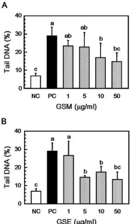
Fig. 1. The effect of Stings of
Gleditsia sinensis
extracts (1, 5, 10, and 50 μg/ml) on 200 μM H2O2induced DNA damage in human leukocytes. NC, PBS-treated normal control (without oxidative stimulus); PC, 200 μM H2O2treated positive control. A: GSM, stings ofGleditsia sinensis
methanol extracts; B: GSE, stings ofGleditsia sinensis
ethanol extracts. Values not sharing the same letter are significantly different from one another (p
<0.05) by Duncan's multiple range test.자 추출물 GSM과 GSE 첨가에 의해 그 손상은 모두 유의적으 로 감소되었으며, GSM은 농도의존적인 경향을 확인할 수 있 었다. 이는 조각자 추출물에 함유되어 있는 다양한 성분들에 의해 산화적 스트레스에 의한 DNA 손상 보호 작용이 나타난 것으로 생각된다. 조각자의 생리활성 성분들로는 ellagic acid 와 flavonoid들이 보고되고 있으며[27], 이러한 페놀류의 성분 들은 프리라디칼을 제거하기 위해 수소 원자를 공여해 줌으로 인해 mammalian cell systems의 in vitro 뿐만 아니라 in vivo에 서 free radical에 의한 DNA 손상을 보호하는 등의 생리 활성 작용을 나타내는 것으로 보고되고 있다[5]. 따라서 산화적 스 트레스에 의한 DNA 손상 유발 시 조각자 추출물의 보호효과 는 조각자 추출물의 폴리페놀 성분과 DPPH 라디칼 소거능과 같은 항산화작용에 의한 결과로 생각된다.
α -glucosidase 저해 활성
다당류를 섭취함으로써 α-amylase를 통해 이당류로 분해되 고 분해된 이당류는 α-glucosidase에 의해 단당류로 분해되어
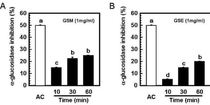
Fig. 2. Anti-diabetes activity of stings of
Gleditsia sinensis
extracts. α-glucosidase inhibitory activity assay of stings ofGleditsia sinensis
extracts. AC, acarbose (1 mg/ml) pos- itive control; GSM, stings ofGleditsia sinensis
methanol extracts; GSE, stings ofGleditsia sinensis
ethanol extracts assay.a-bMeans with the same letter in between acarbose and GSM are not significantly different (p
<0.05).A-BMeans with the same letter in between acarbose and GSE are not significantly different (
p
<0.05).Fig. 3. The case which will increase a control time (10, 30 and 60 min), confirmed that the activity increases of sting of
Gleditsia sinensis
extracts. AC, acarbose (1 mg/ml) positive control time (10 min). A: GSM, sting ofGleditsia si- nensis
methanol extracts, B: GSE, sting ofGleditsia sinensis
ethanol extracts.Values not sharing the same letter are significantly different from one anoth- er (
p
<0.05) by Duncan's multiple range test.소장에 존재하는 융털을 통해 흡수되어 혈관으로 당을 이동시 켜 준다[11]. 그로 인하여 혈당이 증가되어 당뇨병의 환자일 경우 혈당을 조절하지 못하여 여러 가지 합병증을 유발시킨 다. 이당류의 분해효소인 α-glucosidase을 억제시켜 분해되는 이당류의 양을 감소시켜 흡수되는 양을 줄어 급격한 혈당의 증가는 억제시켜 준다. 조각자 추출물인 GSM과 GSE를 이용 하여 이당류의 분해효소인 α-glucosidase 저해 활성을 확인하 여 본 결과는 Fig. 2와 같이 500, 1,000 μg/ml에서 GSM은 10.3%와 17.9%이며, GSE는 5.1%와 10.3%의 억제효과를 나타 내었다. 시간에 따른 α-glucosidase 저해 활성을 확인하기 위 하여 효소처리 시간을 증가시켜 실험을 실시한 결과, 최대 60 min 처리 시 GSM은 25%, GSE는 20%의 저해율을 나타내었으 며, 이 결과로부터 GSM과 GSE의 처리시간에 따라 α-glucosi- dase의 저해 활성이 증가하는 것을 확인할 수 있었다(Fig. 3).
조각자의 α-glucosidase 저해 활성은 acarbose에 비해 낮은 활 성을 보이지만, 추출물인 상태를 감안한다면 충분히 α-gluco- sidase의 억제 활성을 가진다고 보여지고 있다.
위의 결과와 당뇨를 유발시킨 흰쥐에서 소당탕이라는 복합 약물을 처리시 활성산소를 제거하여 항산화 효소의 활성을 증가시켜 췌장, 간, 신장과 같은 기관이 보호가 된다는 연구[9]
와 비교해 보면, 조각자 추출물인 GSM과 GSE는 항산화 효과 를 가지고 있어 췌장, 간, 신장과 같은 기관을 보호할 수 있을 것이라고 생각하며, α-glucosidase 억제효과까지 함유하고 있 으므로 당뇨병 치료제의 소재로 가능성이 있다고 할 수 있다.
감사의 글
이 연구결과물은 2010학년도 경남대학교 학술연구장려금 지원에 의한 것임.
References
1. Ballatori, N., S. M. Krance, S. Notenboom, S. Shi, K. Tieu, and C. L. Hammond. 2009. Glutathione dysregulation and
the etiology and progression of human diseases.
Biol. Chem
. 390, 191-214.2. Blonde, L. 2010. Current antihyperglycemic treatment guidelines and alogrithms patients with type 2 diabetes mellitus.
Am. J. Med
. 123, S12-18.3. Cherdshewasart, W. and W. Sutjit. 2008. Correlation of anti- oxidant activity and major isoflavonoid contents of the phy- toestrogen-rich
Pueraria mirifica
andPueraria lobata tubers
.Phytomedicine
15, 38-43.4. Cowburn, R., J. Hardy, P. Roberts, and R. Briggs. 1988.
Regional distribution of pre- and postsynaptic glutamatergic function in Alzheimer's disease.
Brain Res
. 452, 403-407.5. Dragsted, L. O. 2003. Antioxidant actions of polyphenols in humans.
Int. J. Vitam. Nutr. Res.
73, 112-119.6. Drent, M. L., A. T. Tollefsen, F. H. van Heusden, E. B.
Hoenderdos, J. J. Jonker, and E. A. van der Veen. 2002.
Dose-dependent efficacy of miglitol, an alpha-glucosidase inhibitor, in type 2 diabetic patients on diet alone: results of a 24-week double-blind placebocontrolled study.
Diabetes Nutr. Metab
. 15, 152-159.7. Freeman, J. S. 2009. Role of the incretin pathway in the pathogenesis of type 2 diabetes mellitus.
Clev. Clin. J. Med.
76, S12-19.
8. Kim, H. Y., B. S. Jun, S. K. Kim, J. Y. Cha, and Y. S. Cho.
2000. Polyphenolic compound content and antioxidative by extracts from seed sprout and flower of safflower (
Carthamus tinctorius L
.).J. Korean Soc. Food Sci. Nutr.
29, 1127-1132.9. Jung, J. K. and Y. K. Park. 2010. Antioxidative effect of So-Dang-Tang in streptozotocin-induced diabetic rats.
J. Life Sci.
20, 691-696.10. Kohchi, C., H. Inagawa, T. Nishizawa, and G. Soma. 2009.
ROS and innate immunity.
Anticancer Res
. 29, 817-821.11. van de Laar, F. A., P. L. Lucassen, R. P. Akkermans, E. H.
van de Lisdonk, G. E. Rutten, and C. van Weel. 2005.
Alpha-glucosidase inhibitors for patients with type 2 dia- betes: results from a Cochrane systematic review and meta-analysis.
Diabetes Care
28, 154-163.12. LaSalle, J. R. Reaching HbA1c goals with saxagliptin in com- bination with other oral antidiabetic drugs.
Postgrad. Med
. 122, 144-152.초록:조각자 추출물의 항산화 활성 및 alpha-glucosidase 저해 활성 이정민
1․박재희
2․추원미
2․윤이묵
1․박은주
2․박해룡
1*
(
1경남대학교 식품생명학과,
2경남대학교 식품영양학과)
본 연구는 항산화 활성 및 항당뇨 효과가 있는 조각자 추출물인 GSM (methanol 추출물)과 GSE (ethanol 추출 물)에 대한 연구이다. 추출 용매에 대한 페놀 함량을 측정한 결과 100 g 당 GSM에서는 1.12 g이며, GSE에서는 0.6 g으로 GSM에서 함량이 높은 것으로 나타났다. DPPH 라디칼 소거능의 경우 최대 농도 1,000 μg/ml에서 확인 한 결과 GSM에서는 68.8% 그리고 GSE에서는 70.4%로 두 종류 추출물에서 높은 결과를 확인할 수 있었다. DNA 손상 정도를 확인한 comet assay에서도 200 μM H
2O
2에 의해 나타나는 DNA 손상에 대하여 GSM과 GSE는 DNA 를 보호하는 활성이 있었다. 항당뇨 효과를 알아보는 α-glucosidase 억제 실험에서 GSM은 17.9%, GSE는 10.3%이 었으며, 처리시간을 증가하였을 경우에는 그 억제 정도가 GSM의 경우 25%이며, GSE의 경우 20%로 증가하여 항당뇨 효과가 있다는 것을 확인할 수 있었다. 이 결과로부터 당뇨병 치료에 대한 새로운 소재로써 조각자 추출 물의 가능성을 확인하였다.
13. Lee, S. H., Y. M. Lee, H. S. Lee, and D. K. Kim. 2009.
Anti-oxidative and anti-hyperglycemia effects of Triticum aestivum wheat sprout water extracts on the streptozoto- cin-induced diabetic mice.
Korean J. Pharmacogn
. 40, 408-414.14. Lee, S. J., Y. H. Cho, H. Kim, K. Park, S. K. Park, S. D.
Ha, W. J. Kim, and S. K. Moon. 2009. Inhibitory effects of the ethanol extract of
Gleditsia sinensis
thorns on human co- lon cancer HCT116 cellin vitro
andin vivo
.Oncol. Rep
. 22, 1505-1512.15. Li, W. H., X. M. Zhang, R. R. Tian, Y. T. Zheng, W. M.
Zhao, and M. H. Qiu. 2007. A new anti-HIV lupane acid from
Gleditsia sinensis
Lam.J. Asian Nat. Prod. Res
. 9, 551-555.16. Mattson, M. P. 2008. Glutamate and neurotrophic factors in neuronal plasticity and disease.
Ann. N. Y. Acad. Sci
. 1144, 97-112.17. Mocchegiani, E. and M. Malavolta. 2008. Possible new anti- aging strategies related to neuroendocrine-immune interactions.
Neuroimmunomodulat.
15, 344-350.18. Ron, Y., J. Wainstein, A. Leibovitz, N. Monastirsky, B.
Habot, Y. Avni, and R. Segal. 2002. The effect of acarbose on the colonic transit time of elderly long-term care patients with type 2 diabetes mellitus.
J. Gerontol. A-Biol.
57, M111-114.19. Salman, S., F. Salman, I. Satman, Y. Yilmaz, E. Ozer, A.
Sengül, H. O. Demirel, K. Karşidağ, N. Dinççağ, and M.
T. Yilmaz. 2001. Comparison of acarbose and gliclazide as first-line agents in patients with type 2 diabetes.
Curr. Med.
Res. Opin
. 16, 296-306.20. Santos, F. V., I. M. Colus, M. A. Silva, W. Vilegas, and E.
A. Varanda. 2006. Assessment of DNA damage by extracts
and fractions of
Strychnos pseudoquina
, a Brazilian medicinal plant with antiulcerogenic activity.Food Chem. Toxicol
. 44, 1585-1589.21. Singa, A., M. Rajan, T. Hoerger, and L. Pogach. 2010. Costs and consequences associated with newer medications for glycemic control in type 2 diabetes.
Diabetes Care
33, 695-700.22. Singleton, V. L. and J. A. Rossi. 1965. Colorimetry of total phenolics with phosphomolibidic-phosphotyngstic reagents.
Am. J. Enol. Viticult
. 16, 144-158.23. Thitilertdecha, N., A. Teerawutgulrag, and N.
Rakariyatham. 2008. Antioxidant and antibacterial activities of
Nephelium lappaceum
L. extracts.Lebensm-Wiss. Technol
. 41, 2029-2035.24. Tice, R. R., E. Agurell, D. Anderson, B. Burlinson, A.
Hartmann, H. Kobayashi, Y. Miyamae, E. Rojas, J. C. Ryu, and Y. F. Sasaki. 2000. Single cell gel/comet assay:
Guidelines for
in vitro
andin vivo
genotoxicology testing.Environ. Mol. Mutagen
. 35, 206-221.25. Wei, W., Q. Liu, Y. Tan, L. Liu, X. Li, and L. Cai. 2009.
Oxidative stress, diabetes, and diabetic complications.
Hemoglobin
33, 370-377.26. Wu, J., J. Li, Z. Zhn, J. Li, G. Huang, Y. Tang, and X. Gao.
2010. Protective effects of echinocystic acid isolated from
Gleditsia sinensis
Lam. against acute myocardial ischemia. Fitoterapia
81, 8-10.27. Zhou, L., D. Li, W. Jiang, Z. Qin, S. Zhao, M. Qiu, and J.
Wu. 2007. Two ellagic acid glycosides from