Effects of Post Biopsy Digital Rectal Compression on Improving Prostate Cancer Staging Using Magnetic Resonance Imaging
in Localized Prostate Cancer
Kyung Kgi Park,
1Mun Su Chung,
2Soo Yoon Chung,
3Joo Hee Kim,
4and Byung Ha Chung
5Departments of 1Urology and 3Radiology, Yongin Severance Hospital, Yonsei University College of Medicine, Yongin;
2Department of Urology, Uijeongbu St. Mary’s Hospital, The Catholic University of Korea, Uijeongbu;
Departments of 4Radiology and 5Urology, Gangnam Severance Hospital, Yonsei University College of Medicine, Seoul, Korea.
Received: December 1, 2011 Revised: February 8, 2012 Accepted: February 9, 2012
Corresponding author: Dr. Byung Ha Chung, Department of Urology, Gangnam Severance Hospital, Yonsei University College of Medicine, 211 Eonju-ro, Gangnam-gu, Seoul 135-720, Korea.
Tel: 82-2-2019-3470, Fax: 82-2-3462-8887 E-mail: [email protected]
∙ The authors have no financial conflicts of interest.
© Copyright:
Yonsei University College of Medicine 2013 This is an Open Access article distributed under the terms of the Creative Commons Attribution Non- Commercial License (http://creativecommons.org/
licenses/by-nc/3.0) which permits unrestricted non- commercial use, distribution, and reproduction in any medium, provided the original work is properly cited.
Purpose: To evaluate the effectiveness of digital rectal-compression immediately after transrectal prostate biopsy (P-bx) for improving the accuracy of prostate cancer (PCa) staging. Materials and Methods: Between July 2008 and June 2010, 94 consecutive patients who had a radical prostatectomy were included in our retrospective analysis. The exclusion criteria included a history of previous P-bx and surgery, a biopsy performed in another hospital, a number of biopsy cores different from 12, or a condition interfering with bleeding assessment. The subjects were divided into two groups, compression and non-compression. All enrolled patients took magnetic resonance imaging (MRI) for PCa staging. Re- sults: The compression and non-compression groups were comparable with re- spect to several baseline characteristics. However, the total hemorrhage score of intraprostatic bleeding was significantly different between the groups, even with adjustment for the time from biopsy to MRI (compression:15.4±2.32, non-com- pression: 24.9±2.43, p<0.001). The intra-prostatic cancer location matching rate was higher in the compression group (78.0%) than in the non-compression group (70.2%) (p=0.011). Overall accuracy of staging in compression and non-com- pression groups was 84.7% and 77.3%, respectively. Conclusion: Our results demonstrate that digital rectal compression performed immediately after prostate biopsy to reduce intraprostatic hemorrhage improves the accuracy for detection of PCa using MRI.
Key Words: Needle biopsy, prostate neoplasm, magnetic resonance imaging, hemorrhage, hemostatic technique
INTRODUCTION
Magnetic resonance imaging (MRI) plays an increasingly important role in the lo-
cal staging of prostate cancer (PCa). However, conditions other than PCa, such as
post-biopsy hemorrhaging, post-radiation and hormone therapy, scar/positional/in-
flammatory changes, and dystrophic changes can interfere with accurate MRI in-
warfarin, prostate biopsy was delayed until the Internation- al Normalized Ratio had been corrected to less than 1.5.
Prophylactic oral ciprofloxacin (500 mg) was administrated once daily 30 to 60 minutes before the biopsy and was con- tinued for two to three days after the procedure.
Patients self-administered a cleansing enema the night before the biopsy. No local anesthesia was administered to avoid any possible interference from bleeding.
For the procedure, patients were placed in a right lateral decubitus position. An intra-rectal lidocaine jelly injection was used as a topical analgesic. A spring-driven 18-gauge needle core biopsy gun was used for prostate tissue biopsy via a needle guide that was attached to the ultrasound probe.
A systematic 12-core biopsy was performed for each pa- tient by a urologist with six years of experience. The enrolled subjects were divided into two groups: a compression group (48 patients) and non-compression group (46 patients). Af- ter biopsy, digital rectal compression was immediately per- formed for five minutes in the compression group. This compression was performed only during the second year of the study (July 2009-June 2010).
MRI protocol
All of the patients were imaged using a 3.0T MRI system (Intera Achieva 3.0T, Philips Medical System, Best, the Netherlands) equipped with a six-channel phased-array coil.
No endorectal coil has been used because of discomfort of patients without no significant effectiveness.
8This imaging was performed prior to the radical prostatectomy. All of the patients underwent diffusion-weighted imaging (DWI) in addition to the imaging sequences as part of a routine pros- tate MRI protocol. T2-weighted turbo spin-echo images were acquired in three orthogonal planes (axial, sagittal, and coronal). The T2-weighted imaging scan parameters were TR/TE, 3300 to 3800/80 to 100 milliseconds; slice thickness, 3 mm; interslice gap, 0.3 mm; 512×360 matrix;
field of view (FOV), 20 cm; number of signals acquired, 3;
and sensitivity encoding (SENSE) factor 2. Axial T1-weight- ed THRIVE sequences (3 mm slice thickness; FOV, 20 cm) were acquired to detect artifacts from the biopsy and to as- sess the lymph nodes. Axial DWI was obtained using a sin- gle-shot echo planar imaging technique with the following imaging parameters: TR/TE, 2300 to 3000/63 to 65 milli- seconds; slice thickness, 3 mm; interslice gap, 1 mm; ma- trix, 80×80; FOV, 20 cm; SENSE factor 2; number of sig- nals acquired 4. Diffusion-encoding gradients were applied in the axial directions of motion-probing gradients as bipo- vestigations.
1-3Among these factors, post-biopsy hemor-
rhaging offers substantial limitation to accurate detection of PCa after a prostate biopsy.
4,5Previous studies have shown that a waiting period of 3 weeks after biopsy can reduce the possibility of interference from intraprostatic hemorrhage.
3,6However, this is a passive approach, and our goal was to identify an active method to reduce intraprostatic hemor- rhage after prostate biopsy.
We believe that prostate compression after prostate biop- sy might help more accurately stage PCa because of the re- duction in intraprostatic hemorrhage. Therefore, we evalu- ated the effects of digital rectal compression in patients who underwent ultrasound-guided transrectal prostate biopsy on PCa staging using MRI.
MATERIALS AND METHODS
Patients
Between July 2008 and June 2010, 94 consecutive patients who had undergone a robot-assisted laparoscopic radical prostatectomy (RALRP) after ultrasound-guided prostate biopsy and prostate MRI investigation were enrolled in a retrospective analysis that was approved by our institution’s review board.
All of the enrolled patients had biopsy-proven PCa. The exclusion criteria were a history of previous prostate biopsy and surgery; an interval from prostate biopsy to the MRI in- vestigation of four weeks or more; an interval from MRI to surgery of more than 10 days; a history of use of 5-α reduc- tase inhibitor within three months; a biopsy performed in another hospital; a number of biopsy cores different from 12; a positive nodal or bone metastasis based on preopera- tive imaging; or an additional condition, such as urinary tract stones, renal or bladder tumors, hemorrhoids, anal fis- sure, or rectal inflammatory disease, that could potentially cause hematuria or rectal bleeding and thus interfere with bleeding assessment.
Biopsy protocol
Below biopsy, MRI and image analysis procedures was conducted by previously description
7All anticoagulant therapies, including aspirin/NSAIDs, war-
farin, clopidogrel, and herbal supplements, were discontin-
ued seven to ten days before the prostate biopsy. For pa-
tients with an underlying coagulopathy or for those taking
viewed the pathology and radiology scoring results. Finally, we analyzed for concordance for tumor location between these results by time period between the biopsy and MRI.
Statistical analysis
The relationship between the total hemorrhage score and the period from the biopsy to MRI was assessed using a Pearson’s correlation test. Differences in patient character- istics between the two groups were analyzed using a t-test and Pearson’s chi-square statistical test. Period-adjusted hemorrhage scores for the two groups were compared us- ing the ANCOVA test separately. When the two groups were divided into 4 groups according to the period between prostate biopsy and MRI, separate groups were compared using the t-test. All statistical tests were conducted using SAS (version 9.0), and all tests were two sided, with a p- value <0.05 being considered statistically significant.
RESULTS
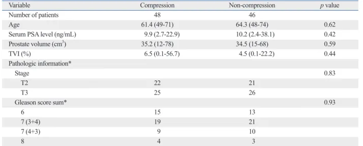
The compression and non-compression groups were compa- rable with regard to age, serum prostate-specific antigen lev- el, prostate volume, tumor volume involvement, pathologic T stage, and Gleason score sum (Table 1). There was a sig- nificant difference in hemorrhage score between the two groups up to 24 days after biopsy after adjusting for the peri- od from biopsy to MRI (ANCOVA test, compression groups:
15.4±2.32, non-compression group: 24.9±2.43, p<0.001).
Total hemorrhage scores were negatively correlated with the time between biopsy and MRI (r=-0.345, p=0.031).
To evaluate the accuracy of PCa stage using MRI, cancer location data obtained through MRI were compared with pathologic data. The intra-prostatic cancer location match- ing rate was significantly increased in the compression group compared to the non-compression group (Fig. 1).
Matching rates for intraprostatic cancer location were 78.0% in the compression groups and 70.2% in the non- compression groups (t-test, p=0.011). Matching rate of ex- tracapsular extension was 82% in the compression group and 75% in the non-compression group (t-test, p=0.032).
Matching rate about seminal vesicle invasion was observed to be 90% in the compression group and 85% in the non- compression group cases (t-test, p=0.046). Overall accura- cy of PCa staging obtained through MRI was 84.7% in the compression groups and 77.3% in the non-compression groups (t-test, p=0.037).
lar pairs at b values of 0 and 1000 s/mm
2. ADC DWI maps were automatically constructed on a pixel-by-pixel basis.
Image analysis and interpretation of hemorrhage and cancer
All images were retrospectively reviewed by two radiolo- gists with 6 and 11 years of experience in interpreting pros- tate MRI results (J.H.K and J.J.J, respectively) and 2 years of experience in DWI. Two radiologists conducted a con- sensus review of the MR images obtained for all patients and determined the degree and number of hemorrhages on three axial MR images for each patient, selected using the following methods. First, the axial image containing the verumontanum area was selected as a standard plane. Sec- ond, a base and apex plane 10 mm from the standard plane was selected. Third, each plane was divided into four pe- ripheral zones and two transitional zones. Each hemorrhag- ic prostate segment was scored according to the diameter of the segment on a scale of 0 to 3 as follows: 0=none, 1=great- er than 0 mm and less than 5 mm, 2=greater than 5 mm and less than 10 mm, 3=greater than 10 mm. In each segment, cancer foci were counted using a two-point scale, where 0=<50% and 1=>50% chance of cancer. A hemorrhage was defined as a high-signal intensity on a T1-weighted image and a low-signal intensity on a T2-weighted image. The ob- server was unaware of the pathology results. These tech- niques were used for similar prostate MRI based study from December 2007.
7Pathology
Previously prepared pathology slides were retrospectively
reviewed by one pathologist (B.J.L.). The slides were cho-
sen from consecutive pathology files using the same criteria
as the radiology interpretations. The pathologist outlined
the region of cancer and hemorrhage on each slide. The
slides were then digitized and saved as a jpeg image. Based
on these digitized slides, each tumor and hemorrhage re-
gions were scored by the pathologist (B.J.L.) in the same
manner as the radiology interpretations. The pathologist
was blinded to the MRI results. We validated the limited
sensitivity of MRI
9in order to make a proper radiologic-
pathologic correlation. Under the cross-sectional area of the
fixed specimen, a region with greater than 0.1 cm diameter
was considered to be a meaningful lesion. Hemorrhage was
defined as the presence of red blood cells outside the identi-
fiable blood vessels, especially in the interstitium and glan-
dular lumen. The urologist (K.K.P.) independently re-
onstrate show low signal intensity within the peripheral zone on T2-weighted images.
1-3In a patient who is diagnosed with PCa via a prostate bi- opsy, post-biopsy hemorrhaging is a major factor that inter- feres with accurate MR image interpretation. Reducing the risk of post-biopsy hemorrhaging, therefore, is necessary in order to minimize this interference. First, all anticoagulant therapy should be discontinued before the biopsy. For those patients coagulation problem such as coagulopathy or tak- ing anticoagulant, a prostate biopsy should be delayed until normal coagulation had been restored.
10Second, several studies have reported that post-biopsy hemorrhaging dimin- ishes with time.
3,6,11We also observed a statistically signifi- cant negative correlation between the sum of the hemor- rhage score and the time from the biopsy in our previous and current study.
7However, some authors argue that post- biopsy hemorrhaging is not significantly reduced until 55 days after the biopsy.
12,13Third, another technique that is less influenced by post-biopsy hemorrhage than conven- tional MRI can be used.
12,14,15We believe that all three of the above-mentioned meth- ods can passively reduce post biopsy hemorrhage. There- fore, we sought a method to reduce post biopsy intrapros- tatic hemorrhage.
Digital rectal compression is a well-known method for reducing rectal bleeding after prostate biopsy.
16However, our results demonstrate that this method can also reduce in- traprostatic hemorrhage and increase the accuracy of MRI.
In this study, we found that compression can reduce the amount of post biopsy hemorrhage. The accuracy of tumor
DISCUSSION
To determine the extent of tumor and the choice of manage- ment, accurate tumor staging is essential. The major role of MRI in PCa is to assist with local staging of the disease.
Prostate carcinoma usually demonstrates lower signal in- tensity of MR images, as compared to the peripheral zone.
The changes of signal intensity are nonspecific. In patiens with tumors, post-biopsy hemorrhage, post-radiation and hormone therapy, scar/positional/inflammatory changes, and dystrophic changes, prostate MR images would dem-
Table 1. Baseline Characteristics of the Compression and Non-Compression Groups
Variable Compression Non-compression p value
Number of patients 48 46
Age 61.4 (49-71) 64.3 (48-74) 0.62
Serum PSA level (ng/mL) 9.9 (2.7-22.9) 10.2 (2.4-38.1) 0.42
Prostate volume (cm3) 35.2 (12-78) 34.5 (15-68) 0.59
TVI (%) 6.5 (0.1-56.7) 4.5 (0.1-22.2) 0.44
Pathologic information*
Stage 0.83
T2 22 21
T3 25 26
Gleason score sum* 0.93
6 15 13
7 (3+4) 19 21
7 (4+3) 9 10
8 4 3
PSA, prostate-specific antigen; TVI (tumor volume percentage involvement)=tumor volume/prostate volume.
*Result according to Pearson’s chi-square statistical test.
Fig. 1. Prostate cancer matching rate for compression and non-compres- sion groups according to the time between biopsy and MRI. Control: non- compression group, *p=<0.05, (p value: 0.023 in 1 week, 0.044 in 2 week, 0.039 in 3 week, 0.031 in 4 week). MRI, magnetic resonance imaging.
0 40 60 80
100 *
* * *
Prostate cancer location matching rate (%)
1
Time from biopsy (weeks)
2 3 4
20
Control Compression
Even if the most similar MRI and pathology sections were selected, there were still some inconsistencies between pa- thology and radiology sections.
In conclusion, this study demonstrates that digital rectal compression immediately after prostate biopsy can reduce intraprostatic hemorrhage. the accuracy in detecting PCa can be improved by performing transrectal digital compres- sion only immediately after biopsy. Therefore, we believe that digital rectal compression should be carried out after prostate biopsy.
REFERENCES
1. Schiebler ML, Tomaszewski JE, Bezzi M, Pollack HM, Kressel HY, Cohen EK, et al. Prostatic carcinoma and benign prostatic hy- perplasia: correlation of high-resolution MR and histopathologic findings. Radiology 1989;172:131-7.
2. Schiebler ML, Schnall MD, Pollack HM, Lenkinski RE, To- maszewski JE, Wein AJ, et al. Current role of MR imaging in the staging of adenocarcinoma of the prostate. Radiology 1993;189:
339-52.
3. White S, Hricak H, Forstner R, Kurhanewicz J, Vigneron DB, Za- loudek CJ, et al. Prostate cancer: effect of postbiopsy hemorrhage on interpretation of MR images. Radiology 1995;195:385-90.
4. Chelsky MJ, Schnall MD, Seidmon EJ, Pollack HM. Use of en- dorectal surface coil magnetic resonance imaging for local staging of prostate cancer. J Urol 1993;150(2 Pt 1):391-5.
5. Schnall MD, Imai Y, Tomaszewski J, Pollack HM, Lenkinski RE, Kressel HY. Prostate cancer: local staging with endorectal surface coil MR imaging. Radiology 1991;178:797-802.
6. Ikonen S, Kivisaari L, Vehmas T, Tervahartiala P, Salo JO, Taari K, et al. Optimal timing of post-biopsy MR imaging of the prostate.
Acta Radiol 2001;42:70-3.
7. Park KK, Lee SH, Lim BJ, Kim JH, Chung BH. The effects of the period between biopsy and diffusion-weighted magnetic reso- nance imaging on cancer staging in localized prostate cancer. BJU Int 2010;106:1148-51.
8. Kim BS, Kim TH, Kwon TG, Yoo ES. Comparison of pelvic phased-array versus endorectal coil magnetic resonance imaging at 3 Tesla for local staging of prostate cancer. Yonsei Med J 2012;53:550-6.
9. Kirkham AP, Emberton M, Allen C. How good is MRI at detect- ing and characterising cancer within the prostate? Eur Urol 2006;50:1163-74.
10. Douketis JD. Perioperative anticoagulation management in pa- tients who are receiving oral anticoagulant therapy: a practical guide for clinicians. Thromb Res 2002;108:3-13.
11. Qayyum A, Coakley FV, Lu Y, Olpin JD, Wu L, Yeh BM, et al.
Organ-confined prostate cancer: effect of prior transrectal biopsy on endorectal MRI and MR spectroscopic imaging. AJR Am J Roentgenol 2004;183:1079-83.
12. Tamada T, Sone T, Jo Y, Yamamoto A, Yamashita T, Egashira N, et al. Prostate cancer: relationships between postbiopsy hemor- rhage and tumor detectability at MR diagnosis. Radiology 2008;
248:531-9.
detection in the compression group was 78.0%, which is significantly superior to the accuracy of detection in the noncompression group (70.2%). Therefore, reducing hem- orrhage through digital rectal compression makes it signifi- cantly more accurate for the detection of PCa. We aimed to develop an approach to actively increase the accuracy of MRI staging for PCa, rather than passively waiting for 2 weeks. Based on our results, we recommend the routine use of digital rectal compression after prostate biopsy to in- crease the accuracy to detect PCa.
We believe that digital rectal compression has a few addi- tional merits. First, rectal compression may reduce surgical difficulty. Some authors have reported that post biopsy hem- orrhage may induce operative difficulty. Hong, et al.
13re- ported that the degree of post biopsy hemorrhage detected by MR image has a significant association with operative outcome such as operative time, estimated blood loss, and return of erectile function in patients who received RALRP.
However, as shown in this study, if digital rectal compres- sion is conducted in patients after prostate biopsy, operative difficulty might decrease because digital rectal compression could reduce intraprostatic hemorrhage. Second, some au- thors have argued that conventional MRI is significantly af- fected by the amount of post biopsy hemorrhage.
3,14Rectal compression may shorten the period during which there is interference in the interpretation of the prostate by conven- tional MRI. In this study, using multiparametric MRI, the timing of MRI did not affect the accuracy of PCa staging.
Because multiparametric MRI usually has a greater sensi- tivity and specificity than conventional MRI, we believe that interpretation of MRI is less influenced by post biopsy hem- orrhage.
7,12,14,15However, if the institute uses conventional MRI rather than multiparametric MRI for PCa staging, rec- tal compression may reduce the development of intrapros- tatic hemorrhage at biopsy. Therefore, the interval between biopsy and prostate MRI can be shortened due to the de- crease in the absolute amount of developing hemorrhage, and postbiopsy hemorrhage will disappear more quickly.
There are some limitations in our study. First, the period between biopsy and MRI was a maximum of 24 days.
Therefore, we did not evaluate the effects of compression
over more than 24 days. Second, in order to achieve an ob-
jective interpretation, three investigators were involved. Be-
cause the results from the radiologist and pathologist were
integrated by the third investigator, it is possible that an in-
creased level of observer error might have been introduced
into the final results. Third, this was a retrospective study.
imaging. Radiology 1998;206:785-90.
15. Morgan VA, Kyriazi S, Ashley SE, DeSouza NM. Evaluation of the potential of diffusion-weighted imaging in prostate cancer de- tection. Acta Radiol 2007;48:695-703.
16. Maatman TJ, Bigham D, Stirling B. Simplified management of post-prostate biopsy rectal bleeding. Urology 2002;60:508.
13. Hong SK, Kim DS, Lee WK, Park H, Kim JK, Doo SH, et al. Sig- nificance of postbiopsy hemorrhage observed on preoperative magnetic resonance imaging in performing robot-assisted laparo- scopic radical prostatectomy. World J Urol 2010;28:721-6.
14. Kaji Y, Kurhanewicz J, Hricak H, Sokolov DL, Huang LR, Nel- son SJ, et al. Localizing prostate cancer in the presence of postbi- opsy changes on MR images: role of proton MR spectroscopic