INTRODUCTION
An estimated 1.8 to 2.5 million venomous snakebites occur worldwide each year and result in at least 100000 to 125000 deaths.1,2 As in other parts of the world, snakebites are common in Korea. Generally, in the treatment of venomous snakebite, focus of treatment is coagulation abnormalities, neuromuscu- lar paralysis and respiratory failure in the elapid bites.3 How- ever, of the various possible complications, cardiac involve-
ment is also an infrequently recognized manifestation of ven- omous snakebites, and is seen mainly associated with viperine bites.4
However, little is known of the adverse cardiovascular events (ACVEs) arising as a result of snakebites in Korea, except one case report on the rare development of acute myocardial in- farction after snakebite.5 In particular, there has been no re- port on myocardial injury in Korea as determined using serum high sensitivity troponin I (hs-TnI) resulting from venomous snakebites.
Accordingly, we studied the prevalence of ACVEs associat- ed with venomous snakebites in Korea and compared the clinical features of patients with and without ACVEs.
MATERIALS AND METHODS
Study design and data
This retrospective, observational study was conducted on con-
Adverse Cardiovascular Events after a Venomous Snakebite in Korea
Oh Hyun Kim, Joon Woo Lee, Hyung Il Kim, KyoungChul Cha, Hyun Kim, Kang Hyun Lee, Sung Oh Hwang, and Yong Sung Cha
Department of Emergency Medicine, Wonju College of Medicine, Yonsei University, Wonju, Korea.
Purpose: Although cardiac involvement is an infrequently recognized manifestation of venomous snakebites, little is known of the adverse cardiovascular events (ACVEs) arising as a result of snakebite in Korea. Accordingly, we studied the prevalence of ACVEs associated with venomous snakebites in Korea and compared the clinical features of patients with and without ACVEs.
Materials and Methods: A retrospective review was conducted on 65 consecutive venomous snakebite cases diagnosed and treated at the emergency department of Wonju Severance Christian Hospital between May 2011 and October 2014. ACVEs were defined as the occurrence of at least one of the following: 1) myocardial injury, 2) shock, 3) ventricular dysrhythmia, or 4) cardiac arrest.
Results: Nine (13.8%) of the 65 patients had ACVEs; myocardial injury (9 patients, 13.8%) included high sensitivity troponin I (hs- TnI) elevation (7 patients, 10.8%) or electrocardiogram (ECG) determined ischemic change (2 patients, 3.1%), and shock (2 patient, 3.1%). Neither ventricular dysrhythmia nor cardiac arrest was observed. The median of elevated hs-TnI levels observed in the pres- ent study were 0.063 ng/mL (maximum: 3.000 ng/mL) and there was no mortality in the ACVEs group. Underlying cardiac diseases were more common in the ACVEs group than in the non-ACVEs group (p=0.017). Regarding complications during hospitalization, 3 patients (5.4%) in the non-ACVEs group and 3 patients (33.3%) in the ACVEs group developed bleeding (p=0.031).
Conclusion: Significant proportion of the patients with venomous snakebite is associated with occurrence of ACVEs. Patients with ACVEs had more underlying cardiac disease and bleeding complication.
Key Words: Snakebite, heart injury, troponin I, shock Yonsei Med J 2016 Mar;57(2):512-517
http://dx.doi.org/10.3349/ymj.2016.57.2.512 pISSN: 0513-5796 · eISSN: 1976-2437
Received: May 13, 2015 Revised: June 20, 2015 Accepted: July 20, 2015
Corresponding author: Dr. Yong Sung Cha, Department of Emergency Medicine, Wonju College of Medicine, Yonsei University, 20 Ilsan-ro, Wonju 26426, Korea.
Tel: +82-33-741-1615, Fax: +82-33-742-3030, E-mail: [email protected]
•The authors have no financial conflicts of interest.
© Copyright: Yonsei University College of Medicine 2016
This is an Open Access article distributed under the terms of the Creative Com- mons Attribution Non-Commercial License (http://creativecommons.org/licenses/
by-nc/3.0) which permits unrestricted non-commercial use, distribution, and repro- duction in any medium, provided the original work is properly cited.
secutive patients who presented with a venomous snakebite at the emergency department (ED) of Wonju Severance Chris- tian Hospital, Wonju College of Medicine, Yonsei University, between May 2011 and October 2014. The study exclusion cri- teria applied were; an age of <18 years, end-stage renal disease, no admittance for treatment at our hospital, ED arrival >48 hrs after being bitten, and the absence of a measurable serum TnI level within 48 hrs of arrival at the ED.
Diagnostic criteria of venomous snakebite included the pres- ence of one or more of the following: 1) a confirmed two fang wound; 2) a triangular head; 3) a venomous snake confirmed by patient’s experience; and 4) abnormal physical and labora- tory findings such as shock, tachypnea, bleeding, thrombocyto- penia, disseminated intravascular coagulation (DIC), or diplo- pia despite insufficient other findings.6 When patients had systemic signs and symptoms or severe local symptoms associ- ated with snakebite, antivenin was administered and, if symp- toms persisted, antivenin was repeated.
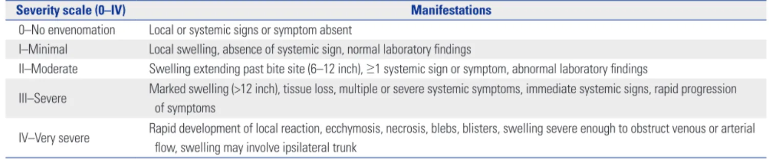
The following information was obtained from medical re- cords: age, gender, site of the bite, ED arrival times, underlying diseases, severity at ED & admission, initial systolic blood pres- sure, and initial symptoms and signs. A traditional snakebite severity grading scale was used (Table 1).7 Complications in- cluding hematologic (anemia,8 thrombocytopenia, prothrom- bin time/partial thromboplastin time prolongation, DIC,9 and bleeding) and neurologic complications (blurred vision or diplopia) occurring during hospitalization, and mortality were investigated.
Cardiac biochemical markers including hs-TnI were investi- gated in the ED. A hs-TnI (Siemens Healthcare Diagnostics Inc., Newark, DE, USA) was used to determine blood TnI levels (ref- erence range: <0.046 ng/mL) and myocardial injury was de- fined as an elevation of hs-TnI within 48 hrs of arrival at the ED.
ACVEs were defined as the occurrence of at least one of the followings: 1) myocardial injury [based on hs-TnI elevation within 48 hrs of presentation or electrocardiogram (ECG) evi- dence of ischemic change, such as ST elevation, ST depression, or T wave inversion]; 2) shock (defined as hypotension requir- ing vasopressors); 3) ventricular dysrhythmia (ventricular tachycardia, ventricular fibrillation, torsades des pointes); and 4) cardiac arrest. We investigated ACVEs that developed with- in 48 hrs of treatment commencement because we wanted to minimize the possibility of ACVEs secondary to malfunctions
of other organs or systems, such as, multiple organ failure.
This study was approved by the Institutional Review Board of Wonju College of Medicine, Yonsei University.
Statistical analysis
Categorical variables are presented as frequencies and percent- ages, and continuous variables are presented as mean and stan- dard deviation or median and interquartile range after assess- ments for normality using the Shapiro-Wilk test. The chi-square test or Fisher’s exact test was used to compare nominal vari- ables, and the two-sample t-test or the Mann-Whitney U test was used to compare continuous variables. p values of <0.05 were considered statistically significant. The analysis was per- formed using IBM SPSS 20 Ver. (IBM, Armonk, NY, USA).
RESULTS
General and laboratory characteristics of patients with venomous snakebite
A total of 65 consecutive patients were found eligible for this study while 33 patients were excluded from the study due to the following exclusion criteria: age less than 18 years (7 patients), end-stage renal disease (1 patient), patients had not been ad- mitted for treatment in our hospital or transfer out (4 patients), ED arrival more than 48 hrs after snakebite (4 patients), and the absence of serum hs-TnI level within 48 hrs of arrival at the ED (15 patients), and insufficient data (2 patients).
General characteristics are shown in Table 2. Forty six of the patients were male (70.8%) and the ages of the 65 study sub- jects ranged from 20 to 90 years with a median of 57 years. All patients were bitten in upper and lower extremities. There were 5 patients (7.7%) with cardiac diseases including coronary ar- tery disease (unstable angina 1 patient and myocardial infarc- tion 1 patient), valvular heart disease (aortic regurgitation 1 patient), hypertensive heart disease (hypertensive heart fail- ure 1 patient), and arrhythmia (atrial fibrillation 1 patient).
Most common grading of severity was grade II. The common symptoms and signs at initial presentation were pain (65 pa- tients, 100%), edema (53 patients, 81.5%), discoloration (18 pa- tients, 27.7%), and dizziness (9 patients, 13.8%). The most com- mon hematologic complication during hospitalization was anemia (39 patients, 60%), and bleeding developed in 6 patients
Table 1. Traditional Snakebite Severity Grading Scale
Severity scale (0–IV) Manifestations
0–No envenomation Local or systemic signs or symptom absent
I–Minimal Local swelling, absence of systemic sign, normal laboratory findings
II–Moderate Swelling extending past bite site (6–12 inch), ≥1 systemic sign or symptom, abnormal laboratory findings
III–Severe Marked swelling (>12 inch), tissue loss, multiple or severe systemic symptoms, immediate systemic signs, rapid progression of symptoms
IV–Very severe Rapid development of local reaction, ecchymosis, necrosis, blebs, blisters, swelling severe enough to obstruct venous or arterial flow, swelling may involve ipsilateral trunk
Table 2. General Characteristics of Venomous Snakebite Victims
Characteristics Total (n=65) Non-ACVEs group
(n=56; 86.2%)
ACVEs group
(n=9; 13.8%) p value
Age (yrs) 57.0 (50.5–67.5)* 56.0 (47.8–66.0)* 64.0 (52.5–71.5)* 0.258
Male gender 46 (70.8%) 40 (71.4%) 6 (66.7%) 0.714
Sites of bite 0.339
Hand 50 (78.1%) 41 (74.5%) 9 (100%)
Foot 11 (17.2%) 11 (20.0%) 0 (0%)
Leg 3 (4.7%) 3 (5.4%) 0 (0%)
ED arrival times (hrs) 3.0 (1.0–10.8)* 4.0 (1.0–15.0)* 3.0 (1.5–3.5)* 0.560
Underlying disease
DM 6 (9.2%) 5 (8.9%) 1 (11.1%) >0.999
HTN 17 (26.2%) 14 (25.0%) 3 (33.3%) 0.687
Cardiac disease 5 (7.7%) 2 (3.6%) 3 (33.3%) 0.017
SBP (mm Hg) 141±27† 143±25† 133±40† 0.308
Severity (ED/admission) 0.170/0.640
I 26/13 (40.0%/20.0%) 22/11 (40.7%/20.8%) 4/2 (44.4%/22.2%)
II 31/44 (47.7%/67.7%) 28/38 (51.9%/71.7%) 3/6 (33.3%/66.7%)
III 4/3 (6.2%/4.6%) 2/2 (3.7%/3.8%) 2/1 (22.2%/11.1%)
IV 2/2 (3.1%/3.1%) 2/2 (3.7%/3.8%) 0/0 (0%/0%)
Antivenin 0.501
None 17 (26.2%) 13 (23.2%) 4 (44.4%)
Single 39 (60%) 34 (60.7%) 5 (55.5%)
Multidose 9 (13.8%) 9 (16.1%) 0 (0%)
Symptoms & signs Local effect
Pain 65 (100%) 55 (100%) 9 (100%)
Blister 7 (10.8%) 7 (26.9%) 0 (0%) >0.999
Edema 53 (81.5%) 46 (88.5%) 7 (87.5%) >0.999
Discoloration 18 (27.7%) 17 (48.6%) 1 (50.0%) >0.999
Systemic effect
Nausea 3 (4.6%) 2 (3.6%) 1 (11.1%) 0.365
Dizziness 9 (13.8%) 6 (10.7%) 3 (33.3%) 0.339
Headache 4 (6.2%) 3 (5.4%) 1 (11.1%) 0.458
Decreased mental 1 (1.5%) 1 (1.8%) 0 (0%) >0.999
Dyspnea 5 (7.7%) 4 (7.1%) 1 (11.1%) 0.538
Complications Hematologic
Anemia 39 (60%) 31 (55.4%) 8 (88.9%) 0.074
Thrombocytopenia 26 (40%) 22 (39.3%) 4 (44.4%) >0.999
PT/PTT prolongation 22 (33.8%) 18 (32.1%) 4 (44.4%) 0.706
DIC 30 (46.2%) 25 (44.6%) 5 (55.6%) 0.722
Bleeding 6 (9.2%) 3 (5.4%) 3 (33.3%) 0.031
Neurologic
Blurred vision/diplopia 2 (3.0%) 2 (3.6%) 0 (0%) >0.999
Rhabdomyolysis 13 (20%) 12 (21.8%) 1 (11.1%) 0.672
TnI levels within 48 hrs (ng/mL) 0.015 (0.015–0.016)* 0.015 (0.015–0.015)* 0.063 (0.033–0.111)* 0.001 ED, emergency department; DM, diabetes mellitus; HTN, hypertension; SBP, systolic blood pressure; PT, prothrombin time; PTT, partial thromboplastin time; DIC, disseminated intravascular coagulation; TnI, troponin I; ACVEs, adverse cardiovascular events.
*Median (interquartile range), †Mean±standard deviation.
at puncture site (2 patients), gastrointestinal tract (3 patients), and brain (1 patient). DIC developed in 46.2% of patients. Out of neurologic complications, blurred vision was developed in 3.0%. Other complication was a rhabdomyolysis (13 patients, 20%) and there was no mortality (Table 2).
ACVEs occurred in 9 patients (13.8%); myocardial injury (9 patients, 13.8%) included hs-TnI elevation (7 patients, 10.8%) or ECG determined ischemic change (2 patients, 3.1%), and shock (2 patient, 3.1%). Neither ventricular dysrhythmia nor cardiac arrest was observed. Cardiac biochemical markers analysis revealed median initial levels of hs-TnI of 0.015 ng/mL.
ECG showed a T wave inversion in 4 patients (6.2%). QT pro- longation was observed in 25 patients (38.5%) (Table 2 and 3).
General characteristics and laboratory findings according to the presence of ACVEs
Underlying cardiac diseases were more common in the ACVEs group than in the non-myocardial injury group (p=0.017). Ho- wever, age, severity, and initial symptoms & signs were not sig- nificantly different between the two groups (Table 2).
Regarding complications during hospitalization, 3 patients (5.4%) in the non-ACVEs group and 3 patients (33.3%) in the ACVEs group developed bleeding (p=0.031). Blurred vision or diplopia developed in 2 patients in non-ACVEs group (Table 2).
DISCUSSION
Cardiac involvement is known mainly to be associated with vi- perine bites.4 There are four types of venomous snakes in Korea.
Three belong to the family Viperidae; that is, Agkistrodon brevi- caudus, Agkistrodon ussuriensis, and Agkistrodon saxatilis. The other Rhabdophis tigrinus belongs to the family Cloubridae.10 In this study, the incidence of ACVEs development within 48 hrs following treatment commencement in a venomous snake- bite cases was greater than expected (13.8%). To the best of our knowledge, this is the first report to address ACVEs after a ven- omous snakebite in Korea. In this study, there was no mortality, irrespective of ACVEs. In the present study, therefore, we can- not predict the prognosis of patients with ACVEs. Nevertheless, previous studies showed that the most severe complications and fatalities are unrelated to cardiac abnormalities.11,12
Serum TnI is used to detect myocardial injury, because it is expressed as a cardio-specific isoforms.13 For this reason there- fore, the occurrence of myocardial injury was checked in the present study by hs-TnI assay and ECG. In fact, all 9 patients who experienced ACVEs exhibited myocardial injury. Fur- thermore, 7 patients showed TnI elevation, but none showed TnI elevation with ECG determined ischemic change. The el- evated hs-TnI levels observed in the present study (median:
0.063 ng/mL, maximum: 3.000 ng/mL) were lower than those of other myocardial injuries reported, such as myocardial in- farction and myocarditis.14
Table 3. Clinical Characteristics and Laboratory Results in Patients of Venomous Snakebite with ACVEs Patients Age (yrs)GenderCardiovascular risk factorsSeverity (ED/admission)ECGHs-TnI within 48 hrsShock Other complicationsTotal hospital admission daysSurvival 177MNoI/IIT wave inversion0.013NoAnemia, PT/PTT prolongation9Alive 271FHTNII/IIQT prolongation0.070NoAnemia5Alive 364MNoI/IT wave inversion0.004NoAnemia5Alive 454FBradyarrhythmiaII/IINormal0.052NoAnemia, PT/PTT prolongation, DIC6Alive 543MNoII/IINormal3.000NoAnemia, thrombocytopenia, DIC, bleeding7Alive 651MNoI/IINormal0.063NoAnemia, thrombocytopenia, PT/PTT prolongation, DIC, bleeding32Alive 772MDM, CADIII/IIQT prolongation0.105Yes Anemia, thrombocytopenia, DIC13Alive 868FHTNI/IQT prolongation0.061NoNo 1Alive 960MHTNIII/IIIQT prolongation0.116YesAnemia, thrombocytopenia, PT/PTT prolongation, DIC, bleeding, rhabdomyolysis, AKI14Alive ED, emergency department; ECG, electrocardiogram; Hs-TnI, high sensitivity troponin I; HTN, hypertension; DM, diabetes mellitus; DIC, disseminated intravascular coagulopathy; AKI, acute kidney injury; PT/PTT, prothrombin time/partial thromboplastin time.
In a study conducted in northeastern Nigeria involving 108 patients with a viperine snakebite, including carpet viper (93%), burrowing asp, and puff adder (4.6%) bites, more than 60% of patients were shown to have electrocardiographic abnormali- ties, but cardiac troponin T was elevated in only 2% of patients.11 The only other systematic study, conducted in Papua New Guinea, using ECG and cardiac troponin T levels, also conclud- ed that myocardial damage is uncommon following elapid sna- kebites.15
In the ACVEs group, two patients had shock requiring vaso- pressors. We thought that shock might be caused by anaphy- laxis because all two patients had skin rash and subsequently recovered after treatment of intravenous epinephrine.
Although the mechanism of myocardial damage from snake- bite remains unclear,16-22 four possible mechanisms have been proposed for the development of acute myocardial infarction after a snakebite: 1) hypovolemic shock due to increased vas- cular permeability; 2) coronary thrombosis resulting from hy- percoagulability; 3) direct cardio-toxicity; and 4) vasospasm caused by snake venom. In this study, shock occurred only in 2 patients and bleeding tendency was rather higher than hy- percoagulability tendency in the ACVEs group. A well designed prospective study is needed to determine the pathogenesis of myocardial injury.
Coagulopathy is a feature of viperin snakebites.3 In Korea, the prevalence of coagulopathy varies from 2.1–82.4% in snake- bite victims.23-26 In the present study, the prevalence of DIC was high (46.2%) in total 65 patients. Although no significant inter- group difference was observed, except for bleeding, hemato- logic complications were more severe in the ACVEs group.
ACVEs group had more cardiac disease history, which result- ed in higher chance of taking antiplatelet or anticoagulant such as aspirin or warfarin. Therefore, it is likely that ACVEs group had more bleeding complication.
This study has some limitations that bear consideration. First, the study was conducted using a retrospective design at one hospital, and thus, some relevant parameters were not includ- ed. Second, we could not conclude for cardiac function in pa- tients with ACVEs because we performed echocardiography on only 2 out of 9 patients with ACVEs. Two patients had nor- mal heart function. Third, our study is limited by lack of serial ECG and serum hs-TnI testing, which would have confirmed any reversal of abnormalities. Fourth, a bias might be caused by exclusions. Therefore, we propose that further prospective stud- ies are needed to investigate serial echocardiography and prog- nosis of cardiac injury in patients with venomous snakebite.
In conclusion, significant proportion of the patients with venomous snakebite is associated with occurrence of ACVEs.
Patients with ACVEs have more underlying cardiac disease and bleeding complication.
REFERENCES
1. Chippaux JP. Snake-bites: appraisal of the global situation. Bull World Health Organ 1998;76:515-24.
2. Kasturiratne A, Wickremasinghe AR, de Silva N, Gunawardena NK, Pathmeswaran A, Premaratna R, et al. The global burden of snake- bite: a literature analysis and modelling based on regional esti- mates of envenoming and deaths. PLoS Med 2008;5:e218.
3. Agarwal R, Singh AP, Aggarwal AN. Pulmonary oedema complicat- ing snake bite due to Bungarus caeruleus. Singapore Med J 2007;
48:e227-30.
4. Nayak KC, Jain AK, Sharda DP, Mishra SN. Profile of cardiac com- plications of snake bite. Indian Heart J 1990;42:185-8.
5. Jang TC, Seo YW, Lee KW. A Case of a acute myocardial infarction complicated by a snake bite. J Korean Soc Emerg Med 2011;22:760-3.
6. Park EJ, Yoon SK, Ahn JH, Choi SC, Kim GW, Min YG, et al. System- ic Complications occurring after Korean Venomous Snake Bite, with focus on Hematologic and Neurologic Complications. J Kore- an Soc Clin Toxicol 2009;7:90-6.
7. Dart RC, Hurlbut KM, Garcia R, Boren J. Validation of a severity score for the assessment of crotalid snakebite. Ann Emerg Med 1996;27:321-6.
8. World Health Organization. Worldwide prevalence of anaemia 1993- 2005: WHO global database on anaemia [accessed on 2015 May 1].
Available at: http://apps.who.int/iris/handle/10665/43894.
9. Gando S. The utility of a diagnostic scoring system for dissemi- nated intravascular coagulation. Crit Care Clin 2012;28:373-88, vi.
10. Shim JH, Son YJ, Lee SS, Park KS, Oh HB, Park YD. Ecological study on poisonous snake and investigation of the venom charac- teristics, snakebiting frequency in Korea. Korean J Environ Ecol 1998;12:58-77.
11. Karaye KM, Mijinyawa MS, Yakasai AM, Kwaghe V, Joseph GA, Ili- yasu G, et al. Cardiac and hemodynamic features following snake- bite in Nigeria. Int J Cardiol 2012;156:326-8.
12. Habib AG, Abubakar SB. Factors affecting snakebite mortality in north-eastern Nigeria. Int Health 2011;3:50-5.
13. Agewall S, Giannitsis E, Jernberg T, Katus H. Troponin elevation in coronary vs. non-coronary disease. Eur Heart J 2011;32:404-11.
14. Mahajan VS, Jarolim P. How to interpret elevated cardiac troponin levels. Circulation 2011;124:2350-4.
15. Lalloo DG, Trevett AJ, Nwokolo N, Laurenson IF, Naraqi S, Kevau I, et al. Electrocardiographic abnormalities in patients bitten by taipans (Oxyuranus scutellatus canni) and other elapid snakes in Papua New Guinea. Trans R Soc Trop Med Hyg 1997;91:53-6.
16. Maheshwari M, Mittal SR. Acute myocardial infarction compli- cating snake bite. J Assoc Physicians India 2004;52:63-4.
17. Aravanis C, Ioannidis PJ, Ktenas J. Acute myocardial infarction and cerebrovascular accident in a young girl after a viper bite. Br Heart J 1982;47:500-3.
18. Blondheim DS, Plich M, Berman M, Khair G, Tzvig L, Ezri J, et al.
Acute myocardial infarction complicating viper bite. Am J Cardiol 1996;78:492-3.
19. Tony JC, Bhat R. Acute myocardial infarction following snake bite.
Trop Doct 1995;25:137.
20. Gaballa M, Taher T, Brodin LA, van der Linden J, O’Reilly K, Hui W, et al. Images in cardiovascular medicine. Myocardial infarction as a rare consequence of a snakebite: diagnosis with novel echocardio- graphic tissue Doppler techniques. Circulation 2005;112:e140-2.
21. Bashir R, Jinkins J. Cerebral infarction in a young female following snake bite. Stroke 1985;16:328-30.
22. Frangides C, Kouni S, Niarchos C, Koutsojannis C. Hypersersen- sitivity and Kounis syndrome due to a viper bite. Eur J Intern Med
2006;17:215-6.
23. Lee BJ, Hong SI, Kim HS, Kim TH, Lee JH, Kim HJ, et al. Hemato- logical features of coagulopathy and the efficacy of antivenin thera- py for a Korean snakebite. J Korean Surg Soc 2007;72:18-26.
24. White J. Snake venoms and coagulopathy. Toxicon 2005;45:951-67.
25. Kim JS, Yang JW, Kim MS, Han ST, Kim BR, Shin MS, et al. Coagu- lopathy in patients who experience snakebite. Korean J Intern Med 2008;23:94-9.
26. Cho NS, Park J. A clinical analysis of snake bite injury. J Korean Soc Emerg Med 1996;7:405-14.