ISSN 2234-3806 • eISSN 2234-3814
288 www.annlabmed.org https://doi.org/10.3343/alm.2017.37.3.288 Ann Lab Med 2017;37:288-289
https://doi.org/10.3343/alm.2017.37.3.288
Letter to the Editor
Clinical Microbiology
Vancomycin Resistance due to vanA Gene Expression in an Aerococcus viridans Isolate: First Case in Korea
Kwangjin Ahn, M.D.1, Gyu Yel Hwang, M.S.1, Young Uh, M.D.1, Kap Jun Yoon, M.D.1, and Shinyoung Hyun, M.D.2
Departments of Laboratory Medicine1 and Internal Medicine2, Yonsei University Wonju College of Medicine, Wonju, Korea
Dear Editor,
Aerococcus viridans is a catalase-negative gram-positive coccus that appears in clusters, tetrads, or irregular arrangements [1].
This organism is generally considered as a contaminant in clini- cal cultures, but is also infrequently reported as a clinically sig- nificant isolate that causes endocarditis, bacteremia, spondylo- discitis, and urinary tract infections [2-5]. Although most A. viri- dans strains were susceptible to penicillin and other commonly used antibiotics, Uh et al [6] described a case of bacteremia caused by an A. viridans strain showing high resistance to peni- cillin, erythromycin, clindamycin, and ceftriaxone. In 2014, Zhou et al [7] reported a peritoneal dialysis-related infection caused by vancomycin-resistant A. viridans harboring the vanA gene.
We report a case of a vancomycin-resistant A. viridans isolate obtained from an excisional biopsy wound. As far as we know, no study regarding vancomycin-resistant A. viridans has yet been published in Korea.
A 77-yr-old farmer visited the emergency room complaining of severe chills. Swelling in an external wound was observed on the inguinal areas where previous excisional biopsy was performed.
He had recently been diagnosed as having colon adenocarci- noma and angioimmunoblastic T-cell lymphoma. On admission, his body temperature was 39.0°C. Hematological investigation revealed a hemoglobin level of 9.2 g/dL, white blood cell (WBC)
count of 18.96 ×10⁹/L (segmented neutrophils; 92.2%), and platelet count of 262 ×109/L. Serum C-reactive protein (CRP) level (14.36 mg/dL, reference range: <0.30 mg/dL) was ele- vated. Two aerobic and anaerobic blood culture sets were incu- bated in the BacT/Alert 3D system (bioMérieux, Durham, NC, USA). The mucus-like whitish aspirate from the wound was plated onto 5% sheep blood agar (BD Diagnostic Systems, Sparks, MD, USA), MacConkey agar (BD Diagnostic Systems), and fluid thio- glycollate medium (BD Diagnostic Systems).
Some colonies grew on the 5% sheep blood agar after 24 hr aerobic incubation at 35°C; these colonies were identified as oxacillin-resistant Staphylococcus hemolyticus by VITEK 2 (bio- Mérieux, Marcy l’Etoile, France). No growth was detected in the blood cultures after five days of incubation. Thirteen days after hospital admission, chemotherapy for lymphoma was initiated.
Although WBC count and CRP level were within reference ranges at that time, mucus-like discharge from the wound remained, and CRP started to rise steadily. A. viridans and S. hemolyticus were repeatedly simultaneously isolated from wound cultures.
The identification probability of A. viridans by VITEK 2 was 98%.
Antimicrobial susceptibility test by a MicroScan MICroSTREP plus panel (Beckman Coulter, Brea, CA, USA) showed that A.
viridans was susceptible to tetracycline, but resistant to vanco- mycin, penicillin, cefotaxime, ceftriaxone, sulfamethoxazole/tri-
Received: October 4, 2016 Revision received: October 11, 2016 Accepted: December 29, 2016 Corresponding author: Young Uh
Department of Laboratory Medicine, Yonsei University Wonju College of Medicine, 20 Ilsan-ro, Wonju 26426, Korea
Tel: +82-33-741-1592, Fax: +82-33-731-0506 E-mail: [email protected]
© Korean Society for Laboratory Medicine.
This is an Open Access article distributed under the terms of the Creative Commons Attribution Non-Commercial License (http://creativecommons.org/licenses/by-nc/4.0) which permits unrestricted non-commercial use, distribution, and reproduction in any medium, provided the original work is properly cited.
Ahn K, et al.
Vancomycin-resistant Aerococcus viridans
https://doi.org/10.3343/alm.2017.37.3.288 www.annlabmed.org 289
methoprim, meropenem, and levofloxacin.
To confirm the species identification and antimicrobial sus- ceptibilities, 16S rRNA sequence analysis, minimal inhibitory concentration (MIC) determination for vancomycin (Daewoong Lilly, Seoul, Korea) and teicoplanin (Narion Merrell Dow, Seoul, Korea), and PCR to detect vanA and vanB genes were performed.
MICs were performed by using the broth microdilution method according to CLSI guidelines [8]. PCR amplification of 16S rRNA was performed by using primers 16SF (5´-TAA YAC ATG CAA GTC GAR CG-3´) and 608R (5´-TAT TAC CGC GGC TGC TGG CA-3´), and sequencing was conducted by using the Big Dye Terminator Cycle Sequencing kit (Applied Biosystems, Foster City, CA, USA) and an ABI PRISM 3730 genetic analyzer (Ap- plied Biosystems). All sequences were analyzed by using the basic local alignment search tool and ribosomal database proj- ect. The 450-bp 16S rRNA gene sequence from our isolate showed 100% similarity to and 99% query coverage with several A. viri- dans strains (GenBank accession no. KR140225.1, LN998006.1, EU169542.1). The primers and PCR procedure for amplification of vanA and vanB genes were previously described [9]. Each DNA product was tested by using gel electrophoresis, where vanA gene was conclusively detected. A. viridans showed a high degree of resistance to vancomycin ( <128 μg/mL) and teico- planin (64 μg/mL), indicating potential acquisition of the vanA
gene.
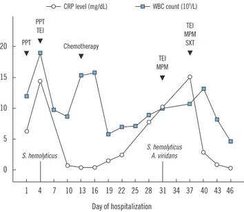
The physician added sulfamethoxazole/trimethoprim to teico- planin and meropenem combination therapy. Consequently, ex- udate lessened, and WBC count and CRP level also decreased (Fig. 1).
It is difficult to distinguish simple colonization from real infec- tion when A. viridans is identified in a wound specimen. How- ever, we paid attention to our A. viridans isolate because this pa- tient struggled with chemotherapy before A. viridans was isolated and because the resistance may spread to other gram-positive cocci through transfer of vanA from A. viridans [10].
Authors’ Disclosures of Potential Conflicts of Interest
No potential conflicts of interest relevant to this article were re- ported.
REFERENCES
1. Rasmussen M. Aerococci and aerococcal infections. J Infect 2013;66:
467-74.
2. Moreno LZ, Matajira CE, Gomes VT, Silva AP, Mesquita RE, Christ AP, et al. Molecular and antibiotic susceptibility characterization of Aerococ- cus viridans isolated from porcine urinary infection. Vet Microbiol 2016;184:7-10.
3. Kerbaugh MA and Evans JB. Aerococcus viridans in the hospital envi- ronment. Appl Microbiol 1968;16:519-23.
4. Gopalachar A, Akins RL, Davis WR, Siddiqui AA. Urinary tract infection caused by Aerococcus viridans, a case report. Med Sci Monit 2004;10:CS73-5.
5. Popescu GA, Benea E, Mitache E, Piper C, Horstkotte D. An unusual bacterium, Aerococcus viridans, and four cases of infective endocardi- tis. J Heart Valve Dis 2005;14:317-9.
6. Uh Y, Son JS, Jang IH, Yoon KJ, Hong SK. Penicillin-resistant Aerococ- cus viridans bacteremia associated with granulocytopenia. J Korean Med Sci 2002;17:113-5.
7. Zhou WQ, Niu DM, Zhang ZZ, Ning MZ, Shen H, Zhang K. Vancomycin resistance due to VanA in an Aerococcus viridans isolate. Indian J Med Microbiol 2014;32:462-5.
8. Clinical and Laboratory Standards Institute. Methods for antimicrobial dilution and disk susceptibility testing of infrequently isolated or fastidi- ous bacteria. CLSI guideline M45. Wayne, PA: Clinical and Laboratory Standards Institute, 2015.
9. Kwon OG, Uh Y, Jang IH, Lee MK, Yoon KJ, Kim HY. Trend of isolation and genotypes of vancomycin-resistant enterococci isolated from tertia- ry care hospital in Wonju area. Korean J Clin Pathol 2000;20:486-93.
10. Périchon B and Courvalin P. VanA-type vancomycin-resistant Staphylo- coccus aureus. Antimicrob Agents Chemother 2009;53:4580-7.
Fig. 1. Changes in white blood cell count and C-reactive protein lev- el during hospitalization.
Abbreviations: CRP, C-reactive protein; MPM, meropenem; PPT, piperacil- lin/tazobactam; SXT, sulfamethoxazole/trimethoprim; TEI, teicoplanin; WBC, white blood cell.
20 15 10 5
0
1 4 7 10 13 16 19 22 25 28 31 34 37 40 43 46 Day of hospitalization
PPT
TEI TEI
MPM SXT TEI MPM
PPT Chemotherapy
S. hemolyticus S. hemolyticus
A. viridans CRP level (mg/dL) WBC count (109/L)