Efficacy of Surgical Treatment for Brain Metastasis in Patients with Non-Small Cell Lung Cancer
Sang Young Kim,
1Chang Ki Hong,
2Tae Hoon Kim,
3Je Beom Hong,
2Chul Hwan Park,
3Yoon Soo Chang,
1Hyung Jung Kim,
1Chul Min Ahn,
1and Min Kwang Byun
11Division of Pulmonology, Department of Internal Medicine, Departments of 2Neurosurgery and 3Radiology, Gangnam Severance Hospital, Yonsei University College of Medicine, Seoul, Korea.
Received: December 24, 2013 Revised: April 17, 2014 Accepted: April 18, 2014
Corresponding author: Dr. Min Kwang Byun, Division of Pulmonology,
Department of Internal Medicine, Gangnam Severance Hospital, Yonsei University College of Medicine, 211 Eonju-ro, Gangnam-gu, Seoul 135-720, Korea.
Tel: 82-2-2019-3454, Fax: 82-2-3463-3882 E-mail: [email protected]
∙ The authors have no financial conflicts of interest.
© Copyright:
Yonsei University College of Medicine 2015 This is an Open Access article distributed under the terms of the Creative Commons Attribution Non- Commercial License (http://creativecommons.org/
licenses/by-nc/3.0) which permits unrestricted non- commercial use, distribution, and reproduction in any medium, provided the original work is properly cited.
Purpose: Patients with non-small cell lung cancer (NSCLC) and simultaneously having brain metastases at the initial diagnosis, presenting symptoms related brain metastasis, survived shorter duration and showed poor quality of life. We analyzed our experiences on surgical treatment of brain metastasis in patients with NSCLC.
Materials and Methods: We performed a single-center, retrospective review of 36 patients with NSCLC and synchronous brain metastases between April 2006 and December 2011. Patients were categorized according to the presence of neuro- logical symptoms and having a brain surgery. As a result, 14 patients did not show neurological symptoms and 22 patients presented neurological symptoms. Symp- tomatic 22 patients were divided into two groups according to undergoing brain surgery (neurosurgery group; n=11, non-neurosurgery group; n=11). We analyzed overall surgery (OS), intracranial progression-free survival (PFS), and quality of life. Results: Survival analysis showed there was no difference between patients with neurosurgery (OS, 12.1 months) and non-neurosurgery (OS, 10.2 months;
p=0.550). Likewise for intracranial PFS, there was no significant difference be- tween patients with neurosurgery (PFS, 6.3 months) and non-neurosurgery (PFS, 5.3 months; p=0.666). Reliable neurological one month follow up by the Medical Research Council neurological function evaluation scale were performed in symp- tomatic 22 patients. The scale improved in eight (73%) patients in the neurosur- gery group, but only in three (27%) patients in the non-neurosurgery group (p=0.0495). Conclusion: Patients with NSCLC and synchronous brain metasta- ses, presenting neurological symptoms showed no survival benefit from neurosur- gical resection, although quality of life was improved due to early control of neu- rological symptoms.
Key Words: Non-small cell lung cancer, brain metastasis, neurosurgery, quality of life, treatment outcome
INTRODUCTION
Brain metastases (BMs) are found in approximately 20‒40% of all patients with
non-small cell lung cancer (NSCLC), especially in adenocarcinoma.
1The overall
tive survival prediction in patients with BM from various primary malignancies. This model has three classes based on age at diagnosis, presence or absence of extracranial me- tastases, KPS, and primary tumor status. A higher RPA class represents a worse prognosis. This study was ap- proved by the Institutional Review Board of Gangnam Sev- erance Hospital, Yonsei University College of Medicine (IRB No: 3-2012-0179), and the necessity for written in- formed consent was waived since this was a retrospective study and patients were anonymous.
Radiologic parameters of brain metastasis
Reviewed radiologic parameters of the brain metastases in- cluded number, size, location, and functional grade.
7In ad- dition, radiologic findings such as edema, hemorrhage, and necrosis were included. BM size was defined as the maxi- mal orthogonal diameter based on T1-weighted gadolini- um-enhanced magnetic resonance imaging (MRI).
Treatment options for brain metastasis
Therapeutic approaches to brain metastases included sur- gery, whole brain radiotherapy (WBRT), stereotactic ra- diosurgery (SRS), and chemotherapy. Many patients were treated with combinations of these modalities. Treatment decisions took into account several parameters such as age, neurologic symptoms, functional status, and intracranial lesions. The decision as to whether intracranial lesions were surgically resected were based on neurologic symp- toms and signs of intracranial hypertension unresponsive to adequate medical therapy (e.g., intravenous corticoste- roid and/or mannitol), insufferable headaches, progressive motor weakness, gait disturbance, or dysarthria. Surgical indications were based on lesion-related relevant symp- toms and radiologic brain imaging showing accessible lo- cation and a mass effect due to edema unresponsive to medical therapy.
Patients with an asymptomatic brain lesion or patients with symptoms showing poor performance status were rec- ommended to undergo WBRT alone without surgery. In these patients, WBRT was delivered with 37.50 gray in 15 fractions by two lateral beams of photons of 6 megavolts or 30.00 gray in 10 fractions in patients with poor general health status and estimated survival less than 3 months.
Once patients received surgery or radiotherapy, systemic platinum-based chemotherapy was started. Initially, two cy- cles of systemic chemotherapy were administered. Follow- ing response assessment after the first two cycles of chemo- survival (OS) of these patients is generally poor, ranging
from 3 to 6 months.
2Neurologically symptomatic patients have significantly shorter survival times than asymptomatic patients, as the median survival times have been estimated to be 4 and 7.5 months, respectively.
3The optimal treatment for patients with neurologically symptomatic, synchronous (simultaneously having brain metastasis at the initial diagnosis), and multiple or solitary brain metastasis from NSCLC still remains controversial.
Surgical resection of these metastatic lesions is considered in only 25% of patients.
4Recently, surgery for stage IV NSCLC (involving an isolated brain or adrenal gland me- tastasis) has been recommended as a treatment option if mediastinal lymph nodes are not involved.
5Brain metasta- tectomy is generally considered for patients who demon- strate good performance status and have a solitary, accessi- ble, symptomatic brain metastasis with significant mass effect, and in addition, for those who need a histologic di- agnosis and for those in whom previous radiation therapy has failed.
4Recently, due to advances in surgical tech- niques, more lesions are now accessible, and surgical com- plications and post-surgical morbidity have been reduced compared to the situation in the past.
In this study, we retrospectively evaluated OS, intracranial progression-free survival (PFS), prognostic factors, and op- timal treatment for brain metastasis in patients with neuro- logically symptomatic, synchronous brain metastasis from NSCLC and compared patients in the neurosurgery group, the non-neurosurgery group, and the asymptomatic group.
MATERIALS AND METHODS
Patient characteristics
Medical records at Gangnam Severance Hospital from
April 2006 through December 2011 were reviewed to iden-
tify patients who presented with synchronous brain metas-
tasis from NSCLC. In all patients, brain metastases were
confirmed at the initial diagnosis of the primary lung can-
cer. The subjects of this study were 36 patients with or
without neurological symptoms. Symptomatic patients
were divided into two groups according to the surgery per-
formed for the brain metastases. The medical records of
each subject were examined for gender, age, smoking histo-
ry, initial symptoms, pathology, Karnofsky Performance
Status (KPS), and Recursive Partitioning Analysis (RPA)
class.
6RPA is the most commonly used model for prospec-
RESULTS
Demographic characteristics
A total of 36 patients were histologically confirmed as NSCLC and radiologically diagnosed as having brain me- tastasis between April 2006 and December 2011. Fourteen patients did not present with neurological symptoms and 22 patients presented with neurological symptoms at admis- sion. Of the neurologically symptomatic 22 patients, 11 pa- tients were treated with neurosurgical treatment and 11 pa- tients were not treated with neurosurgery. Non-neurosurgery group was treated with WBRT or palliative therapy. Table 1 shows the characteristics of 22 patients with neurologically symptomatic, synchronous brain metastasis from NSCLC.
Between the neurosurgery and non-neurosurgery groups, there were no significant differences in any characteristics evaluated. Current or past smoking status, performance sta- tus, prognostic factors, and neurological scale by MRC neu- rological function evaluation at initial diagnosis showed no differences between the two treatment groups.
Pathology, number of brain metastasis and treatment modalities
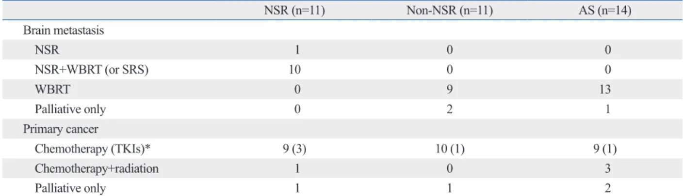
Table 2 shows the pathologic diagnosis and the number of brain metastases and treatment modalities which the symp- tomatic 22 patients and asymptomatic 14 patients under- went. In the neurosurgery group, six patients (55%) showed multiple synchronous metastases and five patients (45%) showed solitary metastasis. In the non-neurosurgery group, 7 patients (64%) showed multiple metastasis and 4 (36%) patients showed single metastasis. These patients were clas- sified for treatment modality. Of the 11 patients in the neu- rosurgery group, 10 patients were followed by WBRT or SRS and AS patients received WBRT or SRS only.
In all patients in the neurosurgery group, the one lesion that was the largest, symptomatic, and surgically accessible was resected. The resected lesions ranged in size from 1.9 to 5.6 cm (median 3.4 cm). The characteristics of multiple metastatic lesions, each patient’s performance scale, and OS are summarized in Table 3.
Treatment outcomes
We analyzed OS and PFS, after excluding 4 patients (3 pa- tients treated with neurosurgery group; 1 patient treated without neurosurgery) harboring sensitive EGFR mutations and treated with EGFR-TKIs following neurosurgery or ra- therapy and in the absence of progression either at the
primary lung tumor or BM, chemotherapy was indicated.
Four patients harboring sensitive epidermal growth factor receptor (EGFR) mutations continuously received EGFR- tyrosine kinase inhibitors (EGFR-TKIs) following surgery or radiotherapy. Palliative treatment only (steroids and anti- convulsants) was administered to patients with poor perfor- mance status or high comorbidity, or to patients who did not agree to undertake invasive treatment.
Follow-up schedules
Follow-up for primary lung cancer was performed with computed tomography, generally every 2 months. Follow- up for brain metastases was conducted by MRI, usually ev- ery two months. Treatment response in patients with BM was evaluated according to the Response Evaluation Crite- ria in Solid Tumors (RECIST) criteria after every two cy- cles in chemotherapy or radiotherapy.
The tumor response in the BM was evaluated by enhanced brain MRI and neurological findings. Neurological status was assessed using the Medical Research Council (MRC) neurological function scale at 1 month after intracranial treat- ment.
8If there were measurable lesions, the tumors were evaluated based on the RECIST version 1.1 criteria. If there were no measurable lesions, the tumor response was classi- fied as partial response, stable disease, or progressive dis- ease based on the enhanced brain MRI findings and neuro- logical findings. Intracranial progression after neurosurgical resection or radiotherapy was defined as either progressive enlargement of the enhancing lesion or no change in tumor size but persistent, symptomatic edema.
Statistical analysis
Data were analyzed with SAS 9.2 version (SAS Institute Inc., Cary, NC, USA). OS was defined as the time elapsed from the starting date of treatment of the brain metastasis or pulmonary lesions to the date of death or the date of last fol- low-up. Intracranial PFS was defined as the time from the date of the start of treatment to the date of documented dis- ease progression or death from any cause. Risk factors af- fecting the survival rate were found using Fisher’s exact test and log-rank test, thus multiple survival analyses with ad- justed and associated risk factors were performed using Cox’s proportional hazard regression model. Median time to intracranial progression and OS were analyzed by the Ka- plan-Meier estimator. Significance was set at a p-value of
<0.05. All tests were two-sided.
Table 1. Demographic and Clinical Characteristics of 22 Patients with Neurologically Symptomatic, Synchronous Brain Me- tastasis from NSCLC
Characteristics NSR (n=11) Non-NSR (n=11) p value
Sex 1.000
Male 9 10
Age 0.553
Median (range) 65.6 (50‒81) 61.9 (40‒76)
<65 5 6
≥65 6 5
Smoking history 1.000
Never 8 8
Former or current 3 3
Pathology 1.000
Adenocarcinoma 8 8
Squamous cell carcinoma 1 1
NSCLC-not otherwise specified 2 2
Number of brain metastases (%) 0.475
Single 5 (45) 4 (36)
Multiple 6 (55) 7 (64)
KPS score 0.635
≥70 8 7
<70 3 4
RPA class 0.325
Class I 3 0
Class II 6 8
Class III 2 3
MRC scale* 0.716
1 0 0
2 or 3 5 6
4 or 5 6 5
NSCLC, non-small cell lung cancer; NSR, neurosurgical resection; KPS, Karnofsky Performance Status; RPA, recursive partitioning analysis.
1. No neurological deficit. 2. Some deficit but adequate function for useful work. 3. Deficits causing moderate functional impairment (e.g., moderate dysphasia, moderate paresis, or visual disturbance such as field defect). 4. Deficit causing major functional impairment (e.g., inability to use limb, gross speech impairment, or visual disturbances). 5. Inability to make conscious responses.
*Medical Research Council (MRC) neurological function scale.
Table 2. Treatment Modality in 36 Patients with Synchronous Brain Metastasis Originating from NSCLC
NSR (n=11) Non-NSR (n=11) AS (n=14)
Brain metastasis
NSR 1 0 0
NSR+WBRT (or SRS) 10 0 0
WBRT 0 9 13
Palliative only 0 2 1
Primary cancer
Chemotherapy (TKIs)* 9 (3) 10 (1) 9 (1)
Chemotherapy+radiation 1 0 3
Palliative only 1 1 2
NSR, neurosurgical resection; AS, asymptomatic; WBRT, whole brain radiotherapy; SRS, stereotactic radiosurgery; TKIs, tyrosine kinase inhibitors; NSCLC, non-small cell lung cancer.
*Patients received epidermal growth factor receptor (EGFR)-TKI treatment after the identification of EGFR mutation on the tissue from brain metastasis le- sion.
for each group. There were no significant differences be- tween the groups in terms of these characteristics. Current- ly, two patients are alive without recurrence after treatment of intracranial metastasis. These patients are from the non- neurosurgery.
Neurological function score
Reliable neurological 1-month follow-up using the MRC neurological function evaluation scale was available for 22 symptomatic patients. The MRC scale improved in 8 (73%) neurosurgery patients and did not change in 3 (27%) neuro- surgery patients, whereas it improved in 3 (27%) non-neu- rosurgery patients, worsened in one (9%) non-neurosurgery diotherapy, because such patients are usually responsive to
EGFR-TKIs and show favorable prognosis.
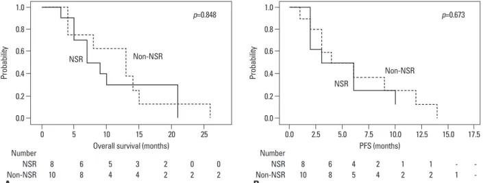
There was no difference in survival between 8 patients with neurosurgery (OS, 12.1 months) and 10 patients with non-neurosurgery (OS, 10.2 months; p=0.550). Likewise for intracranial PFS, there was no significant difference between the neurosurgery group (6.3 months) and the non-neurosur- gery group (5.3 months; p=0.666) (Fig. 1). Additionally, there was no difference in OS (p=0.363) and intracranial PFS between the symptomatic group (neurosurgery group plus non-neurosurgery group) and the asymptomatic group (n=
14, p=0.083) (Table 4). Table 4 also shows the response after BM treatment (p=0.669) and the cause of death (p=0.773)
Table 3. Features of Six Patients with Neurologically Symptomatic, Synchronous and Multiple Brain Metastasis Who Re- ceived Neurosurgery
1 2 3 4 5 6
Brain image
No. of metastasis 5 2 2 3 10 3
No. of resection 1 1 1 1 1 1
Size (cm) 3.1 5.6 1.9 2.5 2.7 4.6
Site Parietal Cerebellum Cortex Cerebellum Parietal Cerebellum
Edema + + + + + +
Hemorrhage + - - - - +
Necrosis + - - - - -
Mass effect + + + + + +
PS
KPS score ≥70 ≥70 ≥70 <70 <70 ≥70
RPA class II II II III III I
OS (month) 13 4 23 4 13 16
EGFR
Mutation analysis - - Mutant Mutant Wild Mutatn
Selected regimen - - Erlotinib Gefitinib - Gefitinib
PS, performance status; KPS, Karnofsky Performance Status; RPA, recursive partitioning analysis status; OS, overall survival; EGFR, epidermal growth fac- tor receptor.
Fig. 1. Kaplan-Meier estimate curves of OS (A) and intracranial PFS (B) for NSR and non-NSR group patients. OS, overall survival; PFS, progression free sur- vival; NSR, neurosurgical resection.
A B
Overall survival (months) 0.0
0.2 0.4 0.6 0.8 1.0
Probability
0 5 10 15 20 25
8 6 5 3 2 0 0
NSR Number
10 8 4 4 2 2 2
Non-NSR
p=0.848
NSR Non-NSR
PFS (months) 0.0
0.2 0.4 0.6 0.8 1.0
Probability
0.0 2.5 5.0 7.5 10.0 12.5 15.0 17.5
8 6 4 2 1 1 - -
NSR Number
10 8 5 4 2 2 1 -
Non-NSR
p=0.673
NSR
Non-NSR
occur in 30‒50% of patients with NSCLC, and confers a poor prognosis and a negative impact on quality of life (QOL) dur- ing the course of their disease.
10-12Patients with NSCLC and synchronous brain metastasis on initial diagnosis, who presented with neurological deficits, had a short median survival and lower QOL.
3Studies have been performed on the effect of neurosurgery for brain metastasis in the pa- tients with neurological symptoms on diagnosis; however, their results were limited to patients with a solitary metasta- sis.
13-16We performed early surgical treatment of brain metas- tasis (including multiple metastases) in patients with lowered performance status (i.e., KPS <70 or RPA class II or III) (Ta- ble 1). Early surgical treatment controlled the progression of brain metastasis and improved neurologic symptoms, and was followed by better performance status that enabled sys- temic chemotherapy (Fig. 2).
patient, and did not change in 7 (64%) non-neurosurgery patients.
The cumulative MRC neurological function scale scores of the initial symptom for the two groups were 26 for the neurosurgery group and 27 for the non-neurosurgery group (i.e., there was no difference in scores). However, the cumu- lative scale scores at 1 month after BM treatment were 16 for the neurosurgery group and 24 for the non-neurosurgery group, indicating a better functional outcome after neurosur- gery (p=0.0495) (Fig. 2).
DISCUSSION
Brain metastases are identified in 7.4‒10.0% of patients with NSCLC upon initial diagnosis.
9Moreover, brain metastases
Table 4. Clinical Outcome of 32 Patients with Synchronous Brain Metastasis from NSCLC
Clinical outcome Symptomatic Asymptomatic
(n=14) p value*
Total (n=18) NSR (n=8) Non-NSR (n=10) p value
Median OS (month) 9.5 12.1 10.2 0.550 13.1 0.363
Intracranial PFS (month) 4.5 6.3 5.3 0.666 8.4 0.083
BM response (No.) 0.827 0.669
PR 9 5 4 5
SD 3 1 2 4
PD 6 2 4 5
Cause of death (No.) 0.775 0.773
Intracranial 4 2 2 3
Extracranial 3 2 1 3
Both 9 4 5 8
Total 16 8 8 14
OS, overall survival; PR, partial response; SD, stable disease; PD, progression of disease; BM, brain metastasis; NSCLC, non-small cell lung cancer; PFS, progression-free survival; NSR, neurosurgical resection.
*p value between symptomatic vs. asymptomatic patients.
Fig. 2. Cumulative change of MRC neurological function scale at 1-month follow-up after therapy for brain metastasis. (A) NSR group (n=11). (B) Non-NSR group (n=11). p=0.0495. *Medical Research Council (MRC) neurological function scale. NSR, neurosurgical resection.
Time Time
0 0
5 5
10 10
15 15
20 20
25 25
30 30
Cumulative scale* Cumulative scale*
Initial diagnosis Follow-up (1 month) Initial diagnosis Follow-up (1 month)
A B
ths.
24For patients with favorable prognostic features [good performance status, younger age (<65 years), well-con- trolled status of the primary lung cancer, and absence of ex- tra-cranial metastasis
6,25-27], treatment with SRS and WBRT could prolong OS to 11.6 months.
28Using RPA as a prog- nostic factor, meta-analyses by the Radiation Therapy On- cology Group trials on brain metastasis treated with SRS showed that the median survival of the best prognosis group (RPA class I: age <65 years, KPS ≥70, and a controlled sys- temic disease) was 7.1 months. The expected survival of the worst prognosis group (RPA class III: KPS <70) was only 2.3 months.
6Until now, treatment for brain metastases includes surgery, SRS, WBRT, or a combination of these modalities, and has achieved median survival ranging from 6.5 to 11.0 months.
28-32In our present study, neurosurgery group showed no sig- nificant differences in survival rate (OS, 12.1 months) com- pared with non-neurosurgery group (OS, 10.2 months) (Ta- ble 4), and showed a favoring trend with 1.9 months, even though patients with a poor prognostic factor (RPA class II or III) were included, compared with the previous data.
28,32A previous study showed a prolonged survival only in patients in the AS group compared to symptomatic synchronous pa- tients.
3Our study showed a similar survival rate between pa- tients in the AS group (OS, 13.1 months) and patients in the neurosurgery group (OS, 12.1 months), and failed to show any statistical significance between the neurosurgery group (OS, 12.1 months) and the non-neurosurgery group (OS, 10.2 months) (Table 4).
In patients with a symptomatic, synchronous brain metas- tasis from NSCLC, the brain lesion mostly has life-threaten- ing mass effects and causes focal neurological deficits that may considerably decrease the functional grade. Thus, ear- ly surgery can immediately provide symptom relief and im- prove QOL in the patients.
There are several limitations in the present study. First, it was a retrospective study, the study population was small, and the research was carried out in a single institution, con- sequently, it is difficult to generalize this result to other set- tings. Second, we compared OS between three treatment groups without considering the control of primary lung can- cer. We could not exclude systemic chemotherapeutic effect for primary lung cancer, and brain metastasis is usually con- sidered most crucial factor in mortality. A large-scale pro- spective randomized study is needed for generalization of this result.
In conclusion, patients with synchronous brain metastasis, Recently, surgical intervention has demonstrated increased
survival rates and decreased complications of post-opera- tion as positive benefits; therefore, resection of a single brain metastasis has become the standard treatment option in pa- tients with good functional status and controlled extra-cra- nial disease.
15,16Advances in surgical techniques have led to lower rates of morbidity and in-hospital mortality, with esti- mates as low as 1.8% in high-volume centers.
17However, in the case of multiple brain metastases, surgery is usually lim- ited to patients with a dominant, symptomatic, or life-threat- ening lesion and to those who require a tissue diagnosis be- fore proceeding with therapy. However, two recent single- center retrospective studies have suggested that patients with good prognostic features and two to three metastases may gain same survival benefit from surgery as patients with one metastasis, if the dominant lesion is resected.
18,19Despite these benefits of surgical resection, surgical candidates can- not clearly be defined. There is especially insufficient evi- dence to make a recommendation for patients with poor performance status, advanced systemic disease, or multiple brain metastases.
In our present study, surgery was performed for accessi- ble brain metastasis causing a mass effects, peri-tumoral ce- rebral edema, and insufferable neurological deficits even if patients had poor performance status or multiple brain me- tastases. There were no complications in 11 patients from the neurosurgery group. Neurological symptomatic com- plaints of neurosurgery group patients were relieved signifi- cantly compared to those of the non-neurosurgery group 1 month after surgery, as indicated by the MRC neurological function evaluation scale (Fig. 2). Furthermore, it is impor- tant to confirm tissue pathology before proceeding with therapy because it is recommended as an option for the first line treatment of NSCLC patients who have a brain metas- tasis and harbor an EGFR mutation with EGFR-TKIs.
20,21Moreover, discordance in the EGFR mutation status be- tween the primary and metastatic brain lesions has been re- cently reported in several studies and the discordance rate was reported to reach 27‒28%.
22,23In our study, early EG- FR-TKI treatment was conducted in three of nine patients receiving systemic chemotherapy, after identification of the EGFR mutation on the metastatic brain tissue (Table 2).
Diverse clinical studies have been carried out to provide a
guideline for the optimal treatment of patients with a symp-
tomatic, synchronous brain metastasis from NSCLC. The
median survival of patients treated with only supportive care
(such as corticosteroids) has been estimated to be 1‒2 mon-
11. Lagerwaard FJ, Levendag PC, Nowak PJ, Eijkenboom WM, Hanssens PE, Schmitz PI. Identification of prognostic factors in patients with brain metastases: a review of 1292 patients. Int J Ra- diat Oncol Biol Phys 1999;43:795-803.
12. Schouten LJ, Rutten J, Huveneers HA, Twijnstra A. Incidence of brain metastases in a cohort of patients with carcinoma of the breast, colon, kidney, and lung and melanoma. Cancer 2002;94:
2698-705.
13. Louie AV, Rodrigues G, Yaremko B, Yu E, Dar AR, Dingle B, et al. Management and prognosis in synchronous solitary resected brain metastasis from non-small-cell lung cancer. Clin Lung Can- cer 2009;10:174-9.
14. Rades D, Raabe A, Bajrovic A, Alberti W. Treatment of solitary brain metastasis. Resection followed by whole brain radiation therapy (WBRT) and a radiation boost to the metastatic site.
Strahlenther Onkol 2004;180:144-7.
15. Granone P, Margaritora S, D’Andrilli A, Cesario A, Kawamukai K, Meacci E. Non-small cell lung cancer with single brain metas- tasis: the role of surgical treatment. Eur J Cardiothorac Surg 2001;
20:361-6.
16. Bonnette P, Puyo P, Gabriel C, Giudicelli R, Regnard JF, Riquet M, et al. Surgical management of non-small cell lung cancer with synchronous brain metastases. Chest 2001;119:1469-75.
17. Barker FG 2nd. Craniotomy for the resection of metastatic brain tumors in the U.S., 1988-2000: decreasing mortality and the effect of provider caseload. Cancer 2004;100:999-1007.
18. Paek SH, Audu PB, Sperling MR, Cho J, Andrews DW. Reevalu- ation of surgery for the treatment of brain metastases: review of 208 patients with single or multiple brain metastases treated at one institution with modern neurosurgical techniques. Neurosurgery 2005;56:1021-34.
19. Stark AM, Tscheslog H, Buhl R, Held-Feindt J, Mehdorn HM.
Surgical treatment for brain metastases: prognostic factors and survival in 177 patients. Neurosurg Rev 2005;28:115-9.
20. Jazieh AR, Bamefleh H, Demirkazik A, Gaafar RM, Geara FB, Ja- vaid M, et al. Modification and implementation of NCCN guide- lines on non-small cell lung cancer in the Middle East and North Africa region. J Natl Compr Canc Netw 2010;8 Suppl 3:S16-21.
21. Park SJ, Kim HT, Lee DH, Kim KP, Kim SW, Suh C, et al. Effi- cacy of epidermal growth factor receptor tyrosine kinase inhibitors for brain metastasis in non-small cell lung cancer patients harbor- ing either exon 19 or 21 mutation. Lung Cancer 2012;77:556-60.
22. Kalikaki A, Koutsopoulos A, Trypaki M, Souglakos J, Stathopou- los E, Georgoulias V, et al. Comparison of EGFR and K-RAS gene status between primary tumours and corresponding metasta- ses in NSCLC. Br J Cancer 2008;99:923-9.
23. Gow CH, Chang YL, Hsu YC, Tsai MF, Wu CT, Yu CJ, et al.
Comparison of epidermal growth factor receptor mutations be- tween primary and corresponding metastatic tumors in tyrosine kinase inhibitor-naive non-small-cell lung cancer. Ann Oncol 2009;20:696-702.
24. Galluzzi S, Payne PM. Brain metastases from primary bronchial carcinoma: a statistical study of 741 necropsies. Br J Cancer 1956;
10:408-14.
25. Penel N, Brichet A, Prevost B, Duhamel A, Assaker R, Dubois F, et al. Pronostic factors of synchronous brain metastases from lung cancer. Lung Cancer 2001;33:143-54.
26. Vogelbaum MA, Suh JH. Resectable brain metastases. J Clin On- col 2006;24:1289-94.
27. Sánchez de Cos J, Sojo González MA, Montero MV, Pérez Calvo
presenting with neurological symptoms, showed no survival benefit from combined treatment with neurosurgical resec- tion, although their QOL was improved due to early resolu- tion of the neurological symptoms. Future studies, incorpo- rating the role of resection for more than one brain metastasis with or without additional adjuvant therapy, will help clarify whether the benefits of resection discussed above apply to multiple lesions. However, prospective clinical trials should be done to confirm the benefits of this treatment.
ACKNOWLEDGEMENTS
The authors are grateful for statistical support provided by the Biostatistics Collaboration Unit of Yonsei University Col- lege of Medicine.
REFERENCES
1. Yawn BP, Wollan PC, Schroeder C, Gazzuola L, Mehta M. Tem- poral and gender-related trends in brain metastases from lung and breast cancer. Minn Med 2003;86:32-7.
2. Patchell RA, Tibbs PA, Walsh JW, Dempsey RJ, Maruyama Y, Kryscio RJ, et al. A randomized trial of surgery in the treatment of single metastases to the brain. N Engl J Med 1990;322:494-500.
3. Billing PS, Miller DL, Allen MS, Deschamps C, Trastek VF, Pai- rolero PC. Surgical treatment of primary lung cancer with syn- chronous brain metastases. J Thorac Cardiovasc Surg 2001;122:
548-53.
4. Cairncross JG, Kim JH, Posner JB. Radiation therapy for brain metastases. Ann Neurol 1980;7:529-41.
5. Shen KR, Meyers BF, Larner JM, Jones DR; American College of Chest Physicians. Special treatment issues in lung cancer: ACCP evidence-based clinical practice guidelines (2nd edition). Chest 2007;132(3 Suppl):290S-305S.
6. Gaspar L, Scott C, Rotman M, Asbell S, Phillips T, Wasserman T, et al. Recursive partitioning analysis (RPA) of prognostic factors in three Radiation Therapy Oncology Group (RTOG) brain metas- tases trials. Int J Radiat Oncol Biol Phys 1997;37:745-51.
7. Sawaya R, Hammoud M, Schoppa D, Hess KR, Wu SZ, Shi WM, et al. Neurosurgical outcomes in a modern series of 400 cranioto- mies for treatment of parenchymal tumors. Neurosurgery 1998;42:
1044-55.
8. van den Bent MJ, Afra D, de Witte O, Ben Hassel M, Schraub S, Hoang-Xuan K, et al. Long-term efficacy of early versus delayed radiotherapy for low-grade astrocytoma and oligodendroglioma in adults: the EORTC 22845 randomised trial. Lancet 2005;366:985- 9. Schuette W. Treatment of brain metastases from lung cancer: che-90.
motherapy. Lung Cancer 2004;45 Suppl 2:S253-7.
10. Sørensen JB, Hansen HH, Hansen M, Dombernowsky P. Brain metastases in adenocarcinoma of the lung: frequency, risk groups, and prognosis. J Clin Oncol 1988;6:1474-80.
Black PM, et al. Stereotactic radiosurgery for the definitive, non- invasive treatment of brain metastases. J Natl Cancer Inst 1995;87:
34-40.
31. Kondziolka D, Patel A, Lunsford LD, Kassam A, Flickinger JC.
Stereotactic radiosurgery plus whole brain radiotherapy versus ra- diotherapy alone for patients with multiple brain metastases. Int J Radiat Oncol Biol Phys 1999;45:427-34.
32. Kocher M, Soffietti R, Abacioglu U, Villà S, Fauchon F, Baumert BG, et al. Adjuvant whole-brain radiotherapy versus observation after radiosurgery or surgical resection of one to three cerebral metastases: results of the EORTC 22952-26001 study. J Clin On- col 2011;29:134-41.
MC, Vicente MJ, Valle MH. Non-small cell lung cancer and silent brain metastasis. Survival and prognostic factors. Lung Cancer 2009;63:140-5.
28. Andrews DW, Scott CB, Sperduto PW, Flanders AE, Gaspar LE, Schell MC, et al. Whole brain radiation therapy with or without stereotactic radiosurgery boost for patients with one to three brain metastases: phase III results of the RTOG 9508 randomised trial.
Lancet 2004;363:1665-72.
29. Noordijk EM, Vecht CJ, Haaxma-Reiche H, Padberg GW, Voor- molen JH, Hoekstra FH, et al. The choice of treatment of single brain metastasis should be based on extracranial tumor activity and age. Int J Radiat Oncol Biol Phys 1994;29:711-7.
30. Alexander E 3rd, Moriarty TM, Davis RB, Wen PY, Fine HA,