• 검색 결과가 없습니다.

A Case of Histiocytoid Variant Eccrine Sweat Gland Carcinoma of the Orbit

N/A
N/A
Protected

Academic year: 2022

Share "A Case of Histiocytoid Variant Eccrine Sweat Gland Carcinoma of the Orbit"

Copied!
3
0
0

로드 중.... (전체 텍스트 보기)

전체 글

(1)

54

Korean J Ophthalmol 2011;25(1):54-56 DOI: 10.3341/kjo.2011.25.1.54 pISSN: 1011-8942 eISSN: 2092-9382

Case Report

A Case of Histiocytoid Variant Eccrine Sweat Gland Carcinoma of the Orbit

Young Min Kim

1

, Jeong Won Kim

2

, Dong-Eun Oh

1

1

Department of Ophthalmology, Seoul Veterans Hospital, Seoul, Korea

2

Department of Pathology, Kangnam Sacred Heart Hospital, Hallym University College of Medicine, Seoul, Korea

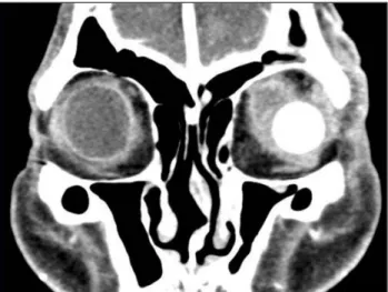
A 79-year-old male presented with left ocular pain. Evisceration and silicone ball implantation were performed after a diagnosis of phthisis. He returned six weeks later because of left facial erythematous swelling, tenderness, mild fever, chills and cough. His condition was diagnosed as orbital cellulitis. Despite two weeks of empirical antibiotic therapy, the symptoms worsened. A subsequent orbital computed tomography scan revealed enhanced soft tissue infiltrations in his left orbit and eyelid. Biopsy showed a diffusely infiltrating tumor of signet ring cell cytology. A sys- temic evaluation revealed multiple bone metastases. Based on this evidence, the patient was diagnosed with a very rare case of histiocytoid variant eccrine sweat gland carcinoma with multiple bone metastases.

Key Words: Eccrine sweat gland carcinoma, Histiocytoid variant, Signet ring cell

ⓒ2011 The Korean Ophthalmological Society

This is an Open Access article distributed under the terms of the Creative Commons Attribution Non-Commercial License (http://creativecommons.org/licenses /by-nc/3.0/) which permits unrestricted non-commercial use, distribution, and reproduction in any medium, provided the original work is properly cited.

Received: July 1, 2010 Accepted: August 24, 2010

Corresponding Author: Dong-Eun Oh, MD. Department of Ophthalmology, Seoul Veterans Hospital, #6-2 Dunchon 2-dong, Gangdong-gu, Seoul 134-791, Korea. Tel: 82-2-2225-1884, Fax: 82-2-2225-1485, E-mail: ezer75

@hanmail.net

Fig. 1. Clinical photograph of the erythematous swelling of the left eyelid.

The histiocytoid variant of eccrine sweat gland carcinoma is very rare, although the histiocytoid variant of eccrine sweat gland carcinoma (specifically, infiltrating signet ring carcinoma) occurring in the eyelid has been reported [1-6].

The histiocytoid variant is a malignant tumor diffusely in- filtrating the dermis and subcutaneous tissue without epi- dermal involvement. Here, we report the first Korean case of a histiocytoid variant of eccrine sweat gland carcinoma, showing infiltrations of signet ring cell cytology and multiple bone metastases. The symptoms were initially misdiagnosed as orbital cellulitis after surgery.

Case Report

A 79-year-old male visited our clinic for the evaluation of left ocular pain that had recently worsened. His left eye was phthitic due to glaucoma. The patient had also previously un- dergone an endoscopic rectal polypectomy. Evisceration and silicone ball (18 mm) implantation were performed. He re- turned six weeks later with symptoms of mild fever, cough, and chills, as well as left facial erythema, swelling, and ten-

derness (Fig. 1). We diagnosed the symptoms as orbital cellu-

litis induced by recent evisceration surgery and the common

cold. However, despite two weeks of empirical antibiotic

therapy, the patient’s symptoms worsened. An orbital com-

puted tomography (CT) scan revealed enhanced soft tissue

infiltrations in his left extraconal and intraconal spaces and a

thickening of the left eyelid (Fig. 2). An incisional biopsy of

the orbital tissue was performed through the superomedial

portion of the conjunctiva. The tumor was gray-white col-

ored and was not separated from the surrounding tissue on

gross examination. Histopathology demonstrated cells with

(2)

YM Kim, et al. Eccrine Sweat Gland Carcinoma

55 Fig. 2. Enhanced extraconal and intraconal infiltrations in the left

orbit, and an inferolateral view of the silicone ball sphere in the eyelid.

Fig. 3. The tumors were composed of cells with hyperchromatic ec- centric nuclei (suggesting signet ring cells), prominent eosinophilic cytoplasm, and intracytoplasmic vacuolations (H&E, ×400).

Fig. 4. A positron emission tomography image revealed the multiple intense hypermetabolic foci on the left orbit and the skull as well as along the spine.

round to oblong nuclei, small distinct nucleoli, and relatively abundant granular amphophilic cytoplasm. Some cells had signet ring cytology, with a dominant cytoplasmic vacuole and an eccentrically placed, crescent-shaped nucleus (Fig.

3). Immunohistochemistry revealed that the tumor tissue was positive for cytokeratin and cytokeratin 7 but negative for cy- tokeratin 20.

Results of an abdominal ultrasound, abdominal-pelvic CT, and a whole body bone scan suggested no systemic involvement. Additionally, tumor marker tests were all with- in the normal ranges. However, brain magnetic resonance imaging and positron emission tomography revealed meta- static lesions on the left frontal and temporal lobes of the skull; nasal bone; left basal skull; right mandibular ramus and sternum; vertebra in the cervical, thoracic, lumbar and sacral regions; and the left ischium (Fig. 4). To rule out a secondary tumor from the previous rectal lesion, rectal biopsy was per-

formed and revealed a benign adenoma confined to the mu- cosal layer. Consequently, the lesion in question was diag- nosed as a primary malignant eccrine sweat gland carcinoma of the eye (a histiocytoid variant of eccrine sweat gland carci- noma) with multiple bone metastases. Although the patient was fully functional on the first visit, his general condition declined during the subsequent three months. The patient be- came bedridden, refused all recommended treatments, and received only conservative treatment.

Discussion

Primary eccrine sweat gland carcinoma is a very rare tu- mor that most typically occurs in the eyelids of the elderly.

There are three variants of this type of carcinoma: mucinous,

adenoid cystic, and ductal [1-6]. The present case is of the

third variant, ductal carcinoma. Ductal carcinomas are fur-

ther classified into two categories based on the type of cell

differentiation: well-differentiated ductal carcinoma featur-

ing numerous signet ring cells are classified as signet ring

carcinoma, while poorly-differentiated ductal carcinoma is

classified as the histiocytoid variant. The tumor in question

showed the characteristics of the latter form [1,2,4,7]. This

histiocytoid variant of eccrine sweat gland carcinoma

showed a histiocyte morphology rather than a signet ring cell

morphology in the tumor burden, although histiocytoid tu-

mors may contain foci of signet ring cell morphology, and

vice versa [1,3]. Tumor cells in this case exhibited eosino-

philic cytoplasm with intracytoplasmic vacuolations, charac-

teristics similar to those of a histiocyte. The majority of tu-

mor cells did not have the distinct, eccentric, scimitar-like

nuclei of signet ring cells.

(3)

Korean J Ophthalmol Vol.25, No.1, 2011

56

A primary histiocytoid variant of eccrine sweat gland car- cinoma is a very rare neoplasm, with only eight cases having been reported in the literature [1,7]. In the cases reported, most tumor cells had abundant eosionophilic cytoplasm with intracytoplasmic vacuolations resembling histiocytes and a signet ring appearance [1,2,4,7,8]. The diagnosis of primary histiocytoid variant of eccrine sweat gland carcinoma is one of exclusion. Common primary foci include the breast, gas- trointestinal tract, lung, skin, and urogenital tract. The most common sites are breast in women and lung in men [1,7,8].

Previously reported cases of the histiocytoid variant of ec- crine sweat gland carcinoma follow an indolent course with slow and painless growth [1,2,7]. Only one of the eight cases showed metastasis, recurrence, and mortality [1]. However, these tumors can exhibit rapid growth with consequent pain.

The case reported here had an unfavorable outcome with rap- id progression, ocular pain, facial pain, and multiple bone metastases.

Most cases of primary eccrine sweat gland carcinoma de- scribed in the literature mainly appeared in the eyelid.

However, Kramer et al. [3] described the eccrine sweat gland carcinoma in the orbit. Like Kramer et al. [3], the imaging studies in this case showed soft tissue infiltrations primarily involved in the orbit. Moreover, the subcutaneous infiltra- tions of the eyelid were also noted on the imaging studies.

These facts suggested that the tumor mass in this case af- fected both the orbit and the eyelid.

The orbital imaging studies were not performed before the evisceration surgery because it was believed that the patient’s accelerated ocular pain was induced by glaucoma. At his pre- operative assessment, the patient’s left eye was phthitic and appeared stable. After the evisceration surgery, the patient’s facial pain and erythema were believed to be caused by orbi- tal cellulitis. He had multiple risk factors for orbital in- fection: advanced age, recent evisceration surgery, silicone ball implantation, and symptoms of the common cold. In hindsight, the increased ocular pain could have been caused by the tumor growth occurring at the time of the evisceration surgery, rather than glaucoma. In other words, the ocular pain could have been cancer-related pain associated with the rapid tumor growth. It is difficult to determine the exact time of tu- mor occurrence because orbital imaging was not performed before the surgical procedure. We presumed that the patient’s pre- and post-operative pain and orbital cellulitis may be re- lated to the tumor growth. Furthermore, the rapid progression of the tumor may have been accelerated by the surgical

procedure. As a result, the interval from the patient’s initial visit with simple left ocular pain to the eventual diagnosis of primary eccrine sweat gland carcinoma was only three months. After the diagnosis, multiple metastases were also detected along the spine.

To our knowledge, this is the first Korean case of a histio- cytoid variant of eccrine sweat gland carcinoma in the eye.

Based on this case, we suggest taking an orbital imaging study prior to evisceration surgery in cases of abnormal ocu- lar pain or rapid progression. In refractory cases of orbital cellulitis, aggressive follow-up may be necessary, including diagnostic confirmation through orbital imaging and tissue biopsy.

Conflict of Interest

No potential conflict of interest relevant to this article was reported.

References

1. Mortensen AL, Heegaard S, Clemmensen O, Prause JU. Signet ring cell carcinoma of the eyelid - the monocle tumour. APMIS 2008;116:326-32.

2. Langel DJ, Yeatts RP, White WL. Primary signet ring cell car- cinoma of the eyelid: report of a case demonstrating further analogy to lobular carcinoma of the breast with a literature review. Am J Dermatopathol 2001;23:444-9.

3. Kramer TR, Grossniklaus HE, McLean IW, et al. Histiocytoid variant of eccrine sweat gland carcinoma of the eyelid and or- bit: report of five cases. Ophthalmology 2002;109:553-9.

4. Swinson B, Ryan F, Barrett AW, et al. Histiocytoid eccrine sweat gland carcinoma of the eyelid: report of a case. Clin Exp Dermatol 2006;31:786-9.

5. Hoppenreijs VP, Reuser TT, Mooy CM, et al. Syringomatous carcinoma of the eyelid and orbit: a clinical and histopatho- logical challenge. Br J Ophthalmol 1997;81:668-72.

6. Krishnakumar S, Mohan ER, Babu K, et al. Eccrine duct carci- noma of the eyelid mimicking meibomian carcinoma: clin- icopathological study of a case. Surv Ophthalmol 2003;48:439-46.

7. Jakobiec FA, Austin P, Iwamoto T, et al. Primary infiltrating signet ring carcinoma of the eyelids. Ophthalmology 1983;90:

291-9.

8. Wollensak G, Witschel H, Böhm N. Signet ring cell carcinoma

of the eccrine sweat glands in the eyelid. Ophthalmology

1996;103:1788-93.

수치

Fig. 1. Clinical photograph of the erythematous swelling of the left  eyelid.
Fig. 4. A positron emission tomography image revealed the multiple  intense hypermetabolic foci on the left orbit and the skull as well as  along the spine

참조

관련 문서

• 이명의 치료에 대한 매커니즘과 디지털 음향 기술에 대한 상업적으로의 급속한 발전으로 인해 치료 옵션은 증가했 지만, 선택 가이드 라인은 거의 없음.. •

To review the distinctive features and potential problems of Korean government plans this report analyzes the case of Power Development Plan and Housing Development Plan. In

The proposal of the cell theory as the birth of contemporary cell biology Microscopic studies of plant tissues by Schleiden and of animal tissues by Microscopic studies of

Since every classical or virtual knot is equivalent to the unknot via a sequence of the extended Reidmeister moves together with the forbidden moves, illustrated in Section 2,

_____ culture appears to be attractive (도시의) to the

It considers the energy use of the different components that are involved in the distribution and viewing of video content: data centres and content delivery networks

After first field tests, we expect electric passenger drones or eVTOL aircraft (short for electric vertical take-off and landing) to start providing commercial mobility

The Joseon government designed and promulgated the Taegeukgi as a national flag for diplomatic and political purposes, but it was the independence movement that made it