• 검색 결과가 없습니다.

Successful Percutaneous Coronary Intervention in an Anomalous Origin of the Right Coronary Artery From the Ascending Aorta Above the Left Sinus of the Valsalva

N/A
N/A
Protected

Academic year: 2022

Share "Successful Percutaneous Coronary Intervention in an Anomalous Origin of the Right Coronary Artery From the Ascending Aorta Above the Left Sinus of the Valsalva"

Copied!
4
0
0

로드 중.... (전체 텍스트 보기)

전체 글

(1)

497 Copyright © 2012 The Korean Society of Cardiology

Korean Circulation Journal

Introduction

Congenital coronary anomalies are present in approximately 1%

of patients referred for cardiac catheterization.

1-3)

However, the in- cidence was found to be 5.6% in a recent prospective angiographic study of 1950 consecutive cases.

4)

Among these, an anomalous or- igin of right coronary artery (RCA) from the left sinus was found in approximately 0.03-0.09% of patients undergoing coronary angi- ography (CAG).
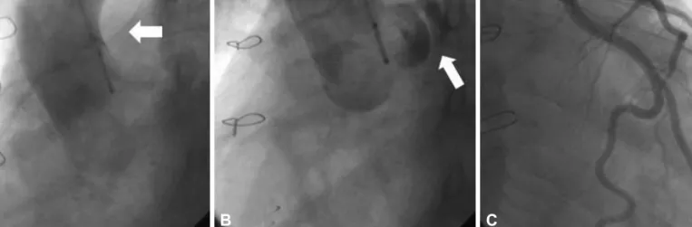
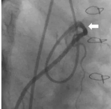
5)

An anomalous origin of RCA from the ascending aorta above the left sinus of valsalva is associated with angina pectoris, acute myocardial infarction or sudden cardiac arrest when it courses between the aorta and the pulmonary trunk.

6)7)

Addition- ally, a case of sinus of valsalva aneurysms with ventricular septal

Case Report

http://dx.doi.org/10.4070/kcj.2012.42.7.497 Print ISSN 1738-5520 • On-line ISSN 1738-5555

Successful Percutaneous Coronary Intervention in an Anomalous Origin of the Right Coronary Artery From the Ascending Aorta Above the Left Sinus of the Valsalva

Seon-Ah Jin, MD 1 , Seok-Woo Seong, MD 1 , Song Soo Kim, MD 2 , Young Dal Lee, MD 1 , Ung Lim Choi, MD 1 , Si-Wan Choi, MD 1 , and Jin-Ok Jeong, MD 1

1

Departments of Internal Medicine and

2

Radiology, Chungnam National University College of Medicine, Daejeon, Korea

The anomalous origin of the right coronary artery (RCA) is a rare condition. Most RCA anomalies are usually found incidentally, but these findings have clinical significance because many patients, particularly young ones, present with sudden death, myocardial ischemia and syncope without other symptoms. We describe a case of a 39-year-old male patient that presented with effort chest pain and was diag- nosed with anomalous RCA that originated from the ascending aorta with prior history of repairing ruptured sinus valsalva and ventricular septal defect. The anomalous origin of RCA was identified by multidetector computed tomography (MDCT). Successful percutaneous coro- nary intervention was performed guided by MDCT coronary images and intravascular ultrasound. (Korean Circ J 2012;42:497-500) KEY WORDS: Coronary vessel anomalies; Angioplasty.

Received: October 11, 2011

Revision Received: December 1, 2011 Accepted: January 12, 2012

Correspondence: Jin-Ok Jeong, MD, Department of Internal Medicine, Chungnam National University College of Medicine, 282 Munhwa-ro, Jung-gu, Daejeon 301-721, Korea

Tel: 82-42-280-8227, Fax: 82-42-280-8238 E-mail: jojeong@cnu.ac.kr

• The authors have no financial conflicts of interest.

This is an Open Access article distributed under the terms of the Creative Commons Attribution Non-Commercial License (http://creativecommons.

org/licenses/by-nc/3.0) which permits unrestricted non-commercial use, distribution, and reproduction in any medium, provided the original work is properly cited.

defect is an extremely rare cardiac anomaly that may be acquired or congenital. We report here a case of successful percutaneous coronary intervention (PCI) by using multidetector computed to- mography (MDCT) and intravascular ultrasound (IVUS) in a patient who had an anomalous RCA arising from the ascending aorta with a history of open heart surgery due to ruptured sinus of valsalva and ventricular septal defect, including a review of the relevant literature.

Case

A 39-year-old man was admitted to the emergency room with ac-

celerating effort chest pain for four months. The patient had past

history in which patch closure was performed for rupture of sinus

of valsalva and ventricular septal defect twenty years ago. The op-

erator could not find the RCA which was at the presumed site ac-

cording to the operation record. The physical examination showed

normal findings. The results of complete blood count, blood chem-

istry, and urinalysis were normal. The electrocardiogram showed

sinus bradycardia and left ventricular hypertrophy. Chest radiogra-

phy revealed borderline cardiomegaly with mild congestion of the

pulmonary vasculature. Transthoracic echocardiography showed

akinesia of basal septum and mid to basal inferior wall with mild left

ventricular systolic dysfunction (ejection fraction=51%). He was sus-

pected of RCA ischemia and referred to the cardiac catheterization

laboratory for CAG. In addition, the left coronary angiogram was

(2)

498 Successful PCI in Anomalous Origin of RCA

http://dx.doi.org/10.4070/kcj.2012.42.7.497 www.e-kcj.org

normal (Fig. 1C). However, we could not easily engage the RCA us- ing several catheters such as 6 French (Fr) sized jukdin 3.5 & 4.0 and amplatz left 1 & 2 (Cordis Corporation, Miami, FL, USA). Therefore, we decided to perform an aortogram, which showed the anomalous

position of the RCA ostium originating from the tubular portion of the ascending aorta above the left coronary cusp (Fig. 1A and B). We decided to delay the PCI and took MDCT CAG images for obtaining information about precise coronary anatomy. The MDCT CAG images

Fig. 1. Aortogram and coronary angiography. A: anomalous right coronary artery ostium (arrow). B: left main coronary artery originated from left coro- nary cusp (arrow). C: normal left coronary artery.

A   B   C

Fig. 2. MDCT coronary images. Volume rendering images and alternative view of coronary MDCT angiography showed ectopic ostium of RCA from the tu- bular portion of ascending aorta (A) and acute angulated tight stenosis (B). RCA originated from the left side and interposited between the pulmonary trunk and ascending aorta (C and D). MDCT: multidetector computed tomography, RCA: right coronary artery.

A  

C  

B  

D  

(3)

499 Seon-Ah Jin, et al.

http://dx.doi.org/10.4070/kcj.2012.42.7.497 www.e-kcj.org

revealed an anomalous origin of RCA from the tubular portion of the ascending aorta with acute angulation and subsequent slit-like luminal narrowing of the proximal RCA between the pulmonary trunk and ascending aorta (Fig. 2). The next day, we tried CAG again and successfully engaged the RCA using 6 Fr Launcher extra-backup 4.5 guiding catheter (Medtronic, Inc., Minneapolis, MN, USA) via a transfemoral approach. The RCA showed a subtotal occlusion in its proximal portion (Fig. 3). We inserted a ChoICE

TM

PT guide wire (Bos- ton Scientific, Heredia, Costa Rica) into RCA and dilated the lesion

using a 3.0×20 mm sized VOYAGER balloon (Abott Vascular, Santa Clara, CA, USA) at 12 atmosphere. Next, an IVUS study was per- formed, which study showed atherosclerotic plaque burden on the proximal RCA with a spindle shaped arterial lumen (a slit-like lu- men) caused by extrinsic compression (Fig. 4A). We implanted a 3.0×38 mm sized Endeavor Resolute stent (Medtronic, Galway, Ire- land) at 12 atmosphere and dilated the stent using a 3.0×12 mm sized Quantum balloon (Boston Scietific, Maple Grove, MN, USA) at 20 atmosphere. The IVUS study then showed a successfully im-

Fig. 3. Right coronary angiography. Baseline coronary angiogram showed an anomalous right coronary artery (RCA) ostium and subtotal occlusion of the proximal RCA (arrow).

Fig. 5. Final right coronary angiography. The final coronary angiogram showed a successfully implanted stent at the proximal right coronary artery (arrow).

Fig. 4. The IVUS study. IVUS of the RCA. A: atherosclerotic plaque burdens on the proximal RCA (arrow). B: successfully implanted stent at the proximal RCA. IVUS: intravascular ultrasound, RCA: right coronary artery.

A   B  

(4)

500 Successful PCI in Anomalous Origin of RCA

http://dx.doi.org/10.4070/kcj.2012.42.7.497 www.e-kcj.org

planted stent at the proximal RCA (Fig. 4B). The final angiogram showed successful revascularization of the RCA (Fig. 5). We finished the procedure without any peri-procedural complications. His symptoms subsided and he was discharged the next day. The pa- tient is still doing well a year after the PCI.

Discussion

Sinus of valsalva aneurysms are very uncommon, with an incid- ence ranging from 0.1% to 3.5% of all congenital heart defects.

8)

Si- nus of valsalva aneurysms have a marked male preponderance (4 : 1), and their incidence is the highest in Asian populations. Most sinus of valsalva aneurysms arise from the right or non-coronary sinuses, and commonly protrude and rupture into the right ventricle or right atrium. Aneurysms may also rupture into the left ventricle, pulmo- nary artery, left artrium, or pericardial cavity.

8)9)

The combination of a sinus of valsava aneurysm and anomalous origin of coronary ar- tery is quite rare, but should be suspected and evaluated. This pa- tient was overlooked on that part when patch closure of ruptured sinus of valsava was performed. An anomalous origin of coronary artery can be classified as follows; 1) absent left main trunk, 2) an- omalous location of coronary ostium within aortic root or near prop- er aortic sinus of valsalva (for each artery), 3) anomalous location of coronary ostium outside normal “coronary” aortic sinuses, 4) an- omalous origination of coronary ostium from opposite, facing “cor- onary” sinus and 5) single coronary artery.

10)

This patient showed an anomalous location of the coronary ostium outside the normal

“coronary” aortic sinuses (i.e., ascending aorta). Anomalous origin of the RCA may have clinical consequences such as angina pectoris, a myocardial infarction, syncope and sudden death. The pathologic mechanism is unclear but the usual explanation is mechanical com- pression of the RCA by great vessels, the aorta and the pulmonary artery. Additionally, an abnormal slit-like RCA ostium in the aortic wall and acute angulation of the RCA can lead to these pathologic results. Hutchins et al.

5)

suggested that the unusual angle of take-off and the more tortuous course of the proximal portion of the anom- alous coronary artery can accelerate the rate of atherosclerosis. Lau- rence et al.

11)

suggested that the junction point of bound portion and the free portion of an anomalous artery as it wraps around the aorta is an area that is susceptible to lipid accumulation. These mecha- nisms can explain the obstructive lesion in the coronary artery of this patients. Clinically, however, angiographic documentation of an- omalous coronary anatomy has not led to any effective or widely agreed recommendations for treatment so far. Though this patient is

still quite well a year after PCI, we should undergo long term observ- ation to clarify the influence of empirical intervention. When per- forming PCI, the operators and catheterization laboratories have to be aware of this anomaly and prepare specialized guiding catheters to facilitate PCI. Because of the technical difficulties during per- forming CAG, we performed 64 channel MDCT. Prior to PCI, MDCT- guided information is helpful in selective cannulation and interven- tion. Also, IVUS investigation of anatomical and functional features of the intussuscepted proximal ectopic segment in such cases may enable the identification of patients who need interventional treat- ment. We report here a case of anomalous origin of RCA arising from the ascending aorta with a history of open heart surgery and suc- cessful PCI guided by MDCT and IVUS.

References

1. Kardos A, Babai L, Rudas L, et al. Epidemiology of congenital coronary artery anomalies: a coronary arteriography study on a central Euro- pean population. Cathet Cardiovasc Diagn 1997;42:270-5.

2. Yamanaka O, Hobbs RE. Coronary artery anomalies in 126,595 pa- tients undergoing coronary arteriography. Cathet Cardiovasc Diagn 1990;21:28-40.

3. Moreno R, Barrera-Ramirez C, Garcia J, Macaya C. Primary stenting of an anomalous left main trunk originating from the right coronary ar- tery during acute myocardial infarction. J Invasive Cardiol 2004;16:

159-61.

4. Angelini P. Coronary artery anomalies--current clinical issues: defini- tions, classification, incidence, clinical relevance, and treatment guide- lines. Tex Heart Inst J 2002;29:271-8.

5. Hutchins GM, Miner MM, Boitnott JK. Vessel caliber and branch-angle of human coronary artery branch-points. Circ Res 1976;38:572-6.

6. Zhang M, Kang WC, Ahn TH, Shin EK. Multidetector computed tomog- raphy for evaluation of ischemic etiology and a post-unroofing pro- cedure for an anomalous origin of the right coronary artery from the left sinus of Valsalva. Korean Circ J 2010;40:251-2.

7. Moon JY, Jeong HC, Cho JY, et al. Anomalous origin of a right coro- nary artery with extrinsic compression between the great vessels: the intravascular ultrasound images. Korean Circ J 2008;38:390-2.

8. Sakakibara S, Konno S. Congenital aneurysm of the sinus of Valsalva:

anatomy and classification. Am Heart J 1962;63:405-24.

9. Scagliotti D, Fisher EA, Deal BJ, Gordon D, Chomka EV, Brundage BH.

Congenital aneurysm of the left sinus of Valsalva with an aortopul- monary tunnel. J Am Coll Cardiol 1986;7:443-5.

10. Angelini P, Velasco JA, Flamm S. Coronary anomalies: incidence, patho- physiology, and clinical relevance. Circulation 2002;105:2449-54.

11. Laurence L, Thurman R, Bruce TC. Atherosclerotic occlusions in anoma- lous left circumflex coronary arteries: a report of two unusual cases &

a review of pertinent literature. Paroi Arterielle 1975;3:55-9.

수치

Fig. 1. Aortogram and coronary angiography. A: anomalous right coronary artery ostium (arrow)
Fig. 3. Right coronary angiography. Baseline coronary angiogram showed  an anomalous right coronary artery (RCA) ostium and subtotal occlusion of  the proximal RCA (arrow).

참조

관련 문서

On his part, CEO of Express Roads Authority, Saud Al-Naqqi said that the heavy rains of the previous day led to clogging parts of the express

Kuwait will celebrate on Sunday the fourth anniversary of the UN honoring and proclamation of His Highness the Amir, Sheikh Sabah Al-Ahmad Al-Jaber Al-Sabah as

The Joseon government designed and promulgated the Taegeukgi as a national flag for diplomatic and political purposes, but it was the independence movement that made it

• 이명의 치료에 대한 매커니즘과 디지털 음향 기술에 대한 상업적으로의 급속한 발전으로 인해 치료 옵션은 증가했 지만, 선택 가이드 라인은 거의 없음.. •

Independently of the question of seduction, our patient, while he was a child, found in his sister a n inconvenient com- petitor for the good opinion of

The proposal of the cell theory as the birth of contemporary cell biology Microscopic studies of plant tissues by Schleiden and of animal tissues by Microscopic studies of

웹 표준을 지원하는 플랫폼에서 큰 수정없이 실행 가능함 패키징을 통해 다양한 기기를 위한 앱을 작성할 수 있음 네이티브 앱과

_____ culture appears to be attractive (도시의) to the