© 2017 The Korean Ophthalmological Society
This is an Open Access article distributed under the terms of the Creative Commons Attribution Non-Commercial License (http://creativecommons.org/licenses /by-nc/3.0/) which permits unrestricted non-commercial use, distribution, and reproduction in any medium, provided the original work is properly cited.
Original Article
Changes in Tear Volume after 3% Diquafosol Treatment in Patients with Dry Eye Syndrome: An Anterior Segment Spectral-domain
Optical Coherence Tomography Study
Kwan Bok Lee, Kyung Min Koh, Young A Kwon, Sang Wroul Song, Byoung Yeop Kim, Jae Lim Chung
Department of Ophthalmology, Kim’s Eye Hospital, Myung-Gok Eye Research Institute, Konyang University College of Medicine, Seoul, Korea
Purpose: To evaluate changes in the tear meniscus area and tear meniscus height over time in patients with dry eye syndrome, using anterior segment spectral-domain optical coherence tomography after the instillation of 3% diquafosol ophthalmic solution.
Methods: Sixty eyes from 30 patients with mild to moderate dry eye syndrome were included. Tear meniscus images acquired by anterior segment spectral-domain optical coherence tomography were analyzed using National Institutes of Health’s image-analysis software (ImageJ 1.44p). Tear meniscus area and tear meniscus height were measured at baseline, 5 minutes, 10 minutes, and 30 minutes after instillation of a drop of diqua- fosol in one eye and normal saline in the other eye. Changes in ocular surface disease index score, tear film break-up time, corneal staining score by Oxford schema, and meibomian expressibility were also evaluated at baseline, and after 1 week and 1 month of a diquafosol daily regimen.
Results: Sixty eyes from 30 subjects (mean age, 29.3 years; 8 men and 22 women) were included. In eyes re- ceiving diquafosol, tear volume was increased at 5 and 10 minutes compared with baseline. It was also higher than saline instilled eyes at 5, 10, and 30 minutes. Changes in tear volume with respect to baseline were not statistically different after the use of diquafosol for 1 month. Ocular surface disease index score, tear film break-up time, and Oxford cornea stain score were significantly improved after 1 week and 1 month of daily diquafosol instillation, but meibomian expressibility did not change.
Conclusions: Topical diquafosol ophthalmic solution effectively increased tear volume for up to 30 minutes, compared to normal saline in patients with dry eye syndrome.
Key Words: Diquafosol, Ophthalmic solutions, Purinergic P2Y receptor agonists, Tear meniscus, Tears secretion
Dry eye syndrome is a multifactorial disease of tears and the ocular surface that results in symptoms of discomfort, visual disturbance, and tear film instability, with potential damage to the ocular surface [1]. The aqueous-deficient type is characterized by reduced lacrimal tear secretion and volume, resulting in symptoms of dry eye. Three per- cent diquafosol tetrasodium ophthalmic solution (Diquas ophthalmic solution 3%; Santen Pharmaceutical, Osaka,
Received: May 30, 2016 Accepted: August 29, 2016
Corresponding Author: Jae Lim Chung, MD. Department of Ophthal- mology, Kim’s Eye Hospital, #136 Yeongsin-ro, Youngdeungpo-gu, Seoul 07301, Korea. Tel: 82-2-2639-7811, Fax: 82-2-2633-3976, E-mail: jaelim.
Japan) acts as a P2Y2 purinergic receptor agonist, stimulat- ing both fluid secretion from conjunctival epithelial cells and mucin secretion from conjunctival goblet cells directly on the ocular surface [2]. Consequently, diquafosol can re- hydrate the ocular surface independent of tear secretion from the lacrimal glands. This action stabilizes tear film on the ocular surface and provides clinical improvement for symptoms of dry eye [3,4].
Anterior segment optical coherence tomography (OCT) is a relatively new noncontact method of imaging the ante- rior segment that can provide detailed information on the status of the tear meniscus. Another benefit of OCT is that it allows noninvasive visualization of the tear meniscus without requiring the use of dyes [5]. Initial anterior seg- ment-OCT imaging devices such as the Visante OCT (Carl Zeiss Meditec, Dublin, CA, USA) and slit-lamp OCT (Hei- delberg Engineering, Dossenheim, Germany) employ time-domain technology, and have been shown to be effec- tive for measurement of tear meniscus [6-8]. Spectral-do- main OCT (SD-OCT) devices have recently become wide- ly available, offering significant advantages over previous time-domain OCT devices, including higher speed, greater sensitivity, and higher resolution [9].
In this study, using SD-OCT, we evaluated changes in tear volume after the instillation of a drop of 3% diquafosol oph- thalmic solution or normal saline in patients with dry eye syndrome.
Materials and Methods
Approval was obtained from the institutional review board of Konyang University Kim’s Eye Hospital before the initiation of the study. Research was conducted in com- pliance with the Declaration of Helsinki, and prior written informed consent was obtained from all subjects after an explanation of the nature of the study and possible conse- quences associated with participation.
Sixty eyes from 30 patients (eight men and 22 women) with mild to moderate dry eye syndrome were included.
Patients were diagnosed based on the following criteria:
tear film break-up time (TBUT) ≤5 seconds and corneal staining score by Oxford schema ≥1 [10]. Exclusion criteria were as follows: systemic disease related to dry eye syn- drome (e.g., Sjögren’s syndrome), acute ocular infection or inflammation not associated with dry eye, drug toxicity,
contact lens wearer, ocular allergy, recent ocular surgery (less than 3 months), conjunctivochalasis, and eyelid or eye- lash disorder.
We used anterior segment SD-OCT (Cirrus 5000, Carl Zeiss Meditec) with a mean value of vertical 5-line raster scan images at the midline of the lower lid margin (Fig. 1).
Tear meniscus area (TMA) and tear meniscus height (TMH) were measured with images acquired from the OCT using the National Institutes of Health’s image-analysis software (ImageJ 1.44p; National Institutes of Health, Bethesda, MD, USA) (Fig. 2A, 2B). TMA and TMH were measured at baseline, 5 minutes, 10 minutes, and 30 minutes after instil- lation of a drop of diquafosol in one eye and normal saline in the other eye (Fig. 3). Solutions were assigned randomly to each eye using a table of random sampling numbers. All examinations were performed by the same blinded exam- iner.
Changes in ocular surface disease index (OSDI) score, TBUT, corneal staining score by Oxford schema, and mei- bomian expressibility were evaluated at baseline, 1 week, and 1 month after the use of diquafosol 6 times a day. Mei- bomian expressibility was measured using the method re- ported by Pflugfelder et al. [11]: compressing the five orific- es of the meibomian glands of the lower lid with a finger, and counting the glands expressing meibum (grade 0, 5 glands; grade 1, 3‒4 glands; grade 2, 1‒2 glands; grade 3, 0 gland). Results were analyzed with a paired t-test using
Fig. 1. Infrared image from anterior segment spectral-domain op- tical coherence tomography illustrating the position of the 5-line raster scan pattern for the tear meniscus image. The distance be- tween each of the 5-line scans was 0.25 mm.
SPSS software ver. 12.0 (SPSS Inc., Chicago, IL, USA).
Results
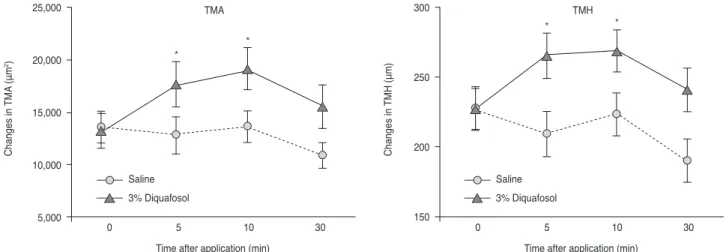
This study included 60 eyes of 30 subjects (mean age, 29.3 years; 8 men and 22 women). In eyes that received diquafosol, tear volume was increased at 5 minutes and 10 minutes com- pared to baseline. Mean TMA was 13,305.53 ± 1,604.78 μm2 at baseline, 17,674.26 ± 2,110.34 μm2 at 5 minutes (p < 0.001), 19,144.52 ± 1,981.22 μm2 at 10 minutes (p = 0.003), and 15,566.55 ± 2,041.95 μm2 at 30 minutes (p = 0.189). Mean TMH was 227.36 ± 15.08 μm at baseline, 265.13 ± 16.22 μm at 5 minutes (p < 0.001), 268.54 ± 15.17 μm at 10 minutes (p <
0.001), and 240.57 ± 15.48 μm at 30 minutes (p < 0.339). In eyes that received normal saline, tear volume was slightly de- creased at 5 minutes and 30 minutes, but this difference was not statistically significant. Tear volume of the eyes that re- ceived diquafosol was higher than that seen in saline-applied control eyes at 5, 10, and 30 minutes (TMA: p = 0.016, p <
0.001, and p = 0.023; TMH: p = 0.004, p < 0.001, and p = 0.023, respectively) (Fig. 4).
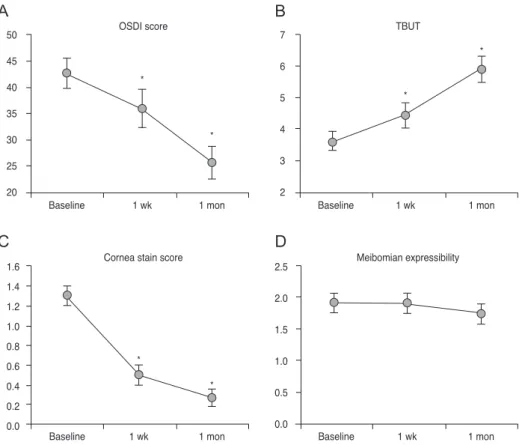
Tear volume after the use of diquafosol six times a day was not changed at 1 week and at 1 month, but tear volume was increased at 10 minutes after the instillation of a drop of diquafosol at each visit (Fig. 5). OSDI score, TBUT, and Oxford cornea stain score improved significantly at 1 week and 1 month after the use of diquafosol, but meibomian expressibility was not changed (Fig. 6A-6D).
Discussion
Slit lamp examination, fluorescein dye staining, TBUT, and the Schirmer test are the traditional diagnostic ap- proaches for dry eye syndrome. However, there have been concerns about the low diagnostic efficiency and reproduc- ibility of these tests [12]. OCT has the advantage of being a noninvasive in vivo technique for quantitative measure-
Fig. 2. Tear meniscus area (A) and tear meniscus height (B) were measured with images acquired from optical coherence tomography using ImageJ software (National Institutes of Health, Bethesda, MD, USA).
A B
Fig. 3. Optical coherence tomography images obtained from the right eye of a 20-year-old woman at baseline (left), and at 10 minutes after the instillation of 3% diquafosol ophthalmic solution (right). The cornea (C), lower eyelids (LL), and lower tear meniscus (TM) were visualized.
The largest increase in tear meniscus volume was observed at 10 minutes after instillation of 3% diquafosol ophthalmic solution.
ment of tear film and tear meniscus that does not require ocular surface contact or dye instillation [13,14].
Yokoi et al. [4] reported increased tear volume after the instillation of a diquafosol ophthalmic solution, using vid- eo meniscometry. However, reflective video meniscometry is not a commonly available method in clinical settings [15].
Koh et al. [3] reported the preliminary long-term efficacy of diquafosol for aqueous-deficient dry eye [3]. They found that diquafosol reduced dry eye symptoms and ocular sur- face staining, and increased TBUT and TMH measured by swept source-OCT (SS-1000; Tomey, Nagoya, Japan).
OCT is a non-invasive optical imaging technique that is clinically utilized for acquiring high-resolution, cross-sec- tional images of the retina, optic nerve, and anterior seg-
ment, aiding in the diagnosis and monitoring of various ocular diseases. Conventional time-domain OCT employs a mechanical scanning reference arm and sequentially measures the echo time delays. In contrast, newer genera- tion spectral or Fourier domain OCT uses a stationary ref- erence arm to obtain an interference spectrum, which then undergoes Fourier transformation to allow for the simulta- neous measurement of all light echo time delays. SD-OCT technology further improves on the time-domain systems, allowing the performance of up to 27,000 axial scans per second. The increased axial scan rate results in an approxi- mately 50 times faster data acquisition in practice. As a re- sult, this new technology significantly improves system speed and sensitivity [16,17].
Fig. 4. Changes in tear meniscus area (TMA, left) and tear meniscus height (TMH, right) with time after the instillation of a drop of diqua- fosol or normal saline (*p < 0.05).
Changes in TMA (μm2)
Time after application (min)
0 5 10 30
25,000
20,000
15,000
10,000
5,000
Changes in TMH (μm)
Time after application (min) Saline
3% Diquafosol
0 5 10 30
300
250
200
150
Saline 3% Diquafosol
⁎ TMA ⁎ ⁎ TMH ⁎
Fig. 5. Changes in tear meniscus area (TMA, left) and tear meniscus height (TMH, right) at baseline and 10 minutes after the use of diqua- fosol (DQ) for 1 month (*p < 0.05).
Initial 1 wk 1 mon
Baseline 10 min DQ Baseline 10 min DQ Baseline 10 min DQ 25,000
20,000 15,000 10,000 5,000 0
⁎ ⁎
⁎
TMH (μm)
TMA (μm2)
350 300 250 200 150 100 50 0
⁎ ⁎ ⁎
Initial 1 wk 1 mon
Baseline 10 min DQ Baseline 10 min DQ Baseline 10 min DQ
To date, most tear meniscus studies measured TMA or TMH using Fourier-domain OCT (RTVue-100; Optovue, Fremont, CA, USA) [18-20] or time-domain OCT [6,7].
RTVue offers more anterior than posterior segment OCT features. In the current study, we used SD-OCT (Cirrus 5000, Carl Zeiss Meditec), which is the most commonly used model, for the evaluation of tear meniscus volume in patients with dry eye syndrome and we were able to quan- tify tear meniscus quickly and accurately.
In this study, we demonstrated the facilitation of tear se- cretion by a 3% diquafosol ophthalmic solution using ante- rior segment SD-OCT. Tear volume of the eyes receiving diquafosol was increased at 5 and 10 minutes compared to baseline, and was significantly higher than in saline in- stilled control eyes at 5, 10, and 30 minutes.
A previous report found that the increase in tear meniscus lasted for only 5 minutes after artificial tear instillation, and for only 10 minutes after 0.1% sodium hyaluronate instilla- tion [21]. Another study on the topical instillation of 0.5%
carboxymethylcellulose in healthy eyes also reported that meniscus height and cross-sectional area returned to base- line levels at 5 minutes after instillation [22]. According to
our findings, 3% diquafosol ophthalmic solution sustained the increase in tear volume for 10 minutes compared to baseline.
Our results showed a statistically significant improvement in OSDI, TBUT, and Oxford cornea staining scores after 1 week and 1 month of daily instillation of diquafosol, but meibomian expressibility was not improved. In this study, meibomian expressibility was measured with a method re- ported by Pflugfelder et al. [11] that used digital pressure.
There are various means for measuring meibomian express- ibility, and more recently instruments that can standardize the pressure (meibomian gland evaluator; TearScience Inc., Morrisville, NC, USA) have been developed. With this small handheld instrument, a defined pressure is applied to the lateral, middle, and nasal third of the lower eyelid, and the number of secreting glands is counted [23]. Further stud- ies implementing such tools might be necessary to obtain more objective and accurate results.
This study has several limitations regarding placebo drug use. Because 3% diquafosol has higher viscosity than nor- mal saline, OCT variables might be influenced by its differ- ent viscosity and hydrophilicity. We recognized this prob-
Fig. 6. Changes in ocular surface disease index (OSDI) score (A), tear-film break up time (TBUT) (B), cornea stain score (C), and meibo- mian expressibility (D) at baseline, after 1 week, and 1 month of daily treatment regimen with diquafosol ophthalmic solution (*p < 0.05).
A
C
B
D
OSDI score
Baseline 1 wk 1 mon
50 45 40 35 30 25 20
⁎
⁎
Cornea stain score
1 wk 1 mon
1.6 1.4 1.2 1.0 0.8 0.6 0.4 0.2 0.0
⁎
⁎
Baseline
Baseline 1 wk 1 mon
1 wk 1 mon
Baseline
Meibomian expressibility 2.5
2.0 1.5 1.0 0.5 0.0
7 TBUT
6 5 4 3 2
⁎
⁎ OSDI score
Baseline 1 wk 1 mon
50 45 40 35 30 25 20
⁎
⁎
Cornea stain score
1 wk 1 mon
1.6 1.4 1.2 1.0 0.8 0.6 0.4 0.2 0.0
⁎
⁎
Baseline
Baseline 1 wk 1 mon
1 wk 1 mon
Baseline
Meibomian expressibility 2.5
2.0 1.5 1.0 0.5 0.0
7 TBUT
6 5 4 3 2
⁎
⁎
lem when we designed the study. However, the proper drug vehicle, with identical constituents except the active ingredient (3% diquafosol), was not available. Therefore, we had to conduct this study with normal saline. Further study comparing the effects of diquafosol with an appro- priate placebo drug might be necessary.
In conclusion, tear meniscus evaluation by anterior seg- ment SD-OCT demonstrated the enhancement of aqueous tear secretion by 3% diquafosol ophthalmic solution. There- fore, 3% diquafosol might be helpful in the treatment of dry eye syndrome, particularly in the aqueous-deficient type.
Conflict of Interest
No potential conflict of interest relevant to this article was reported.
Acknowledgements
This study was funded by Santen Pharmaceutical Co., Ltd.
References
1. The definition and classification of dry eye disease: report of the definition and classification Subcommittee of the In- ternational Dry Eye WorkShop (2007). Ocul Surf 2007;5:75- 92.
2. Nichols KK, Yerxa B, Kellerman DJ. Diquafosol tetrasodi- um: a novel dry eye therapy. Expert Opin Investig Drugs 2004;13:47-54.
3. Koh S, Ikeda C, Takai Y, et al. Long-term results of treat- ment with diquafosol ophthalmic solution for aqueous-de- ficient dry eye. Jpn J Ophthalmol 2013;57:440-6.
4. Yokoi N, Kato H, Kinoshita S. Facilitation of tear fluid se- cretion by 3% diquafosol ophthalmic solution in normal human eyes. Am J Ophthalmol 2014;157:85-92.e1.
5. Bitton E, Jones L, Simpson T, Woods C. Influence of the blink interval on tear meniscus height in soft contact lens and nonlens wearers. Eye Contact Lens 2010;36:156-63.
6. Ibrahim OM, Dogru M, Kawashima S, et al. Visante optical coherence tomography and tear function test evaluation of cholinergic treatment response in patients with sjogren syn- drome. Cornea 2013;32:653-7.
7. Ibrahim OM, Dogru M, Takano Y, et al. Application of visante optical coherence tomography tear meniscus height measurement in the diagnosis of dry eye disease. Ophthal- mology 2010;117:1923-9.
8. Shen M, Wang J, Tao A, et al. Diurnal variation of upper and lower tear menisci. Am J Ophthalmol 2008;145:801-6.
9. Keech A, Flanagan J, Simpson T, Jones L. Tear meniscus height determination using the OCT2 and the RTVue-100.
Optom Vis Sci 2009;86:1154-9.
10. Bron AJ, Evans VE, Smith JA. Grading of corneal and con- junctival staining in the context of other dry eye tests. Cor- nea 2003;22:640-50.
11. Pflugfelder SC, Tseng SC, Sanabria O, et al. Evaluation of subjective assessments and objective diagnostic tests for diagnosing tear-film disorders known to cause ocular irri- tation. Cornea 1998;17:38-56.
12. Nichols KK, Mitchell GL, Zadnik K. The repeatability of clinical measurements of dry eye. Cornea 2004;23:272-85.
13. Fukuda R, Usui T, Miyai T, et al. Tear meniscus evaluation by anterior segment swept-source optical coherence tomog- raphy. Am J Ophthalmol 2013;155:620-4.e1-2.
14. Wang J, Palakuru JR, Aquavella JV. Correlations among upper and lower tear menisci, noninvasive tear break-up time, and the Schirmer test. Am J Ophthalmol 2008;145:795- 800.
15. Yokoi N, Bron A, Tiffany J, et al. Reflective meniscometry:
a non-invasive method to measure tear meniscus curvature.
Br J Ophthalmol 1999;83:92-7.
16. Brennen PM, Kagemann L, Friberg TR. Comparison of StratusOCT and Cirrus HD-OCT imaging in macular dis- eases. Ophthalmic Surg Lasers Imaging 2009;40:25-31.
17. Ho J, Sull AC, Vuong LN, et al. Assessment of artifacts and reproducibility across spectral- and time-domain optical co- herence tomography devices. Ophthalmology 2009;116:1960- 70.
18. Nguyen P, Huang D, Li Y, et al. Correlation between optical coherence tomography-derived assessments of lower tear meniscus parameters and clinical features of dry eye dis- ease. Cornea 2012;31:680-5.
19. Qiu X, Gong L, Lu Y, et al. The diagnostic significance of Fourier-domain optical coherence tomography in Sjogren syndrome, aqueous tear deficiency and lipid tear deficiency patients. Acta Ophthalmol 2012;90:e359-66.
20. Tung CI, Perin AF, Gumus K, Pflugfelder SC. Tear menis- cus dimensions in tear dysfunction and their correlation with clinical parameters. Am J Ophthalmol 2014;157:301-10.e1.
21. Yokoi N, Komuro A. Non-invasive methods of assessing the tear film. Exp Eye Res 2004;78:399-407.
22. Wang Y, Zhuang H, Xu J, et al. Dynamic changes in the lower tear meniscus after instillation of artificial tears. Cor- nea 2010;29:404-8.
23. Finis D, Pischel N, Schrader S, Geerling G. Evaluation of lipid layer thickness measurement of the tear film as a di- agnostic tool for Meibomian gland dysfunction. Cornea 2013;32:1549-53.