기관 - 기관지의 수, 길이, 직경 및 위치에 다양한 변이가 있 다. 기관 - 기관지 분지 기형 (Tracheobronchial branching anomalies)에는 기관지의 분기(division)기형이 가장 흔하며 폐의 발달과정에서 발생하는 여러 기형과 동반된다. 또한, 선 천성 기형 중 비정상적인 혈관 형태와 전 폐의 발달 장애가 복 합된 dysmorphic lung과도 연관된다. 기관 - 기관지 분지 기 형은 크게 기관지 분기 기형과 dysmorphic lung으로 나뉠 수 있다. 다양한 기관지 분기 기형이 알려져 있으며, 일부는 독립 된 이상이며 일부는 다양한 이상과 동반되어 신생아기에서 발 견되거나 사산된다. 흔하지만 대부분 증상이 없는 분절 기관지 와 아분절 기관지의 분지 이상 외에, 주기관지의 분지 이상은 대엽 또는 분절 기관지 개수의 이상, 대엽기관지의 비정상적 기원, 기관지 이성체 등을 포함하며 대표적인 예로 기관 - 기관 지(tracheal bronchus), 부심장 기관지(cardiac bronchus), 기관게실, 이성체 현상(isomerism) 등이 있다(1).
저자들은 기관 - 기관지 분지 기형의 다양한 형태를 분류하 고 CT 영상을 보고하고자 한다.
기관지 분기 기형
발생학
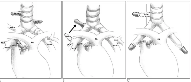
기관지 기형은 정복, 이동, 선택의 세 가지 가설로 설명하는 데(Fig. 1), 첫 번째의 정복 가설은 양측의 대칭성과 양측성 폐동맥 상부 기관지(eparterial bronchus)등을 포함하는 현 존하는 포유동물의 기관지 형태를 설명하는 기본이 되는 가설 이다. 기관의 양측과 중간기관지 내측 벽에서 기원하는 비정상 적인 기관지 형태는 태생 발달 동안에 완전히 수축 혹은 억제
되지만, 일부가 남아서 게실, 맹관의 기관지, 기관지 형태로 보 일 수 있다. 이때 맹관의 기관지는 폐실질의 흔적과 연관이 있 을 수 있으며, 기관지도 환기가 되는 폐 실질과 연관될 수 있 다.
두 번째의 이동가설은 신장(extension) 가설이라고도 불리 는데, 일정한 파생 분지 수를 가진 대칭적인 좌폐동맥 하부 기 관지(hyparterial bronchial)를 가설로 한다. 이러한 파생물 이 처음의 위치에서 기관이나 기관지의 새로운 위치로 이동한 다는 것으로 그 예로 우상엽 기관지가 폐동맥 아래에서 정상적 인 위치인 폐동맥의 위로 이동할 수 있으며, 비정상적으로 기 관까지 이동할 수도 있다.
세 번째의 선택가설은 형태형성(morphogenesis)의 국소적 장애로 기관지 기형이 일어난다는 가설로, 기관지 간엽 (mesenchyme)의 영향으로 기관지의 발아(budding) 과정이 일어난다. 예를 들면, 기관지 간엽이 기관 상피로 이식되면 기 관에서 budding이 이루어지는데, 정상적으로 기관 상피는 상 피의 budding을 막는 기관 간엽에 의해 보호되지만, 기관 간 엽이 약한 부위에 비정상적인 budding이 발생하게 되어 우측 기관 기관지를 형성하게 된다(2).
기관-기관지
기관 - 기관지는 원래 분기부보다 상부 기관에서 기원하는 기 관지로 처음 기술하였으나 최근에는 기관과 주기관지에서 기원 하여 상엽으로 가는 다양한 형태의 기관지 기형(Figs. 1-6)을 표현하는데 쓰인다(Figs. 2-4) (2, 3). 기관 - 기관지는 기관의 어느 부위에서나 발생할 수 있지만 대개 기관용골(carina)의 2 cm 이내에서 생기며 6 cm까지 생길 수 있다. Posteparterial bronchus는 상엽 기관지의 기시부보다 원위부에서 기원하는 기관지를 말하는 것으로(2), 넓은 의미의 기관 - 기관지에 포함 된다(Fig. 7). 상엽으로 가는 기관지 기형은 우측이 좌측보다
기관 - 기관지 분지 기형1
홍민지∙김영통∙조성식∙박아영2
기관-기관지의 수, 길이, 직경 및 위치에 다양한 변이들이 있다. 기관-기관지 분지 기형은 두 형태로 나뉜다. 첫째는 기관지 분기 기형으로 기관-기관지, 부심장 기관지, 기관게실, 이성체 현 상 그리고 소수 변이 등이 있다. 두 번째 형태인 dysmorphic lung은 폐 무발생-형성부전복합 체와 엽무발생-무형성복합체를 포함한다.
1순천향대학교 천안병원 영상의학과
2순천향대학교병원 가정의학과
이 논문은 2009년 12월 11일 접수하여 2010년 3월 21일에 채택되었음.
A B C Fig. 1. Three major theories of congenital bronchial anomaly.
A. Reduction theory.
Perforated lines mean major potential abnormal bronchial patterns. These abnormal bronchi shrink during embryologic develop- ment normally. The persistence of such bronchi results in a tracheal diverticulum or a tracheal bronchus or a cardiac bronchus.
B. Migration theory.
Right upper lobe bronchus (RUL br) has migrated from its hyparterial location to eparterial location on the right main bronchus.
C. Selection theory.
The interaction has happened between bronchial epithelium and bronchial mesenchyme (gray area demarcated by perforated lines). The tracheal epithelium is protected by tracheal mesenchyme (black line) originally. But there may be epithelial budding on a weak point of tracheal mesenchyme due to epitheliomesenchymal interaction and abnormal organogenesis. A right tracheal bronchus (Rt TB) is an example.
PA = pulmonary artery, LLL br = left lower lobe bronchus, Lt TB = left tracheal bronchus, Rt TB = right tracheal bronchus, LUL br = left upper lobe bronchus, ML br = middle lobe bronchus, RLL br = right lower lobe bronchus, Rt TB = right tracheal bronchus, RULB = right upper lobe bronchus. AC br = accessory cardiac bronchus.
A B
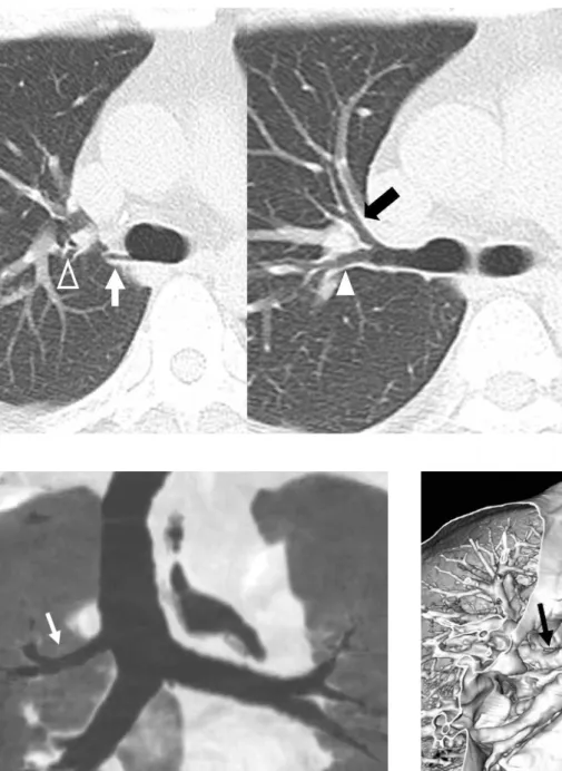
Fig. 2. Tracheal bronchus: An anomalous bronchus from lower trachea.
A. Axial scan shows an anomalous bronchus (arrow) from the right lateral wall of lower trachea, which supply the apical segment
of right upper lobe.B. Reformatted coronal images demonstrates the small tracheal bronchus (arrow) and right upper bronchus normally arising from
the right main bronchus (arrowhead).약 7배가량 많다. 기관에서 기원하는 우상엽기관지를“pig bronchus” 라 하며 약 0.2%의 빈도를 보인다(Fig. 4). 대부분 의 기관 기관지는 우측에 있고 대부분 무증상이며, 반복적 감 염, 무기폐, 그리고 기관지확장증에 의해 증상이 발현되어 발 견된다.
부심장 기관지
부심장 기관지는 주로 중간기관지의 내측 벽에서 기원하며 (Figs. 8-10) 중격동을 향해서 꼬리 쪽으로 주행하기 때문에 부심장 기관지라 불린다(3). 끝부분은 맹점으로 끝나거나
(Fig. 8) 일부의 비정상적 폐실질과 연관되며(Fig. 9), 정상적 인 기관지 점막과 연골을 가지는 점이 기관지게실이나 누공과 구별된다(4). 부심장 기관지의 빈도는 약 0.08%이며 14개 중 10개는 맹관으로 보이며 4개는 폐실질과 연관이 있으며 비정 상적인 열(fissure)에 의해 우하엽과 분리가 된다. 부심장 기 관지는 대부분이 증상이 없지만 감염(Fig. 10), 국균종 (aspergilloma), 종양에 의해 기침이나 객혈의 증상을 보일 수 있다.
Fig. 4. Tracheal bronchus: Right upper lobe bronchus from
lower trachea.Coronal thin-slab MinIP image shows the right upper lobe bronchus (arrow) arising from the trachea 1 cm proximal to the carina. It is called as “pig bronchus”.
Fig. 5. Tracheal bronchus: Right upper lobe bronchus from tra-
cheal bifurcation.Volume rendered image shows that the right upper lobe bronchus (arrow) arising from the trachea near the bifurcation site, instead of normal position.
Fig. 3. Tracheal bronchus: An anom-
alous bronchus from lower trachea.Serial axial images show a small anom- alous bronchus (white arrow) from lower trachea, which supply some portion of apical segment of right up- per lobe.
And also note apical (open arrow- head), anterior (black arrow) and pos- terior (arrowhead) segmental bronchi arising from the right upper lobe bronchus.
기관게실
기관게실은 기관의 벽이 단수 혹은 복수로 함입되는 양성 질 환으로 선천성과 후천성으로 나뉘며 위치와 특성, 그리고 조직
학적으로 다양하다. 기관게실의 빈도는 약 2% 정도이다(5).
여자보다 남자에서 더 흔하며, 주로 기관의 우외측에서 성대 하방 4-5 cm, 혹은 기관 분기부에서 수 센티미터 상방에 생긴 다(Figs. 11, 12). 기관게실이 크기는 작으며 기관과의 교통은 좁다. 기관게실의 원인을 설명하는 가설로는 첫째, 과잉 폐의 흔적(vestigial supernumerary lung)이거나 비정상적으로
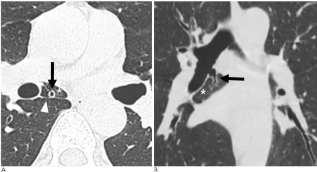
A B
Fig. 8. Cardiac bronchus.
Reformed volume rendered image clearly shows a diverticu- lum (arrow) with blind pouch, arising from the medial wall of the bronchus intermedius.
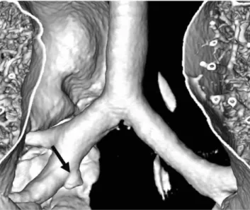
Fig. 6. Tracheal bronchus: Double right upper lobe bronchi
from right main bronchus.Volume rendered image shows two right upper lobe bronchi (arrow, arrowhead) arising from the right main bronchus.
Fig. 7. Tracheal bronchus: Postepartrial right upper lobe bronchus.
A. B. Axial (A) and reformatted coronal image (B) show the right upper lobe bronchus (arrow) arising posterior to and at the same
level of the right pulmonary artery (open arrow), described as posteparterial. Normally, the right main bronchus is eparterial as arising above right pulmonary artery. Postepaterial right upper lobe bronchus is included in the wide spectrum of tracheal bronchus.높게 위치한 원시 폐발아가 발육이 저지되어 생긴다는 것이나, 둘째, 기관의 후방 근섬유막의 발생과정 중 내배엽 분화에 결 손이 생기거나 태생 6주에 기관 연골의 발생과정의 결손에 의 해 발생한다는 것이다(6).
기관게실은 호흡 상피와 평활근의 완전한 기관 해부학을 가
지고 있으며 보통 점액으로 차 있다. 종종 기관식도루공과 같 은 다른 선천 기형과 동반되기도 한다.
이성체 현상
폐의 이성체 현상은 흉부나 복부 장기의 좌우가 같은 모양으
A B
Fig. 9. Cardiac bronchus with own accessary lobe.
A, B. Axial (A) and reformatted coronal images (B) show an accessory cardiac bronchus (arrow) that originates from the medial as-
pect of the right bronchus intermedius. There is an anomalous accessory lobe (*) with apparent pleural fissure (arrowhead), sepa- rating from right lower lobe.A B
Fig. 10. Cardiac bronchus with own accessory lobe.
A 43-year-old man has complained of cough and sputum.
A. Axial scan with lung setting shows cardiac bronchus (arrowhead) and bronchiolitis in right lower lobe.
B. Axial scan shows accessary lobe with increased attenuation (arrow), suggesting combined infection.
로 대칭적으로 배열되는 것으로 좌우측이 모두 우측 장기의 특 징을 가진 양측 우이성체 현상, 또는 좌측 장기의 특징을 가진 양측 좌이성체 현상으로 나뉜다. 우이성체 현상은 양측성 세 개의 엽을 가진 폐와 양측성 소열, 폐동맥 상부 기관지, 중앙에 있는 간과 정해지지 않은 위치의 위를 포함하며 양측성 전신의 심방(atria)을 갖는다. 복부 대동맥과 하대정맥은 같은 쪽에 위치한다. 이 증후군은 남자에게서 더 자주 발생하며, 환자들 은 종종 청색증과 심각한 호흡부전을 일으키기도 한다. 흉부 사진에서 심첨부가 위, 간과 일치하지 않는 특징적인 양상을 보인다. 일반적으로 동반하는 심기형은 공통 심방 캐널 (commom AV canal), 단심방, 대혈관 전위, 전 폐정맥환류 이상을 비롯한 기형적인 정맥 유입 등이 있다. 이러한 심기형 이 비정상적인 면역 상태와 밀접하게 연관되어 이 그룹 환자들 의 예후는 좋지 않으며 생후 1년 이내 80%가 사망한다.
좌이성체 현상(Fig. 13)은 양측성 이엽성 폐 및 양측성 이엽 성 기관지, 양측성 폐 심방, 중앙에 있는 간, 정해지지 않은 위 치의 위장과, 다발성 비장을 포함한다. 하대정맥이 간으로 들 어가기 전에 단절되어 기정맥이나 반기정맥과 연결되는 것이 흔히 나타난다. 다비증은 여성에게 더 흔하며, 다양한 임상증
Fig. 11. Tracheal diverticulum.
Axial image shows a round outpouching (arrow) from the right posterior-lateral wall of the trachea.
Fig. 12. Tracheal diverticulum.
Volume rendered image shows a round outpouching (arrow) from the right posterior-lateral wall of the trachea.
A B
Fig. 13. Left isomerism with heterotaxy syndrome in a 34-year-old man.
A. Axial image demonstrates morphologically similating both atria (*).
B. Coronal thin-slab MinIP image shows bilateral main bronchus with bilobar branching pattern. PA= pulmonary artery.
상과 예후를 갖는다. 소수의 다비증 환자들은 청색증과 좌우 단락에서 오는 심부전의 증상을 나타낸다. 측면사진에서 하대 정맥이 보이지 않고, 전면사진에서 기정맥과의 연결이 관찰되 며, 심첨부가 복부 내장과 일치하지 않는 소견을 보이면 진단 에 도움이 된다. 심기형은 우이성체 현상에 비해 흔하지 않으 며 많은 환자가 심질환이 없거나 경미하다. 몇몇 양측성 이엽 성 폐를 가진 환자에서는 정상적인 복부 장기 위치에 있으며 심기형도 없다(7).
그 외 기관지 분지 변이들
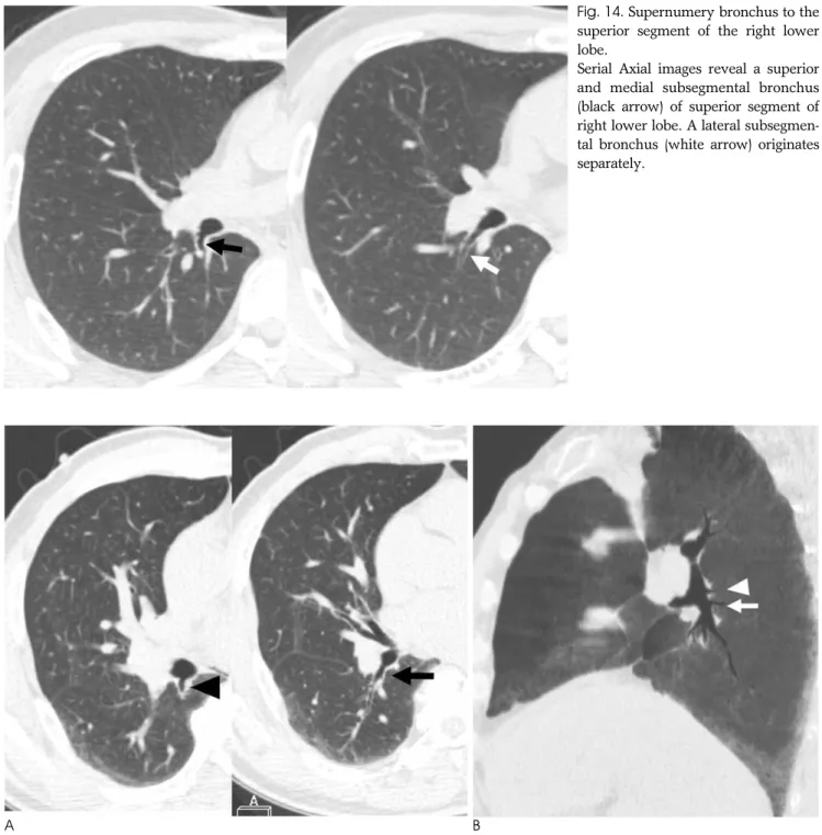
우리가 흔히 접하는 변이 중 하나는 우하엽의 첨분절로 가는 과잉의 기관지이다. 우하엽의 상분절 기관지는 상, 내측, 외측 세구역기관지(subsegmental bronchus)로 나뉘는데 상과 내 측 구역기관지와 외측 구역기관지로 분기하지만, 상 세구역기 관지가 따로 분지하거나 세구역기관지가 서로 다른 위치에서 분지하기도 한다(Figs. 14, 15). 좌하엽의 내측폐저지(medial basal segmental bronchus)는 전폐저지와 총관을 만들어 분
A B
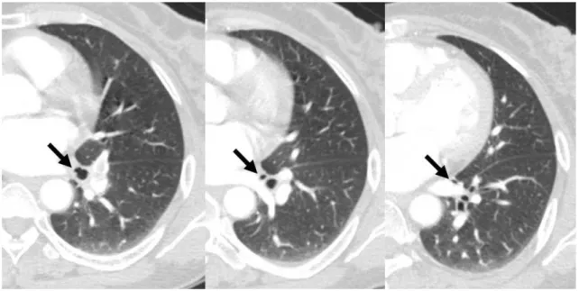
Fig. 15. Supernumery bronchus to the superior segment of the right lower lobe.
A. Serial axial images show a small bronchus (arrowhead) supplying superior subsegment of superior segment of right lower lobe.
And also note the main portion of the superior segment bronchus (arrow) at the level of right middle lobe.
B. Reformatted slap minIP image shows two bronchi (arrowhead, arrow) supplying superior segment of right lower lobe.
Fig. 14. Supernumery bronchus to the
superior segment of the right lower lobe.Serial Axial images reveal a superior and medial subsegmental bronchus (black arrow) of superior segment of right lower lobe. A lateral subsegmen- tal bronchus (white arrow) originates separately.
지하지만 약 4%에서는 따로 분지하기도 한다(Fig. 16). 그 외 우중엽기관지에서 기시하는 비정상적인 기관지가 있을 수 있 다(Fig. 17). 부적절한 기관지 해부학, 부기관지, 소실 기관지 등으로 분류되는 양측성 선천성 해부학적 기형은 비흡연자에 서 자발성 기흉을 일으킬 수 있다. bridging bronchus는 우하 엽을 공급하기 위해 중격동을 가로지르는 왼쪽 주기관지에서 기원하는 이소성 기관지이다(3). 그 외에도 우상엽 기관지가 삼분되거나 우상엽 첨분절 기관지가 소실되거나, 우중엽 기관 지가 설분절 처럼 상,하 분절로 나뉘거나, 반대로 설분절 기관 지가 우중엽처럼 내, 외분절로 분지할 수 있다.
Dysmorphic lung
선천성 폐 기형은 복잡하고 다양하여 여러 가지로 분류할 수 있는데, 첫 번째는 태생학을 이용한 분류로 기형이 발생하는 자궁 내 발달 단계에 따라 기형을 분류하는 것이다. 이는 기관 지 낭종이나 선청성 기관지 폐쇄와 같은 단순한 기형의 기원을 설명하는 데는 매우 유용하지만, 혈관 기형과 폐의 기형을 동 반하는 venolobar syndrome와 같은 복합 기형을 충분히 설 명하기에는 미흡하다.
두 번째는 형태학에 따른 분류로 폐 기형을 순수한 폐 기형, 단순 혈관이상을 가진 기형 그리고 그것들의 연속체인 중간 그 룹으로 나눈 것이다.
세 번째는 형태학적-영상학적 틀에 따라 전폐 기형과 국소적 기형의 두 그룹으로 나눈 것으로 전폐의 기형으로는 dysmorphic lung이 있으며 국소적 기형으로는 선천성 기관 지폐쇄증, 기관지낭, 선천성 낭성선종양기형, 허파분리증 그리 고 systemic irrigation of healthy lung parenchyma 등이 포함된다. 이 분류는 폐의 병리를 진단하는 첫 단계인 흉부사
진에서의 구분이 쉽다는 장점이 있으며, 대부분의 국소적 기형 은 수술적 치료가, 대부분의 전폐 기형은 보존적 치료가 이루 어진다.
전폐 기형인 dysmorphic lung 에 대해 살펴보면, 비정상적 인 혈관 형태와 폐의 발달 장애가 복합되어 있는 기형을 말하 며, 주로는 언월도증후군과 그와 관련된 질환을 의미한다.
흉부사진에서 모양에 따라 두 가지 그룹으로 나눌 수 있는데 폐의 무발생, 무형성, 형성부전을 포함하는, 폐무발생-형성부 전복합체(lung agenesis-hypoplasia complex)와 엽무발생- 무형성복합체(lobar agenesis-aplasia complex)로 나눌 수 있다.
폐무발생-형성부전복합체
폐의 발달이 멈추는 것은 흔한 질환은 아니며 주로 양측 흉 곽에 동일하게 나타난다. 폐의 무발생, 무형성, 형성부전은 다 른 기형을 의미하지만 여기서는 폐무발생 - 형성부전복합체라 는 용어 하에 표현하였는데, 그 이유는 흉부사진 상 유사한 영 상 소견을 보이기 때문이다. 임상적으로 대부분 무증상이고, 대부분 치료되지 않는 선천성 폐기형을 이유로 흉부사진 촬영 을 하여 발견된다. 근골격, 소화기관, 심정, 비뇨기 혹은 다른 폐의 기형과 관련이 되기도 한다(8).
폐 무발생에서는 분기부가 없고 하나의 기관지 내강만 있지 만, 폐 무형성은 주기관지 중 하나가 맹점으로 끝난다는 차이 점이 있다(3).
엽무발생-무형성복합체
앞서 말한 바와 같이 엽무발생 - 무형성복합체는 주로 우측 흉곽에 영향을 주는 흉곽 내 기형을 말하며, 엽 무발생, 엽 무 형성, venolobar syndrome, 마제폐, 부횡격막과 기형적 일측
Fig. 16. Medial basal segmental bronchus of left lower lobe: an independent origin.
Serial axial images show medial basal bronchus (arrow) arising from left lower lobe basal bronchus independently. Normally, the medial and anterior basal bronchi originate together as a common trunk.
Fig. 17. Supranumery bronchus from right middle lobe bronchus.
Serial axial images shows an anomalous bronchus (white arrow) arising from inferior portion of right middle lobe bronchus (black arrow), which traverses the minor fissure to medial basal segment of right lower lobe.
A B
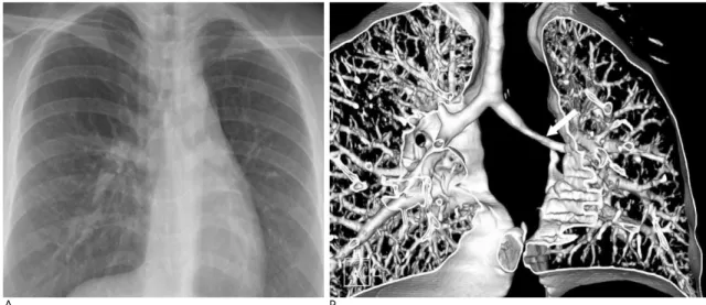
Fig. 18. Agenesis of left upper lobe in a 35-year-old woman.
A. Chest radiograph shows slightly decreased left lung volume with mediastinal shifting.
B. Volume rendered image shows the absence of the left upper lobe bronchus (arrow: normal origin site of left upper lobe
bronchus) from main bronchus, and small left lung with the mediastinal shift to the left side.성 단일 폐정맥 등을 포함한다. 이 기형들은 엽이 한 개 혹은 그 이상이 없거나 발달이 덜 된 폐엽이 있는 폐 기형이다. 폐 기형만 있으면 저발생폐 증후군(hypogenetic lung syndrome)이라고 하며(Fig. 18), 폐정맥이 전신정맥으로 가 는 기형을 동반하면 congenital pulmonary venolobar syndrome이라고 한다(Fig. 19).
저발생폐 증후군은 주로 우측 상엽과 중엽에 자주 발생하며 폐동맥이 정상 혹은 없거나 작아져 있으며, 비정상적으로 흉부 대동맥에서 혈액공급을 받을 수 있다.
Congenital pulmonary venolobar syndrome은 아주 다 양하다. 저발생 폐와 부분정맥환류이상이 있으면 언월도증후 군이라 한다(8). 정확한 발생 기전은 알려져 있지 않으나 발생
초기 폐아의 전반적인 발생 장애에 의한 것으로 생각한다. 폐 동맥의 저형성으로 인해 폐실질의 발생 이상이 생긴다는 주장 도 있다. 이유는 잘 알려져 있지 않지만 주로 우측에 생기며 형 태학적으로 다양한 양상을 보인다. 형성이 덜 된 우측 폐는 일 부 또는 전부를 체동맥에 의해 공급받아 좌우 단락을 형성하는 데, 체동맥은 주로 대동맥에서 기원하며 하엽을 향해 주행한 다. 기관지 이상, 특히 이성체가 흔하며, 반복되는 감염에 의해 게실이 생길 수도 있다. 정맥 배출은 일정치 않으나, 대부분 우 측 폐 전체가 기형 정맥에 의해 하대정맥, 간문맥, 또는 간정맥 으로 배출되며, 이것은 흉부사진에서 심장의 우측으로 넓고 휘 어진 음영이 횡격막 쪽으로 주행하는 특징적 소견으로 보여 언 월도 증후군이라는 이름을 갖게 되었다. 동반된 심혈관계 질환
A C
B
Fig. 19. Scimitar syndrome in a 56-year-old woman.
A. Volume rendered image shows scimitar vein (open arrowheads) in the right lung.
B. Serial axial images show anomalous fissure (arrows), separating medial and lateral portion of right lung. Also note scimitar vein
(arrowheads) near the anomalous fissureC. Coronal thin-slab minIP image shows anomalous branching of right bronchi.
이 흔하며 심방중격 결손이 25% 정도로 가장 흔하다. 그 외 폐엽형성 이상, 척추 기형 등이 잘 동반된다. 환자의 일부는 심 장과 관련된 증상으로, 일부는 반복된 기관지폐 감염 또는 객 혈로 나타난다. 흉부사진에서 언월도 징후, 작은 우폐, 심장 및 종격동의 우측이동 등의 진단적인 소견을 볼 수 있으나, CT는 기관지조영술이나 혈관조영술과 같은 침습적 검사를 하지 않 고도 저형성 폐실질, 저형성 폐동맥, 기형적 폐정맥 배출, 기관 지 이상 등을 잘 보여주어 확진하는데 유용하다(Fig. 19). CT 는 흉부사진에서 잘 보이지 않는 작은 기형 정맥이나 다수의 기형정맥을 찾는데도 도움이 된다(3, 9).
언월도증후군과 밀접한 연관이 있는 질환으로 마제폐가 있 으며, 양측 폐가 심장 뒤에 서로 붙어 있다. 이 협부는 형성저 하증을 보이는 폐에서 동맥 및 기관지 연결을 보이는 것이 특 징이다. 간혹 두 폐가 만나는 부위가 흉막으로 나누어지기도 하며 이는 좌폐에 하부에 비스듬한 줄로 보인다(10). 그 외 우 측에 있는 심장, 하피분리증, 하대정맥의 결손, accessory diaphragm, 횡격막 결손, 좌측 심막결손 등을 동반할 수 있 다.
참 고 문 헌
1. Yedururi S, Guillerman RP, Chung T, Braverman RM, Dishop MK, Giannoni CM, et al. Multimodality imaging of tracheobron-
chial disorders in children. Radiographics 2008;28:E29
2. Ghaye B, Szapiro D, Fanchamps JM, Dondelinger RF. Congenital bronchial abnormalities revisited. Radiographics 2001;21:105-119 3. Beigelman C, Howarth NR, Chartrand-Lefebvre C, Grenier P.
Congenital anomalies of tracheobronchial branching patterns: spi- ral CT aspects in adults. Eur J Radiol 1998;8:79-85
4. Zylak CJ, Eyler WR, Spizarny DL, Stone CH. Developmental lung anomalies in the adult: radiologic-pathologic correlation.
Radiographics 2002:S25-S43
5. Goo JM, Im JG, Ahn JM, Moon WK, Chung JW, Park JH, Seo JB, Han MC. Right paratracheal air cysts in the thoracic inlet: clinical and radiologic significance. AJR Am J Roentgenol 1999;173:65-70 6. Soto-Hurtado EJ, Peñuela-Ruiz L, Rivera-Sa′nchez I, Torres-
Jime′nez J. Tracheal diverticulum: a review of the literature. Lung 2006;184: 303-307
7. Applegate KE, Goske MJ, Pierce G, Murphy D. Situs revisited:
imaging of the heterotaxy syndrome. Radiographics 1999;19:837- 852
8. Mata JM, Ca′ceres J. The dysmorphic lung: imaging findings. Eur J
Radiol 1996;6:403-414
9. Godwin JD, Tarver RD. Scimitar syndrome: four new cases exam- ined with CT. Radiology 1986;159:15-20
10. Frank JL, Poole CA, Rosas G. Horseshoe lung: clinical, pathologic, and radiologic features and a new plain film finding. AJR Am J
Roentgenol 1986;146:217-226
J Korean Soc Radiol 2010;63:149-159
Address reprint requests to : Young Tong Kim, M.D., Department of Radiology, Soonchunhyang University, Cheonan Hospital 23-20 Bongmyung-dong, Cheonan 330-721, Korea.
Tel. 82-41-570-3515 Fax. 82-41-579-9026 E-mail: [email protected]
Tracheobronchial Branching Anomalies
1Min Ji Hong, M.D., Young Tong Kim, M.D., Sung Shick Jou, M.D., A Young Park, M.D.2
1
Department of Radiology, Soonchunhyang University Cheonan Hospital
2
Department of Family Medicine, Soonchunhyang University Hospital
There are various congenital anomalies with respect to the number, length, diameter, and location of tra- cheobronchial branching patterns. The tracheobronchial anomalies are classified into two groups. The first one, anomalies of division, includes tracheal bronchus, cardiac bronchus, tracheal diverticulum, pulmonary isomerism, and minor variations. The second one, dysmorphic lung, includes lung agenesis-hypoplasia com- plex and lobar agenesis-aplasia complex.
Index words :
Trachea BronchiRespiratory System Abnormalities Tomography, X-Ray Computed
′