A CASE OF TUBO-OVARIAN ABSCESS IN A PREGNANT WOMAN
5
0
0
전체 글
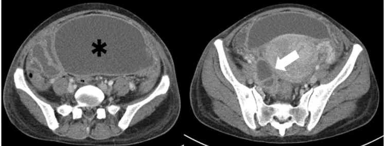
(2) KJOG Vol. 55, No. 11, 2012. fetus of 18 weeks of gestation. Lower edge of the posteriorly located placenta, with the appearance of placenta previa totalis, was covering the whole internal os of cervix (Fig. 1). No adnexal mass was noted on ultrasonography. Clinical progress: Empirical intravenous antibiotics therapy with 3rd cephalosporin and magnesium sulfate and ritodrine therapy for tocolysis were begun immediately. On the seventh day of hospitalization at pregnancy 19 weeks and 4 days, continuous vaginal bleeding was noted. Pelvic examination revealed a cervical os with 3 cm dilation with protruding placenta through the cervix and a 280 g male fetus was delivered spontaneously. We underwent blood test and hemoglobin was 5.4 g/dL. She got transfusion of. packed red cell and fresh frozen plasma. After delivery of the fetus we could find about 15 cm-sized mixed echogenic mass around uterine fundus by ultrasonography (Fig. 2). She underwent abdominal computed tomography and not only abscess in lower abdominal and pelvic cavity but also right tuboovarian abscess were detected (Fig. 3). We decided to perform an emergency laparotomy. Laparotomy was performed with a median incision. There was severe adhesion between great omentum and both ovaries covered by pus like material with old inflammation. After adhesiolysis we could find huge abscess around uterus and small bowel. General surgery team came in and omentectomy, appendectomy, adhesiolysis was done. Recovering normal anatomy we found ruptured right tuboovarian abscess and right salpingo-oophorectomy was carried out; the operation was completed with complete irrigation and suction of the entire abdominopelvic cavity. Pus culture which was done during surgery detected Staphylococcus hominis (Fig. 4). Intravenous antibiotic treatment with 3rd cephalosporin and metronidazole were administered after the operation. The patient recovered well and was discharged 10 days later. Histologic examination confirmed a tubo-ovarian abscess with periappendiceal abscess.. Discussion Fig. 1. Transvaginal ultrasonography at pregnancy 18+5 weeks showed placenta previa totalis (arrow) covering the whole internal os of cervix (asterisk).. The development of a tubo-ovarian abscess during pregnancy is uncommon. Although a patient with a pelvic abscess in pregnancy usually shows symptoms of infection ranging from fever, lower ab-. Fig. 2. Transabdominal ultrasonography after termination showed about 15 cm-sized echogenic mass around uterus fundus.. 844. WWW.KJOG.ORG.
(3) Hyo Jin Lee, et al. Tubo-ovarian abscess in a pregnant woman. Fig. 3. Abdominal computed tomography showed pelvic abscess (asterisk) and right tubo-ovarian abscess (arrow).. Fig. 4. Operation findings which showed ruptured right tubo-ovarian abscess.. dominal pain to sepsis and acute abdomen, tubo-ovarian abscess has rarely been considered as the cause of fever and abdominal pain because of its rare occurrence. Thus, a pelvic abscess during pregnancy must be differentiated from various other conditions. Abdominal pain encountered during pregnancy should be differentiated from various possible causes, including physiologic effects of pregnancy and pathologic conditions related or not related to pregnancy [9]. Physiologic conditions in pregnancy include round ligament pain, uterine torsion and Braxton-Hicks contraction. Pathologic conditions related to pregnancy include spontaneous miscarriage, ischemic uterine leiomyoma, placental abruption, chorioamnionitis, preterm labor, ectopic pregnancy, ovarian hem-. WWW.KJOG.ORG. orrhagic cyst, acute fatty liver of pregnancy, and severe gestational hypertension. Pathologic conditions not related to pregnancy include appendicitis, intestine obstruction, cholecystitis, ovarian torsion, TOA, inflammatory bowel disease, peptic ulcer, acute pancreatitis, urinary tract pathology, sickle cell crisis, porphyria, malaria, arteriovenous hemorrhage, and tuberculosis. Unlike most reported cases, no signs or symptoms attributable to pelvic abscess throughout the pregnancy were observed in our patient. The proposed mechanisms of tubo-ovarian abscess formation in pregnancy are hematogenous spread from a distant focus; lymphatic spread from an infected cervix; infection of an ovarian cyst; flare-up of a previous infection; an infection after fertilization and. 845.
(4) KJOG Vol. 55, No. 11, 2012. before fusion of the decidua parietalis and capsularis; or instrumentation and contiguous spread from adjacent organs [2,9]. All of these possible mechanisms have recently been discussed in a case of ruptured tubo-ovarian abscess in late pregnancy reported by Laohaburanakit and colleauges [6]. Ultrasonography might not be as useful in late gestation as in early gestation or the non-pregnant state for detection of adnexal pathologies because of an enlarged uterus. Tubo-ovarian abscess may not be detected on routine obstetric ultrasonography unless the diagnosis is suspected, as in our case. To ensure accurate diagnosis, laparotomy or laparoscopic intervention should be considered. In recent years, laparoscopy in pregnancy has been a feasible option. But its use is limited in the third trimester owing to an increased risk of injury to the enlarged uterus. It is advisable to make a midline laparotomy incision that can be extended more easily. If obvious abscess is found using radiographic modalities, image-guided percutaneous transcatheter drainage has potential advantages to avoid surgical drainage in some patients. In our case, we did not perform percutaneous transcatheter drainage because of the possible development of toxic signs of sepsis with further delay [10]. The pathogens of tubo-ovarian abscess are variable. It is known that tubo-ovarian abscesses are usually polymicrobial in origin; organisms isolated from tubo-obarian abscesses seem to belong to the facultative anaerobe Enterobacteriaceae family (Escherichia coli, Proteus, Klebsiella ) and anaerobic Peptostreptococcus , Streptococcus or Actinomyces [11-14]. In present case, only Staphylococcus hominis was detected. Most of the cases of tubo-ovarian abscess in pregnancy reported in the literature were managed conservatively as soon as they were recognized during surgery. Pregnant women with unilateral involvement managed with the preservation of the contralateral ovary and the tube had a favorable outcome [15]. In this case, we performed only right salpingo-oophorectomy to preserve reproductive organs as the patient was young and the contralateral adnexa seemed to be free of disease. Tubo-ovarian abscess in pregnancy is an extremely rare condition. Delayed diagnosis and intervention may cause maternal death or fetal loss. Surgical intervention should be considered if the condition progresses or persists after conservative medical treatment [10]. In this report we have presented a case of tubo-ovarian abscess with no signs or symptoms which was diagnosed incidentally after delivery. This was treated with right salpingo-oophorectomy. 846. and antibiotics treatment.. References 1. Jafari K, Vilovick-Kos J, Webster A, Stepto RC. Tubo-ovarian abscess in pregnancy. Obstet Gynecol Surv 1977;32:585-7. 2. Cummin RC. Ovarian abscess during pregnancy. J Obstet Gynaecol Br Emp 1951;58:1025-7. 3. James AN, Knox JM, Williams RP. Attachment of gonococci to sperm. Influence of physical and chemical factors. Br J Vener Dis 1976;52:128-35. 4. Sherer DM, Schwartz BM, Abulafia O. Management of pelvic abscess during pregnancy: a case and review of the literature. Obstet Gynecol Surv 1999;54:655-62. 5. Sogaard Andersen E, Nielsen GL. The combination of pregnancy and acute salpingitis in a case of uterus didelphys. Acta Obstet Gynecol Scand 1988;67:175-6. 6. Laohaburanakit P, Treevijitsilp P, Tantawichian T, Bunyavejchevin S. Ruptured tuboovarian abscess in late pregnancy. A case report. J Reprod Med 1999;44:551-5. 7. Fuselier P, Alam A. Pregnancy complicated by pelvic abscess. J Reprod Med 1978;21:257-8. 8. Sukcharoen N, Witoonpanich P. Pelvic actinomycosis in pregnancy: a case report and review of the literature. J Med Assoc Thai 1992;75:66-71. 9. Blanchard AC, Pastorek JG 2nd, Weeks T. Pelvic inflammatory disease during pregnancy. South Med J 1987;80:1363-5. 10. Chen YF, Hsu ST, Ho ES, Chou MM, Tseng JJ. Tuboovarian abscess in pregnancy. Taiwan J Obstet Gynecol 2008;47:370-1. 11. Myers SA, Benavides E, Alrenga DP, Freese U. Ovarian abscess in mid-trimester. Colo Med 1980;77:133-4. 12. Davey MM, Guidozzi F. Ruptured tubo-ovarian abscess late in pregnancy. A case report. S Afr Med J 1987;71:120-1. 13. de Clercq AG, Bogaerts J, Thiery M, Claeys G. Ovarian actinomycosis during first-trimester pregnancy. Adv Contracept 1987;3:167-71. 14. Dashow EE, Cotterill R, BeMiller D. Ruptured tuboovarian abscess in early gestation. A case report. J Reprod Med 1990;35:418-9. 15. Baydoun AB, Sarram M. Ovarian abscess in pregnancy. Obstet Gynecol 1961;18:739-43.. WWW.KJOG.ORG.
(5) Hyo Jin Lee, et al. Tubo-ovarian abscess in a pregnant woman. 임신부에서 발생한 자궁관난소농양: 증례보고 1. 전남대학교 의과대학 산부인과학교실, 2서남대학교 의과대학 산부인과학교실. 이효진1, 김윤하1, 김종운1, 송태복1, 이기호2 임신부에서 자궁관난소농양은 매우 드문 질환으로서 우리는 임신 중 무증상 자궁관난소농양이 있었던 1예를 보고하고자 한다. 이는 질 식분만 직후 우연히 진단되었으며, 응급개복술을 시행하였다. 수술 소견으로는 4 cm 정도 크기의 우측 자궁관난소농양이 있었으며 이는 파열되어 골반강 전체를 심한 화농성 감염을 초래한 상태였다. 따라서 우측 난소난관절제술과 수 차례 세척 후 수술을 마쳤다. 중심단어: 골반농양, 자궁관난소농양, 임신. WWW.KJOG.ORG. 847.
(6)
수치

관련 문서
It considers the energy use of the different components that are involved in the distribution and viewing of video content: data centres and content delivery networks
After first field tests, we expect electric passenger drones or eVTOL aircraft (short for electric vertical take-off and landing) to start providing commercial mobility
1 John Owen, Justification by Faith Alone, in The Works of John Owen, ed. John Bolt, trans. Scott Clark, "Do This and Live: Christ's Active Obedience as the
- distributes indexes across multiple computers and/or multiple sites - essential for fast query processing with large numbers of documents - many variations:
※ In case of students who received their degree from a Korean university, there is no need for official verification (Submit your copy of college
○ In the case of returning students who have not applied for return to school after paying tuition fees, it is written only in the temporary attendance register as a student on
(in case of partially miscible and form a low-boiling azeotrope).
3) A comparison of the stoichiometric equation with the experimental kinetic expression can suggest whether or not we are dealing with an elementary reaction. 4) If one
