Endovascular Treatment of Multilevel Chronic Total Occlusion Using a Stent Puncture Technique in Buerger’s Disease
4
0
0
전체 글
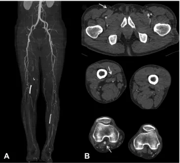
(2) 418 Endovascular Treatment Using Stent Puncture. B. A . C. D. Fig. 1. Angiography of the patient’s right lower extremity at the time Buerger’s disease was first diagnosed. (A) Proximal part of superficial femoral artery was grossly normal without evidence of atherosclerosis while distal part of superficial femoral artery and popliteal artery were totally occluded with collateral vessels. (B) Below-the knee level angiogram showed total occlusion of popliteal artery, anterior tibial artery, and posterior tibial artery with collateral vessels. (C) Foot level angiogram showed the typical angiographic findings of corkscrew collateral vessel. (D) Final angiogram after endovascular treatment shows that that there was no residual stenosis in femoropopliteal artery.. A . B. Fig. 2. The patient’s lower extremities, as observed via computed tomography angiography. (A) A maximum intensity projection reconstruction image shows total occlusion of the entire native femoropopliteal artery, the bypass graft, and below-the-knee arteries. (B) Axial images; white arrows indicate total occlusion of the surgical bypass graft and the stent that was implanted in the popliteal artery. http://dx.doi.org/10.4070/kcj.2016.46.3.417. were performed. On examination, no arterial pulse was palpable below the common femoral artery level. Computed tomography (CT) angiography showed total occlusion of the entire native femoropopliteal artery, the bypass graft, and below-the-knee arteries (Fig. 2). The previously implanted stent in the right popliteal artery was also totally occluded. The ankle brachial index was 0.56 for the right leg. Surgical recanalization was not eligible due to the lack of distal targets, so an endovascular procedure was planned as an alternative option. By using the antegrade femoral approach with a 6-Fr sheath, angiography revealed the same findings as the CT angiography (Fig. 3). There was no visible proximal stump of the native superficial femoral artery or the bypass graft. Distal vessels could not be visualized on the angiogram for a potential retrograde vascular access. To overcome this access problem, direct retrograde puncture of the popliteal stent was performed with an 18-gauge puncture needle under fluoroscopic guidance (Fig. 4A). A 0.035inch hydrophilic guide wire (Terumo, Tokyo, Japan) was gently introduced and advanced through the stent to the level of the proximal femoropopliteal bypass graft. The guide wire, which was supported by a 4-Fr diagnostic Judkins right catheter (Cook Medical, Bloomington, IN, USA), was passed to the common femoral artery and carefully withdrawn through the femoral access using the Rendezvous technique. Once a sufficient length of guide wire was withdrawn through the femoral sheath, the 6-Fr shuttle sheath was advanced over the same guide wire to the previously inserted stent in the popliteal artery after repeated thrombosuction (Fig. 4B). Then, the wire-loop technique with a balloon support was used in. A . B. C. Fig. 3. Baseline angiography before endovascular treatment showed no visualized distal vessel to access. (A) Total occlusion of the previous femoral popliteal graft. (B) Below-the knee level angiogram showed total occlusion of the popliteal stent, anterior tibial artery, and posterior tibial artery. (C) Foot level angiogram showed total occlusion of the popliteal stent, anterior tibial artery, and posterior tibial artery. www.e-kcj.org.
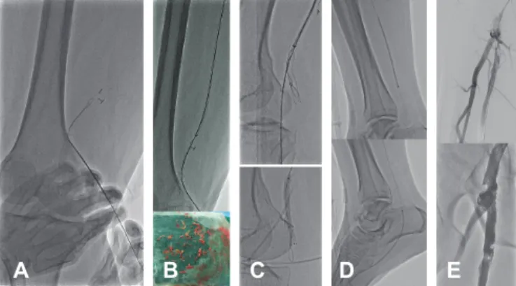
(3) Jung-Hee Lee, et al.. 419. final angiography revealed that there was no residual stenosis without any dissection (Fig. 5). A significant improvement in the ankle brachial index for the right leg was observed after the procedure (from 0.56 to 1.00).. Discussion A . B. C. D. E. Fig. 4. Step-by-step procedure scheme. (A) Direct retrograde puncture of the previous popliteal stent and advance of the 0.035-inch guide wire. (B) Thrombosuction with the 6-Fr shuttle sheath and an image of the retrieved material. (C) Wire-loop technique with a balloon support. (D) Balloon angioplasty of the posterior tibial artery and dorsal-plantar loop arteries. (E) Stenting at the proximal anastomosis site of the previous femoral popliteal bypass graft.. A . B. C. D. Fig. 5. Final angiogram. (A) Previous femoral popliteal bypass graft. (B) Image of stent in the popliteal artery. (C) Posterior tibial artery. (D) Dorsalplantar loop arteries.. a reattempt to crack the occluded in-stent segment in the popliteal artery (Fig. 4C). This time, a 0.014-inch guide wire (Hi-Torque CommandTM, Abbott Vascular, IL, USA) was able to pass through the occluded stent and advance into the posterior tibial artery (PTA) by the subintimal method. After the guide wire was passed through the lesion, balloon angioplasty was performed with a 3.0×250-mm balloon. At that time, the middle part of the 3.0×250-mm balloon was broken at the PTA, because this balloon passed through the previous popliteal stent. Fortunately, the remnant balloon material was successfully retrieved by snare (Amplatz GooseNeck® Microsnare, ev3 Endovascular, Plymouth, MN, USA). Subsequently, balloon angioplasty was done with another 2.5×210-mm balloon in the PTA and dorsal-plantar loop arteries (Fig. 4D), followed by stenting at the proximal anastomosis site of the previous femoral popliteal bypass graft (Fig. 4E). After complete recanalization of the previous femoral popliteal bypass graft, occluded popliteal stent, and infrapopliteal lesions, www.e-kcj.org. Although the exact pathophysiology of Buerger’s disease is unknown, the inflammatory process is possibly initiated by an unknown intimal antigen. In contrast to atherosclerotic peripheral arterial diseases, Buerger’s disease is characterized by the segmental occlusion of not only the distal lower extremity arteries but also the upper extremity arteries.6) The long-term prognosis of patients with Buerger’s disease is reported to be significantly worse than that of patients with atherosclerotic peripheral arterial disease. A recent report showed that about 25% of patients are likely to have an amputation, and the risk of amputation increases to 45% after 10 years.7)8) Several clinical diagnostic criteria of Buerger’s disease have been suggested, including an age of <45 years, a history of heavy smoking, the presence of distal-extremity ischemia, and exclusion of autoimmune disease, hypercoagulable state, and diabetes mellitus.1) Our patient fulfilled all these criteria at the time Buerger’s disease was first diagnosed. For the treatment of Buerger’s disease, surgical revascularization is a reasonable option when bypass is technically feasible. However, surgical treatment for Buerger’s disease is often not feasible, due to the lack of distal targets for bypass. Although endovascular treatment may be considered as a therapeutic choice in patients with Buerger’s disease, it is also technically challenging because of the diffuse segmental involvement of small distal arteries.9) A small case report series on 17 patients with Buerger’s disease showed the clinical benefit of endovascular revascularization in patients with Buerger’s disease and CLI.3) With extensive endovascular treatment, technical success was achieved in 95% of patients, and almost 84% of patients had sustained clinical improvement at the time of the 2-year follow-up. In addition, no mortality or complication was observed, and major amputation was not needed.3) Furthermore, with successful revascularization, patients could have an improved quality of life owing to improvements in claudication, resting pain, or wound healing. Nevertheless, in-stent occlusion is also a major problem in the endovascular treatment of patients with Buerger’s disease. The therapeutic options to solve this problem are surgical bypass and new endovascular treatment for the occluded stent. In our patient, revascularization therapy was necessary, due to aggravated gangrene in the toes. Unfortunately, there were no distal targets for bypass, and surgical revascularization was not http://dx.doi.org/10.4070/kcj.2016.46.3.417.
(4) 420 Endovascular Treatment Using Stent Puncture. suitable. A major technical problem was how to gain access for endovascular treatment. By directly puncturing the stent at the popliteal artery, we overcame access difficulty and were able to introduce a guide wire into the intraluminal space. A previous case report showed successful recanalization of occluded superficial femoral artery stents by stent puncture in patients without distal flow.10) In a prospective clinical analysis of diabetics with CLI, the direct stent puncture technique showed a high rate of technical success (98.2%) and clinical success (94.4%).11) Furthermore, it provided favorable clinical outcomes during follow-up periods.11) In patients with Buerger’s disease, endovascular approaches using various techniques should be considered for effective recanalization if a surgical treatment option is not available. For patients with completely occluded stents, direct stent puncture provided easy access to the intravascular space and allowed for successful recanalization to occur without major trauma or fractures of the stent. In conclusion, the stent puncture technique is a feasible and safe option for overcoming the limitations of vascular access in patients with multilevel total occlusions. Endovascular treatment could be a useful tool for clinically improving CLI in Buerger’s disease.. Research Group in Japan. Surg Today 2000;30:600-5. 3. Graziani L, Morelli L, Parini F, et al. Clinical outcome after extended endovascular recanalization in Buerger’s disease in 20 consecutive cases. Ann Vasc Surg 2012;26:387-95. 4. Laird JR, Katzen BT, Scheinert D, et al. Nitinol stent implantation versus balloon angioplasty for lesions in the superficial femoral artery and proximal popliteal artery: twelve-month results from the RESILIENT randomized trial. Circ Cardiovasc Interv 2010;3:267-76. 5. Schlager O, Dick P, Sabeti S, et al. Long-segment SFA stenting--the dark sides: in-stent restenosis, clinical deterioration, and stent fractures. J Endovasc Ther 2005;12:676-84. 6. Del Conde I, Peña C. Buerger disease (thromboangiitis obliterans). Tech Vasc Interv Radiol 2014;17:234-40. 7. Puechal X, Fiessinger JN. Thromboangiitis obliterans or Buerger’s disease: challenges for the rheumatologist. Rheumatology (Oxford) 2007;46:192-9. 8. Cooper LT, Tse TS, Mikhail MA, McBane RD, Stanson AW, Ballman KV. Long-term survival and amputation risk in thromboangiitis obliterans (Buerger’s disease). J Am Coll Cardiol 2004;44:2410-1. 9. Graziani L, Piaggesi A. Indications and clinical outcomes for below knee endovascular therapy: review article. Catheter Cardiovasc Interv 2010;75:433-43. 10. Gandini R, Del Giudice C, Assako Ondo EP, Spano S, Stefanini M,. References. Simonetti G. Stent puncture for recanalization of occluded superficial femoral artery stents. J Endovasc Ther 2012;19:30-3.. 1. Olin JW. Thromboangiitis obliterans (Buerger’s disease). N Engl J Med 2000;343:864-9.. 11. Palena LM, Manzi M. Direct stent puncture technique for intraluminal stent recanalization in the superficial femoral and. 2. Sasaki S, Sakuma M, Kunihara T, Yasuda K. Distribution of arterial. popliteal arteries in-stent occlusion: outcomes from a prospective. involvement in thromboangiitis obliterans (Buerger’s disease): results. clinical analysis of diabetics with critical limb ischemia. Cardiovasc. of a study conducted by the Intractable Vasculitis Syndromes. Revasc Med 2013;14:203-6.. http://dx.doi.org/10.4070/kcj.2016.46.3.417. www.e-kcj.org.
(5)
수치
관련 문서
The locations of aneurysms were middle cerebral artery in 15 patients, cerebral artery in 15 patients, cerebral artery in 15 patients, cerebral artery in
7.2 Turbulent Flow and Eddy Viscosity 7.3 Fluid Flow Past Solid Boundaries 7.4 Characteristics of Boundary Layers 7.5 The Laminar Boundary Layer*.. 7.6 The
- Turbulence is generated primarily by friction effects at solid boundaries or by the interaction of fluid streams that are moving past each other with different velocities
7.2 Turbulent Flow and Eddy Viscosity 7.3 Fluid Flow Past Solid Boundaries 7.4 Characteristics of Boundary Layers 7.5 The Laminar Boundary Layer*.. 7.6 The
Grunder U, Hürzeler MB, Schüpbach P, Strub JR: Treatment of ligature-induced peri-implantitis using guided tissue regeneration: A clinical and histologic study
Conclusion: In Neer type II distal clavicle fracture treatment, both groups showed satisfactory result in clinical and radiological outcomes, but the hook plate
To assess the clinical usefulness of performing Q-PCR in practice as a diagnostic technique, we compared blindly the Q-PCR results using blood samples of the
We report a rare case of pericatheter abscess formation after scalp acupuncture in a 25-year-old woman who had a history of meningitis and hydrocephalus, which were treated
