Introduction
Osteosarcoma is highly metastatic tumor that mainly occurs in ju- venile patients. Although the prognosis of these patients have im- proved substantially through the development of effective adjuvant and neoadjuvant regimens of chemotherapy, >20% of patients still die as a result of tumor metastasis and unresectable tumor.1) To im- prove the long-term survival rate of osteosarcoma, more efficacious therapeutic targets are needed to reduce or eliminate primary and recurrent osteosarcoma, as well as metastatic disease.
The latest member of the nuclear hormone receptor superfam- ily to be identified is peroxisome proliferator-activated receptor γ (PPARγ), which expresse in normal monocytes, various leukemias, and epithelial malignancies. Recently, the role of PPARγ in tumors
has been extensively studied, and PPARγ agonists have been shown to have direct effects on tumor cells, including breast, colon, lung, stomach, and liver cancer.2)
Thiazolidinedione (TZD) groups are synthetic ligands of PPARγ, including troglitazone, rosiglitazone, pioglitazone, and ciglitazone, which have been used widely in patient with insulin-resistant diabetes mellitus.3) It has been reported that TZD inhibits the cell proliferation and colony formation via inducing apoptosis in sev- eral human cancer cells.4) However, those effects of troglitazone on osteosarcoma cells are poorly understood.
The tumor suppressor gene, phosphatase and tensin homologue deleted on chromosome 10 gene (PTEN), controls a variety of cel- lular functions, including cell proliferation, migration, and survival, as well as tumor cell invasion and tumor directed angiogenesis. It has been suggested that the loss of PTEN could stimulate cell prolifera- tion, reduce apoptosis, and induce tumor angiogenesis.5) Previous studies have shown that PPARγ activation by troglitazone could upregulate the transcription of PTEN in human osteosarcoma cell lines.6) However, how the level of PTEN expression of human os-
Copyrights © 2011 by The Korean Bone and Joint Tumor Society
“This is an Open Access article distributed under the terms of the Creative Commons Attribution Non-Commercial License (http://creativecommons.org/licenses/by-nc/3.0/) which permits unrestricted non-commercial use, distribution, and reproduction in any medium, provided the original work is properly cited.”
대한골관절종양학회지:제17권 제1호 2011
Received February 28, 2011 Revised May 8, 2011 Accepted May 30, 2011 Correspondence to: Jung Ryul Kim, M.D., Ph.D.
Department of Orthopedic Surgery, Chonbuk National University Hospital, 634-18, Geumam-dong, Dugjin-gu, Jeonju 561-712, Korea
TEL: +82-63-250-1767 FAX: +82-63-271-6538 E-mail: [email protected]
Over-expression of PTEN Involved in Troglitazone- induced Apoptosis in Human Osteosarcoma Cells
Sun Jung Yoon, M.D., Ph.D., Lu Zhou, M.D., and Jung Ryul Kim, M.D., Ph.D.
Department of Orthopaedic Surgery, Medical School and Research Institute for Endocrine Science, Chonbuk National University, Jeonju, Korea
Purpose: We investigated the effects of phosphatase and tensin homologue deleted on chromosome 10 gene phosphatase and tensin ho- mologue deleted on chromosome 10 gene (PTEN) expression on the cell proliferation and on the responsiveness of troglitazone in osteosar- coma cells.
Materials and Methods: Western blotting alnalysis was performed to detect the expression of PTEN in U-2OS cells treated with trogli- tazone. WST (water-soluble tetrazolium) assay was used to evaluate cell proliferation. Flow cytometry was used to determine cell apopto- sis. Further, transfection of wild-type PTEN plasmid DNA was used to upregulate PTEN expression.
Results: Troglitazone treatment induced growth inhibition of U2-OS cells in a dose- and time-dependent manner. Troglitazone increased the expression of PTEN in a dose-dependent manner. PTEN upregulation induced by troglitazone treatment resulted in cell growth inhibition and apoptosis in U-2OS cells. PTEN over-expression by plasmid transfection enhanced these effects of troglitazone. Moreover, no changes were observed in the mutant type-PTEN group.
Conclusion: Upregulation of PTEN is involved in the inhibition of cell growth and induction of cell apoptosis by troglitazone. Further, PTEN over-expression can cause cell growth inhibition in osteosarcoma cells and these cell growth inhibitions could be enhance by troglitazone treatment.
Key words: osteosarcoma, apoptosis, PTEN (phosphatase and tensin homologue deleted on chromosome 10 gene), PPARγ (peroxisome proliferator-activated receptor γ) agonist, troglitazone
teosarcoma cells affects the response of cell growth to troglitazone has not been elucidated.
Thus, we hypothesized that the upregulation of PTEN by trans- fection could enhance tumor cell growth inhibition by troglitazone.
In present study, we investigated the effects of PTEN expression level on the cell growth, and the response of troglitazone in osteo- sarcoma cells.
Materials and Methods
1. Cell culture
The human osteosarcoma cell lines (U-2OS) were purchased from the American Type Culture Collection. The cells were maintained into 75-cm2 tissue culture flasks and grown at 37oC under a humidi- fied, 5% CO2 atmosphere in the Dulbecco's modified Eagle's me- dium (DMEM, Gibco BRL, Grand Island, NY) supplemented with 10% (v/v) fetal bovine serum and 2 mM glutamine, 100 units/ml of penicillin, 10 μg/ml of streptomycin, and 2.5 μg/ml of amphotericin B.
PPARγ agonists, troglitazone was as obtained from Sigma-Aldrich, Inc (St. Louis, MO).
2. Cell proliferation assay
The viability of cultured cells was determined by reducing WST-8 (water-soluble tetrazolium salt, (2-(2-methoxy-4-nitrophenyl)-3- (4-nitrophenyl)-5-(2,4-disulfophenyl)-2H-tetrazolium, monosodi- um salt) to highly water-soluble formazan as described previously.7) After treatment, the cells in 96-well plates were washed twice with PBS and Cell counting kit-8 (CCK-8, Dojindo Lab, Kumamoto, Japan) solution (10 μl) was added to each well. Cells were then in- cubated at 37oC for 1hr. Absorbance was measured at 450 nm with model Spectra MAX Plus (Molecular Devices, Sunnyvale, CA).
3. Apoptosis assay
Cells were cultured in FBS-free DMEM for 24 hours to induce apoptosis by troglitazone. Annexin V-fluorescein isothiocyanate (FITC) apoptosis detection kit (BD Pharmingen, San Diego, CA) was used to determine the apoptosis in osteosarcoma cells. The assay was performed according to the manufacturer's protocol. Briefly, cells (1×10⁶/well) were seeded overnight in 6-well tissue culture plates and treated with various concentrations of TGZ for 48 hours, washed with PBS, resuspended in 1 ml binding buffer, stained with 5 μl An- nexin V-FICT and 5 μl propidium iodide (PI) at the room tempera- ture for 15 min, and added 400 μl of 1x binding buffer to each tube.
Histogram patterns of U2-OS cells treated with various concentra-
tions (10, 20, 30, 40, and 50 μM) of troglitazone for 48 hours were obtained by FACS. The fluorescence of FITC and PI was analyzed by flow cytometry (Cell Quest, Becton Dickinson) and software (FCS Express, De Novo Software). Cell cycle distribution was analyzed by flow cytometry after coupled staining with Annexin V conjugated to FITC and PI.
4. Western blot analysis
Cells were homogenized in 100 μl of ice-cold lysis buffer (20 μM HEPES, pH 7.2, 1% Triton X-100, 10% glycerol, 1mM phenyl- methylsulfonyl fluoride (PMSF), 10 μg/ml leupeptin, 10 μg/ml aprotinin). Homogenates containing 20 μg of protein were separated by SDS-PAGE with 10 % resolving gel and 3% acrylamide stacking gel, and transferred to nitrocellulose membranes (Millipore, Bedford, MA). The nitrocellulose membranes were blocked with 2% bovine serum albumin and then probed overnight with primary antibodies for PTEN, and β-actin(Santa Cruz Biochemical, Santa Cruz, CA).
Horseradish peroxidase-conjugated IgGs (Zymed, San Francisco, CA) were used as secondary antibodies and protein expression levels were determined by analyzing the signals captured on the nitrocel- lulose membranes using a Chemi-doc image analyzer (Bio-Rad, Hercules, CA).
5. Construction of recombinant plasmids
GFP expression plasmids based on pCDNA with full-length wild- type PTEN and the point mutant C124A (Cys124-Ala) PTEN were purchased from Invitrogen (Carlsbad, CA). The plasmids were re- strictively digested with HindIII and XbaI, then the full length wild- type and mutant type PTEN mRNA were reclaimed and purified, and then cloned into pCDNA-C1 form pCDNA-C1-WT-PTEN and pCDNA-C124A-PTEN, pCDNA-C1 was treated as a control.
6. Plasmid transfections
For transfection, U-2 OS cells were seeded in six well plates at 3×10⁵/well and incubated in 5% CO2, 95% air incubator at 37oC.
When cells were 50-60% confluent, cells were transfected using TransIT-LT1 transfection reagent according to the manufacturer's protocol. Recombinant plasmids (2.5 μg were mixed with transfec- tion reagent and pre-incubated for 20 min at room temperature in serum-free and antibiotic-free DMEM. U-2OS cells transfected with pCDNA-C1-WT-PTEN, pCDNA-C124A-PTEN and pCD- NA empty vector.
7. Statistical analysis
Statistical analysis of the date was performed with ANOVA and the Student's t-test. Differences of p<0.05 were considered statistically different.
Results
1. Troglitazone induced cell death of human osteo
sarcoma cells
The WST assay was performed to determine the growth inhibition effects of troglitazone on the human osteosarcoma cells at various
Figure 1. Growth inhibition effects of troglitazone in the human osteosarcoma U-2OS cell line in a dose (A) and time (B) -dependent manner. Cells (1×10⁵) were treated with various concentrations of troglitazone. Cell viability was determined by the WST assay and was presented as a calculated percentage of viable cells between troglitazone-treated and -untreated control cells. Each points represents the mean SEM of three determinations.
†p<0.01 vs. untreated control.
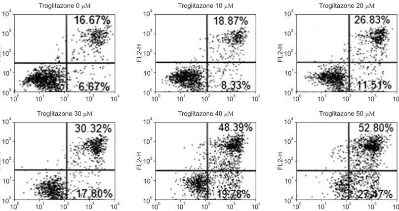
Figure 2. Effect of troglitazone on the cell survival of osteosarcoma cells by flow cytometry. Histogram patterns of U-2OS cells treated with various concentrations (0, 10, 20, 30, 40, and 50 μM) of troglitazone for 48 hrs by FACS. Cell cycle distribution was analyzed by flow cytometry after coupled staining with Annexin V conjugated to fluorescein isothiocyanate (FITC) and Propidium iodide (PI) as described in Materials and Methods.
concentrations. Troglitazone (20 μM) significantly (p<0.05) inhibited the proliferation of U-2OS cell line, when compared with untreated cells after 48 hrs (Fig. 1A). The specific concentrations were selected for more detailed studies of troglitazone time-dependence (Fig. 1B).
Troglitazone treatment decreased U-2OS cell numbers significantly within 24 hours and this inhibition was maintained for 72 hours. As shown in Fig. 1, troglitazone inhibited U-2OS cell proliferation in a concentration and time-dependent manner.
2. Troglitazone caused apoptotic death of U2OS cells
We investigated the possible involvement of apoptosis induction by troglitazone that might have contributed to the cell necrosis. After 48 hr treatment, there were primarily three populations of cells: cells that were viable and not undergoing apoptosis (Annexin V-FICT and PI negative) and cells undergoing apoptosis (Annexin V-FITC positive and PI negative). Another population of cells was observed to be Annexin V-FITC and PI positive, indicating that they were in end stage apoptosis or already dead (Fig. 2). By flow cytometric analysis, troglitazone increased the apoptosis and necrosis of cells
that peak to 27.2%, 48.12%, 68.17% and 80.27%, respectively.
3. Troglitazone stimulated PTEN expression of U2OS cells in a dose dependent manner
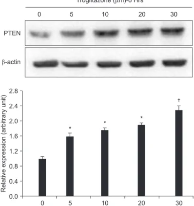
To evaluate the effect of troglitazone on PTEN expression in osteo- sarcoma cells, the U-2OS cells were treated for 6 hours with vari- ous concentration of troglitazone. Results of Western blot analysis showed that troglitazone treatment (0, 5, 10, 20, and 30 μM for 6 hr) of U-2OS cells markedly induced protein levels of PTEN in a dose- dependent manner (Fig. 3). This result suggested that troglitazone enhanced PTEN expression in a dose-dependent manner.
4. Establishment of PTEN overexpression osteo
sarcoma cells using transfection
To elucidate the mechanism of troglitazone induced apoptosis, we established PTEN-over expression U2-OS cells line through trans- fection with wild and mutant plasmid DNA. Morphologically, the two pools (wild and mutant cells) were similar to the native cells. We first confirmed activities of wild and mutant PTEN expressing cells by Western blotting (Fig. 4). However, the PTEN expression in the U-2OS cells declined gradually after being treated with troglitazone for a longer time period (e.g. 72 hrs), since the activity of plasmid transfection was decreased with time.
Figure 3. Dose-dependent effect of troglitazone on the PTEN expression. U2-OS cells (5×10⁶) were treated with 5, 10, 20, and 30 μM of troglitazone for the indicated concentrations. A representative result was presented from among at least three separate experiments yielding similar results. Each points represents the mean SEM of three determinations. *p<0.05 vs. untreated control. †p<0.01 vs. untreated control.
Figure 4. Establishment of PTEN over-expression osteosarcoma cells using transfection. The PTEN expression in the U-2OS cells declined gradually after being treated with troglitazone for a longer time period (eg 72 hrs), since the activity of plasmid trasfection was decreased with time.
5. Transfection of active PTEN in U2OS cells en
hanced the inhibitory effect of troglitazone on cell growth
Transfection with wild type-PTEN caused cell growth inhibition, however transfection with mutant type-PTEN did not affect U-2OS cell proliferation. To further evaluation the role of PTEN over- expression in U-2OS cell growth, pCDNA-C1-WT-PTEN was used to enhance the upregulation of PTEN. To evaluate whether PTEN upregulation played a crucial role in the effect of troglitazone on cell growth, we mutated the PTEN gene and the transfected cells were exposed to troglitazone (30 μM) for 48 hrs. The effect of troglitazone-decreased cell viability was enhanced in the wild type- PTEN group, whereas no changes were observed in the mutant type-PTEN group (Fig. 5).
Discussion
PTEN is frequently deleted or mutated in a wide range of human and tumor cell lines such as glioblastoma and melanoma, and lym- phoid, lung, and endometrial cancers. As a tumor suppressor gene, PTEN expression is downregulated in tumors and tumor cell lines by genetic and epigenetic mechanisms.8,9) To our knowledge, PTEN mutations have not been identified in human osteosarcoma. The abnormal expressions of PTEN by differential compartmentalization may play a role in the development and progression of osteosar- coma, instead of genetic alterations of PTEN.10)
Our results show that PTEN over-expression can cause cell
growth inhibition in osteosarcoma cell line. We further found that the cell growth inhibition could be enhanced by troglitazone treat- ment. The finding of enhancement of troglitazone inhibitory effect PTEN over-expression has not been reported previously in human osteosarcoma cells. Interestingly, the functional disabling of PTEN blocked the inductive effect of troglitazone on cell apoptosis, indi- cating that PTEN was required for troglitazone-induced apoptosis of U-OS cells.
Activated PTEN affects a dephosphorylation of PIP3, gener- ates PIP2, and decreases the phosphorylation level of Akt, which results in cell growth arrest and apoptosis.11) The oncogene Akt has been confirmed as the key cell survival kinase of the PI3K pathway and is negatively regulated by the tumor suppressor gene PTEN.12,13) Functional PTEN decrease the Akt phosphorylation level to inhibit the PI3K/Akt pathway.13) Previous studies have demonstrated that the phosphorylation level of Akt (ser-473) phosphorylation was markedly reduced when expression of PTEN was increased by TZD treatment, indicating that the PTEN overexpression resulted in the inhibition of the PI3K/Akt pathway activation.14,15) PPARγ has been shown to inhibit the transcription of genes related to tumor progres- sion such as COX-2 and NF-κB.16,17)
Peroxisome proliferator activated receptors originally were iden- tified as nuclear receptors that mediate the biologic effects of group of synthetic compounds called peroxisome proliferators.18) Three subtypes of PPARs have been identified in mammalian cells: PPARα, PPARγ, and PPARδ.19) PPARγ is expressed mainly in adipose tissue, and to a lesser extent in colon and other tissues. Adipocytes and os- teoblasts are derived from the same bone marrow stromal progenitor cells.20) When the osteosarcoma cell lines were treated with various concentrations of PPARγ agonists, troglitazone, and ciglitazone, retinoic acid receptor ligand, and 9-cis retinoic acid, all three ligands exhibited the ability to inhibit cell proliferation and to induce apop- tosis, with troglitazone as the most potent of these agents.21) Recent reports have shown that the activation of PPARγ strongly induced cell apoptosis of osteosarcoma both in vitro and in vivo.22) How- ever, these mechanisms of action have still remained obscure. This study showed that troglitazone decreased U-2OS cell viability via cell apoptosis, and the cell growth inhibition by troglitazone could be enhance when PTEN expression is upregulated. Because PTEN- disabling blocked the effect of troglitazone-induced apoptosis in U- 2OS cells, suggesting that apoptosis induction was not only through the upregulation of PTEN, but also through other signaling pathway, which required further investigation.
In summary, our results demonstrated that troglitazone, a syn- Figure 5. Transfection of active PTEN in U2-OS cells enhanced the
inhibitory effect of troglitazone on cell growth . The effect of troglitazone- decreased cell viability was enhanced in the wild type-PTEN group, whereas no changes were observed in the mutant type-PTEN group.
Each points represents the mean SEM of three determinations. *p<0.05 vs. untreated control. †p<0.01 vs. untreated control.
thetic ligand of PPARγ, increases PTEN expression, which in turn inhibits cell growth and induces apoptosis of osteosarcoma cells.
Moreover, our results showed that the effect of troglitazone-de- creased cell viability was enhanced by PTEN overexpression. These observations could represent a new target for the development of improved targeting drugs or prevention of osteosarcoma.
References
1. Bramwell VH. Osteosarcomas and other cancers of bone. Curr Opin Oncol. 2000;12:330-6.
2. Koeffler HP. Peroxisome proliferator-activated receptor gam- ma and cancers. Clin Cancer Res. 2003;9:1-9.
3. Young PW, Buckle DR, Cantello BC, et al. Identification of high-affinity binding sites for the insulin sensitizer rosigli- tazone (BRL-49653) in rodent and human adipocytes using a radioiodinated ligand for peroxisomal proliferator-activated receptor gamma. J Pharmacol Exp Ther. 1998;284:751-9.
4. Kim KY, Kim SS, Cheon HG. Differential anti-proliferative actions of peroxisome proliferator-activated receptor-gamma agonists in MCF-7 breast cancer cells. Biochem Pharmacol.
2006;72:530-40.
5. Leslie NR, Bennett D, Lindsay YE, Stewart H, Gray A, Downes CP. Redox regulation of PI 3-kinase signalling via inactivation of PTEN. EMBO J. 2003;22:5501-10.
6. Kim JY, Kim TK, Park JY, Kim HJ, Lee JW. Effects of the per- oxisome proliferator-activated receptor ligand troglitazone in osteosarcoma cell lines. J Korean Orthop Assoc. 2005;40:591- 7.
7. Ishiyama M, Miyazono Y, Sasamoto K, Ohkura Y, Ueno K. A highly water-soluble disulfonated tetrazolium salt as a chro- mogenic indicator for NADH as well as cell viability. Talanta 1997;44:1299-305.
8. Ali IU, Schriml LM, Dean M. Mutational spectra of PTEN/
MMAC1 gene: a tumor suppressor with lipid phosphatase ac- tivity. J Natl Cancer Inst. 1999;91:1922-32.
9. Zhou XP, Gimm O, Hampel H, Niemann T, Walker MJ, Eng C.
Epigenetic PTEN silencing in malignant melanomas without PTEN mutation. Am J Pathol. 2000;157:1123-8.
10. Moon SH, Lee SH, Kim HS, Kim CH, Chung TW. Phospha- tase and Tensin Homologue Deleted on Chromosome 10) in Osteosarcoma. J Korean Orthop Assoc. 2003;38: 39-46.
11. Maehama T, Dixon JE. The tumor suppressor, PTEN/MMAC1 dephosphorylates the lipid second messenger, phosphatidyl- inositol 3,4,5-triphosphate. J Biol Chem. 1998;273:13375-8.
12. Stoll V, Calleja V, Vassaux G, Downward J, Lemoine NR.
Dominant negative inhibitors of signalling through the phos- phoinositol 3-kinase pathway for gene therapy of pancreatic cancer. Gut. 2005;54:109-16.
13. Pedrero JM, Carracedo DG, Pinto CM, et al. Frequent genetic and biochemical alterations of the PI 3-K/AKT/PTEN path- way in head and neck squamous cell carcinoma. Int J Cancer.
2005;114:242-8.
14. Kreisberg JI, Malik SN, Prihoda TJ, et al. Phosphorylation of Akt (Ser473) is an excellent predictor of poor clinical outcome in prostate cancer. Cancer Res. 2004;64:5232-6.
15. Cao LQ, Chen XL, Wang Q, et al. Upregulation of PTEN in- volved in rosiglitazone-induced apoptosis in human hepato- cellular carcinoma cells. Acta Pharmacol Sin. 2007;28:879-87.
16. Yim HW, Jong HS, Kim TY, et al. Cyclooxygenase-2 inhibits novel ginseng metabolite-mediated apoptosis. Cancer Res.
2005;65:1952-60.
17. Okano H, Shiraki K, Inoue H, et al. 15-deoxy-delta-12-14- PGJ2 regulates apoptosis induction and nuclear factor-kappaB activation via a peroxisome proliferator-activated receptor- gamma-independent mechanism in hepatocellular carcinoma.
Lab Invest. 2003;83:1529-39.
18. Lemberger T, Desvergne B, Wahli W. Peroxisome proliferator- activated receptors: a nuclear receptor signaling pathway in lipid physiology. Annu Rev Cell Dev Biol. 1996;12:335-63.
19. Jow L, Mukherjee R. The human peroxisome proliferator-acti- vated receptor (PPAR) subtype NUC1 represses the activation of hPPAR alpha and thyroid hormone receptors. J Biol Chem.
1995;270:3836-40.
20. Caplan AI, Bruder SP. Mesenchymal stem cells: building blocks for molecular medicine in the 21st century. Trends Mol Med. 2001;7:259-64.
21. Haydon RC, Zhou L, Feng T, Breyer B, et al. Nuclear receptor agonists as potential differentiation therapy agents for human osteosarcoma. Clin Cancer Res. 2002;8:1288-94.
22. Haydon RC, Luu HH, He TC. Osteosarcoma and osteoblastic differentiation: a new perspective on oncogenesis. Clin Orthop Relat Res. 2007;454:237-46.
사람골육종세포주의 트로글리타존 유도 세포사에서 PTEN의 역할
윤선중 • 주 로 • 김정렬
전북대학교 의학전문대학원 정형외과학교실, 내분비 연구소
목적: 본 연구에서는 골육종 세포내 PTEN 발현정도가 세포 성장과 트로글리타존에 대한 반응도에 미치는 영향에 대해 알아보고자 하
였다.
대상 및 방법: 웨스턴 블롯 분석을 통해 트로글리타존 처리 후 PTEN 발현 정도를 관찰하였고, WST를 통해 세포 증식정도를 측정하였
다. 야생형 PTEN 및 돌연변이형 PTEN 발현시키는 플라스미드 DNA를 트랜스펙션하여 PTEN 발현 정도를 측정하였다.
결과: 사람골육종 세포주 U-2OS는 트로글리타존 처리 농도 및 시간에 비례하여 증식 억제를 보였고, 세포내 PTEN 발현 정도는 트로
글리타존 처리 농도에 비례하여 증가하였다. 트로글리타존을 이용하여 U-2OS세포 내 PTEN 발현을 증가시키면 세포 성장 억제와 세 포사 유도가 나타났다. 또한 플라스미드 트랜스펙션에 의한 PTEN 과발현은 트로글리타존의 세포증식 억제 효과를 증가 시키며 돌연 변이형 PTEN을 트랜스펙션 시키는 경우 세포증식효과는 관찰되지 않았다.
결론: 골육종 세포내 PTEN 과발현이 트로글리타존에 의한 골육종 세포의 증식 억제 및 세포사 유도와 관련 되어있음을 알 수 있으며,
세포내에 PTEN이 과발현 된 상태에서 트로글리타존의 효과가 증가됨을 알 수 있었다.
색인단어: 골육종, 세포사, PTEN, PPAR-γ, 트로글리타존
접수일 2011년 2월 28일 심사수정일 2011년 5월 8일 게재확정일 2011년 5월 30일 교신저자 김정렬
전북 전주시 덕진구 금암동 634-18, 전북대학교 의학전문대학원 정형외과학교실 TEL 063-250-1767, FAX 063-271-6538, E-mail [email protected]