Indian Journal of Dermatology, Venereology and Leprology | Volume 86 | Issue 3 | May-June 2020 308
Letters to the Editor
A case of metastatic malignant melanoma in congenital systemic dermal melanocytosis
Sir, Dermal melanocytoses are characterized by the presence and increased number of melanin‑producing dendritic melanocytes in the dermis.
1Malignant transformation of dermal melanocytosis is very rare and there has been only one prior report of malignant melanoma arising in a case of disseminated dermal melanocytosis.
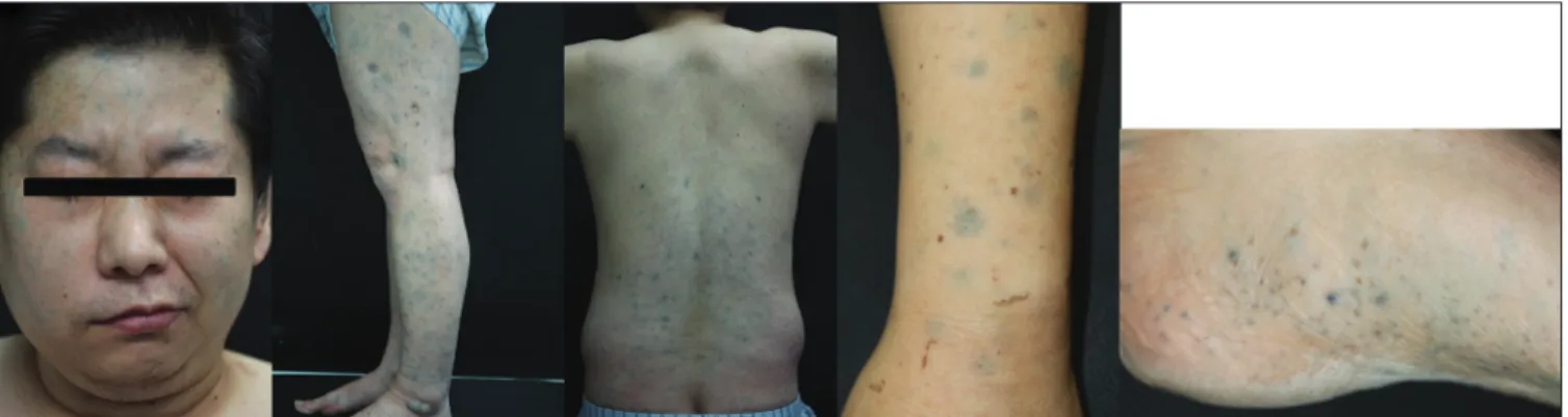
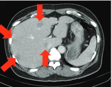
2A 42‑year‑old man presented with numerous bluish‑grey hyperpigmented macules and patches on the face, trunk and both extremities since birth [Figure 1a]. He had fever and abdominal pain for the past 4 days. On physical examination, he had abdominal distension and right sided facial palsy. Abdominal computed tomography scan showed hypointense nodules in the liver which were biopsied [Figure 2a]. Esophagogastroduodenoscopy and colonoscopy showed bluish grey macules in the mucosa of the stomach, ileum and rectum. Biopsies were done from each site [Figure 2b]. On histopathology, melanin pigment was seen in the submucosal layer [Figure 3a]. Liver biopsy showed distorted hepatic tissues and irregular, abnormal proliferation of melanocytes with atypia [Figure 3b].
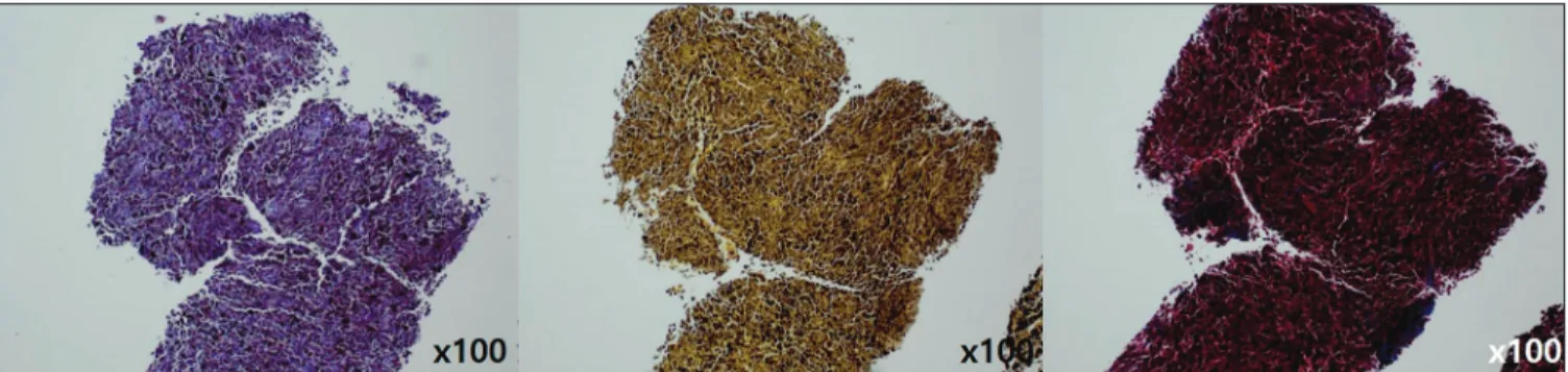
Immunohistochemistry was positive for HMB‑45,
S‑100, MiTF and Melan‑A [Figure 3c] and negative for periodic‑acid‑S chiff, mucicarmine and Masson’s trichrome [Figure 3d].
Magnetic resonance imaging of the brain showed no intracranial metastasis, except for the old microhemorrhages in the pons and subacute infarction with a hemorrhagic change in the right occipital lobe [Figure 2c]. His right facial palsy was considered to have been contributed by multiple old microhemorrhages.
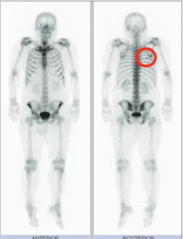
Positron emission tomography scan showed hypermetabolic lesions in the right lung and lower peribronchial area [Figure 2d]. Bone scan showed a hot spot on the right sixth rib [Figure 2e]. He was diagnosed with metastatic malignant melanoma and referred to our department to rule out primary cutaneous malignant melanoma.
Skin lesions were asymptomatic and there was no recent change in size, color or number of lesions. On examination there was no evidence of malignant change like ugly‑duckling sign. Skin biopsy was done from the five most suspicious and variable lesions (on the forehead, left thigh, back, right wrist
Figure 1a: Numerous grey‑bluish hyperpigmented macules and patches over the face, trunk and extremities
Figure 1b: Biopsy from forehead, left thigh, back, right wrist and ankle showed spindle shaped melanocytes and melanophases and no atypia (H and E,
×400, ×100)
309 Indian Journal of Dermatology, Venereology and Leprology | Volume 86 | Issue 3 | May-June 2020
Letters to the Editor
and left ankle). Biopsy showed pigmented spindle‑shaped melanocytes with no atypia and melanophages in all five lesions [Figure 1b]. He was finally diagnosed with metastatic malignant melanoma with the unknown primary site. He underwent nivolumab chemotherapy but died 2 months later due to hepatic failure.
The etiology of dermal melanocytosis is not fully understood.
1,3The arrest of melanocyte migration from neural
crest to interfollicular and follicular epidermis is believed to be responsible.
1While malignant transformation has been reported in blue nevus, nevus of Ota and Ito; malignant melanoma arising from other types of dermal melanocytosis has never been reported.
2,4Except by Levene, where no primary site was discovered.
2In our case also, we did not find the primary focus of malignant melanoma in the skin lesions. However, we consider it is reasonable to believe
Figure 2c: Magnetic resonance imaging of brain showed old hemorrhages
in pons and right occipital lobe Figure 2d: Position emission tomography scan showed hypermetabolic lesion in right lung
Figure 2a: Abdominal computed topography scan showed hypo intense
nodules in liver Figure 2b: Esophagogastroduodenoscopy showed bluish grey macules in
mucosa of stomach ileum
the malignant transformation arose from the skin lesions of melanocytosis.
In 1948, Carleton and Biggs described a 14‑year‑old girl with
profuse bluish spots covering her entire skin surface since
the age of 3 years.
5Later, in 1979, the evolution of this case
was reported by Levene.
2At the age of 43, she had developed
metastatic malignant melanoma involving the lymph nodes
Indian Journal of Dermatology, Venereology and Leprology | Volume 86 | Issue 3 | May-June 2020 310
Letters to the Editor
and liver but no primary site was identified. She died soon after admission and autopsy demonstrated widespread dermal, visceral and cranial melanosis. This is the only case in the literature of disseminated dermal melanocytosis with malignant melanoma.
2Mutations in the guanine nucleotide‑binding protein q polypeptide gene (GNAQ) and guanine nucleotide‑binding protein alpha 11 genes (GNA11) have been described in mice with dermal melanocytosis and mutations in GNAQ also have been demonstrated in humans with dermal melanocytosis and uveal melanoma.
3CDKN2A, BRAF, N‑RAS and KIT mutations are found in malignant melanoma.
3Since no direct relation or common gene mutation has been demonstrated between dermal melanocytosis and malignant melanoma, further study of the pathogenesis of dermal melanocytosis and association with malignant melanoma will be required.
Our patient did not undergo any genetic studies except for the negative BRAF mutation.
Through careful consideration, we reached a diagnosis of
“congenital systemic dermal melanocytosis” since skin lesions were present since birth and were disseminated throughout the skin. Systemic involvement was diagnosed in view of the grey‑bluish macules in hollow organs such as the stomach, small and large intestines. Finally, a skin biopsy showed dermal melanocytosis.
In conclusion, it is important to look for malignant transformation in cutaneous and extracutaneous sites in a patient of systemic dermal melanocytosis.
Declaration of patient consent
The authors certify that they have obtained all appropriate patient consent forms.
Financial support and sponsorship
This work was supported by a National Research Foundation of Korea (NRF) grant funded by the Korean Government (MSIP) (No. 2014R1A5A2010008).
Conflicts of interest
There are no conflicts of interest.
Figure 2e: Bone scan showed hot spot in the sixth rib
Figure 3a: Melanin pigment in submucosal layer of stomach, (H and E, ×200) ileum and rectum (H and E, ×100)
Figure 3b: Liver biopsy showed distorted hepatic tissues and irregular proliferation of melanocytes with atypia (H and E, ×200, ×400)
Figure 3c: Positive HMB‑45, S‑100, MiTF and Melan‑A (×100)
311 Indian Journal of Dermatology, Venereology and Leprology | Volume 86 | Issue 3 | May-June 2020
Letters to the Editor
Won‑Oh Kim, Young‑Wook Ryoo, Sung‑Ae Kim
Department of Dermatology, School of Medicine, Keimyung University, Daegu, Korea Correspondence: Dr. Sung‑Ae Kim, Department of Dermatology, School of Medicine, Keimyung University, 1095 Dalgubeol‑Daero, Dalseo‑Gu, Daegu 42601, Korea.
E‑mail: [email protected]
References
1. Franceschini D, Dinulos JG. Dermal melanocytosis and associated disorders. Curr Opin Pediatr 2015;27:480‑5.
2. Levene A. Disseminated dermal melanocytosis terminating in melanoma. A human condition resembling equine melanotic disease.
Br J Dermatol 1979;101:197‑205.
3. Pessach Y, Goldberg I, Sprecher E, Gat A, Harel A. An unusual presentation of congenital dermal melanocytosis fitting the rare diagnosis of dermal melanocyte hamartoma. Cutis 2014;94:E16‑7.
4. Tse JY, Walls BE, Pomerantz H, Yoon CH, Buchbinder EI, Werchniak AE, et al. Melanoma arising in a nevus of Ito: Novel genetic mutations and a review of the literature on cutaneous malignant transformation of dermal melanocytosis. J Cutan Pathol 2016;43:57‑63.
5. Carleton A, Biggs R. Diffuse mesodermal pigmentation with congenital cranial abnormality. Br J Dermatol Syph 1948;60:10‑3.
Figure 3d: Negative PAS, mucicarmine and trichrome (×100)
How to cite this article: Kim WO, Ryoo YW, Kim SA. A case of metastatic malignant melanoma in congenital systemic dermal melanocytosis. Indian J Dermatol Venereol Leprol 2020;86:308‑11.
Received: September, 2019. Accepted: December, 2019.
© 2020 Indian Journal of Dermatology, Venereology and Leprology | Published by Wolters Kluwer ‑ Medknow
This is an open access journal, and articles are distributed under the terms of the Creative Commons Attribution‑NonCommercial‑ShareAlike 4.0 License, which allows others to remix, tweak, and build upon the work non‑commercially, as long as appropriate credit is given and the new creations are licensed under the identical terms.
Access this article online Quick Response Code: Website:
www.ijdvl.com DOI:
10.4103/ijdvl.IJDVL_747_19 PMID:
*****