• 검색 결과가 없습니다.

Invasive Pulmonary Aspergillosis after Influenza A Infection in an Immunocompetent Patient

N/A
N/A
Protected

Academic year: 2021

Share "Invasive Pulmonary Aspergillosis after Influenza A Infection in an Immunocompetent Patient"

Copied!
4
0
0

로드 중.... (전체 텍스트 보기)

전체 글

(1)

Invasive Pulmonary Aspergillosis after Influenza A Infection in an Immunocompetent Patient

Oh Kyung Kwon, M.D.

1

, Myung Goo Lee, M.D., Ph.D.

2

, Hyo Sun Kim, M.D.

1

, Min Sun Park, M.D.

1

, Kyoung Min Kwak, M.D.

1

and So Young Park, M.D.

3

1

Department of Internal Medicine,

2

Division of Pulmonary, Allergy and Critical Care Medicine, Department of Internal Medicine, Hallym University Chuncheon Sacred Heart Hospital, Hallym University College of Medicine, Chuncheon,

3

Division of Pulmonary, Allergy and Critical Care Medicine, Department of Internal Medicine, Hallym University Kangdong Sacred Heart Hospital, Hallym University College of Medicine, Seoul, Korea

Invasive aspergillosis has emerged as a major cause of life-threatening infections in immunocompromised patients.

Recently, patients with chronic obstructive pulmonary disease, who have been receiving corticosteroids for a long period, and immunocompetent patients in the intensive care unit have been identified as nontraditional hosts at risk for invasive aspergillosis. Here, we report a case of invasive pulmonary aspergillosis after influenza in an immunocompetent patient.

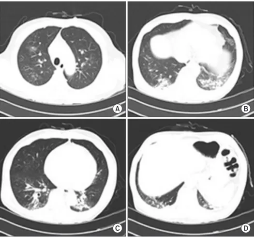
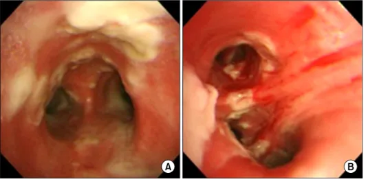
The patient’s symptoms were nonspecific, and the patient was unresponsive to treatments for pulmonary bacterial infection. Bronchoscopy revealed mucosa hyperemia, and wide, raised and cream-colored plaques throughout the trachea and both the main bronchi. Histologic examination revealed aspergillosis. The patient recovered quickly when treated systemically with voriconazole, although the reported mortality rates for aspergillosis are extremely high. This study showed that invasive aspergillosis should be considered in immunocompetent patients who are unresponsive to antibiotic treatments; further, early extensive use of all available diagnostic tools, especially bronchoscopy, is mandatory.

Keywords: Invasive Pulmonary Aspergillosis; Influenza A Virus; Immunocompetence

of morbidity and mortality in immunocompromised patients.

However, during recent years, several reports have described a rising incidence of invasive aspergillosis in critically ill patients admitted to the intensive care unit, even in the absence of an apparent predisposing immunodeficiency. In this situation is related to difficulties in timely diagnosis, caused by insensitive and non-specific clinical signs and lack of unequivocal diag- nostic criteria

1

.

After the 2009 A/H1N1 influenza pandemic, it has been shown that invasive aspergillosis is a much more frequent complication in critically ill H1N1 patients and that use of sys- temic steroids contribute to this superinfection

2

.

The authors report this case study, along with a literature re- view, which involves invasive pulmonary aspergillosis occur- ring after influenza infection in an immunocompetent patient who did not use systemic steroids.

Copyright © 2013

The Korean Academy of Tuberculosis and Respiratory Diseases.

All rights reserved.

Introduction

Invasive aspergillosis is known to have an important cause

CASE REPORT

http://dx.doi.org/10.4046/trd.2013.75.6.260

ISSN: 1738-3536(Print)/2005-6184(Online) • Tuberc Respir Dis 2013;75:260-263

260

Address for correspondence: So Young Park, M.D.

Department of Internal Medicine, Hallym University Kangdong Sacred Heart Hospital, Hallym University College of Medicine, 150 Seongan-ro, Gangdong-gu, Seoul 134-701, Korea

Phone: 82-2-2224-2213, Fax: 82-2-2224-2213 E-mail: p0702@chol.net

Received: Jul. 30, 2013 Revised: Aug. 27, 2013 Accepted: Sep. 10, 2013

cc

It is identical to the Creative Commons Attribution Non-Commercial

License (http://creativecommons.org/licenses/by-nc/3.0/).

(2)

Pulmonary aspergillosis in an immunocompetent patient

http://dx.doi.org/10.4046/trd.2013.75.6.260 261

www.e-trd.org

Case Report

A 60-year-old man was admitted chiefly with shortness of breath, pleural chest pain. The patient presented with fever, cough, and whitish sputum 10 days prior to consultation.

Treatment was received from a local hospital for upper respi- ratory tract infection, but no improvements were observed.

The patient was transferred to our hospital presenting with shortness of breath and pleural chest pain.

Underlying disease includes hypertension, and the patient has been taking medication for 20 years.

He was a non-smoker and did not have alcohol abuse his- tory.

1. Physical examination findings

At the time of consultation, the blood pressure was 120/70 mm Hg; heart rate, 102 beats/min; respiratory rate, 24 breaths/

min; and body temperature, 38.1

o

C. The patient was conscious but showed acute ill-looking appearance. Physical examina- tion showed normal conjunctiva with no palpable lymph nodes around the neck. On auscultation of the chest, coarse breathing sounds without wheezing in both lower lung fields were heard. The abdomen was soft, and bowel sounds were

normal; no hepatomegaly and splenomegaly were noted. Skin lesions were not observed.

2. Laboratory findings

In the peripheral blood test, leukocytes were 7,650/mm

3

(neutrophils, 79.3%; lymphocytes, 12.3%; and eosinophils, 0.8%); hemoglobin, 14.9 g/dL; platelets, 124,000/mm

3

; and erythrocyte sedimentation rate, 20 mm/hr (reference range, 0−20 mm/hr). C-reactive protein was 175 mg/dL (reference range, 0−3 mg/dL). In serum biochemistry, blood urea nitro- gen/creatinine was 11.9/1.1 mg/dL; total protein/albumin, 6.3/3.6 g/dL; Ca/P, 7.7/2.7 mg/dL; aspartate aminotransferase/

alanine aminotransferase, 43/36 IU/L; and total bilirubin, 0.83 mg/dL. For the hepatitis test in autoimmune serology screen- ing, surface antigen of the hepatitis B virus and anti-hepatitis C virus were negative; anti-hepatitis B surface antigen was positive; in the thyroid function test results, T3 was 70.8 ng/dL (reference range, 58−159 ng/dL), whereas thyroid stimulating hormone was 0.701 mIU/mL (reference range, 0.35−4.94 IU/

mL) and free T4 was 1.25 ng/dL (reference range, 0.7−1.48 ng/dL); and human immunodeficiency virus and venereal disease research laboratory results were negative. Serum level of IgA, IgG and IgM were within normal range. CD4 count was

Figure 1. (A−D) Chest computed tomog-

raphy findings. Multifocal ground glass

opacities and peribronchial infiltration in

both the lungs.

(3)

OK Kwon et al.

262 Tuberc Respir Dis 2013;75:260-263 www.e-trd.org

within normal. Although urinalysis did not show pyuria, mi- croscopic hematuria and proteinuria were observed.

3. Chest radiography

Chest radiography revealed focal consolidation in the right lower lung field, but no cardiomegaly.

4. Chest computed tomography (CT) scan

Multifocal ground glass opacities and peribronchial infiltra- tion in both lungs were observed (Figure 1).

5. Clinical outcomes

The patient presented shortness of breath and pleural chest pain. Chest radiographic findings showed focal con- solidation in the lung. Therefore, ceftriaxone+azithromycin were administered since community-acquired pneumonia was suspected. However, fever persisted and hypoxia wors- ened, and thus, the medication was changed to piperacillin/

tazobactam+levofloxacin. However, fever, worsening hypoxia (SpO

2

, 82.3%; PO

2

, 43.7 mm Hg), and chest radiographic find- ings worsened in addition to worsening of the leukocytosis (13,890/mm

3

). Since community-acquired methicillin-resis- tant Staphylococcus aureus infection was suspected, vanco- mycin was administered, and bronchoscopy was performed.

6. Bronchoscopy

Diffuse mucosal hyperemia and wide, raised, and cream- colored plaques (pseudomembrane formation) were observed throughout the trachea and both main bronchi, without significant airway obstruction (Figure 2). Polymerase chain reaction test for influenza A from bronchoalveolar la- vage was positive.

7. Biopsy

Numerous branching septate hyphae with acute angles less than 45

o

were observed (Figure 3).

8. Treatment and progress

The characteristic septate hyphae of Aspergillus were ob- served in biopsy. Voriconazole was administered, and the patient was discharged as improvements were observed in the patients fever, hypoxia (SpO

2

, 97.7%; PO

2

, 91.0 mm Hg), and leukocytosis (6,270/mm

3

). However, we were not able to further test for patient’s financial reasons.

Figure 2. Bronchoscopic examination showing mucosa hyperemia and wide, raised and cream-colored plaques (pseu- domembrane formation) through the carina (A) and the right bronchus inter- medius (B).

Figure 3. Histologic examination of bronchoscopic biopsy show-

ing numerous branching septate hyphae (PAS stain, ×200).

(4)

Pulmonary aspergillosis in an immunocompetent patient

http://dx.doi.org/10.4046/trd.2013.75.6.260 263

www.e-trd.org

Discussion

Aspergillus is widely distributed in the soil, and people normally inhale hundreds of spores daily, although healthy in- dividuals are able to filter airborne fungi through the epithelial mucociliary defense mechanism and resident phagocyte ac- tions

3

. Pulmonary aspergillosis is influenced by the immune status of the host and is categorized into aspergillosis species, allergic bronchopulmonary aspergillosis, chronic necrotiz- ing aspergillosis, and invasive aspergillosis

4

. Among these, invasive aspergillosis typically occur in patients with seriously compromised immune system, which easily occurs in pa- tients with hematologic malignancies, transplant recipients, and patients undergoing long-term steroid administration or with genetic immunodeficiency

3

. Tracheobronchial aspergil- losis such as that described in this case study is an uncommon clinical form of invasive aspergillosis infection limited to the tracheobronchial tree

5

. In many cases of invasive aspergillosis patients, the observation of the halo sign in the initial chest CT scan helps initiate early treatment

6

; in the case of tracheobron- chial aspergillosis, characteristic lesions are not observed in the chest radiographic and CT scans, and bronchoscopy may help by biopsy and characterization of the lesions

7

.

In the case of our patient, false membrane tracheobronchial aspergillosis was diagnosed, which is an advanced stage of disease that almost never occurs in people with normal im- mune system. Nonspecific symptoms include fever and cough, and wheezing occurs only when airways are obstructed by inflammation, mucus and fungal masses. Because of atypical clinical symptoms and radiological examination, diagnosis may be incorrect in the case of intensive care unit patients, but the characteristic lesions of bronchoscopy such as that described in this case study is the most sensitive diagnostic test

8

. The inci- dence of invasive aspergillosis has recently increased in chronic obstructive pulmonary disease patients with long-term steroid administration and non-immunocompromised intensive care unit patients with very high mortality rate. The infection due to the influenza virus is caused by cell-mediated defects, disrup- tion of normal ciliary clearance, and leukopenia, and these may be the leading factors of invasive fungal disease. Lat et al.

9

repoted 2 patients with invasive aspergillosis after infection with pandemic (H1N1). They suggested that high-dose corticoste- roids may be novel risk factors predisposing immunocompe- tent patients to invasive aspergillosis

9

. However, several reports have described invasive aspergillsosis occurring after influenza in a normal immune patients who has not received systemic steroids treatment. Hasejiima et al.

10

reported a case of invasive aspergillosis, which was associated with influenza B.

Voriconazole is recommended as initial therapy in the treat- ment of tracheobronchial aspergillosis

11

. Multi-drug combi- nation therapy can also be considered because effective out- comes have been shown

12

, although there is lack of evidence with respect to large-scale studies

1

.

For ICU patients with normal immune system, bronchos- copy is necessary for early diagnosis, when patients do not re- spond to common antibiotics and when invasive aspergillosis may be the leading causative factor.

References

1. Trof RJ, Beishuizen A, Debets-Ossenkopp YJ, Girbes AR, Groeneveld AB. Management of invasive pulmonary asper- gillosis in non-neutropenic critically ill patients. Intensive Care Med 2007;33:1694-703.

2. Wauters J, Baar I, Meersseman P, Meersseman W, Dams K, De Paep R, et al. Invasive pulmonary aspergillosis is a frequent complication of critically ill H1N1 patients: a retrospective study. Intensive Care Med 2012;38:1761-8.

3. Lass-Florl C, Roilides E, Loffler J, Wilflingseder D, Romani L.

Minireview: host defence in invasive aspergillosis. Mycoses 2013;56:403-13.

4. Xu XY, Sun HM, Zhao BL, Shi Y. Diagnosis of airway-invasive pulmonary aspergillosis by tree-in-bud sign in an immuno- competent patient: case report and literature review. J Mycol Med 2013;23:64-9.

5. Lee HY, Kang HH, Kang JY, Kim SK, Lee SH, Chung YY, et al.

A case of tracheobronchial aspergillosis resolved sponta- neously in an immunocompetent host. Tuberc Respir Dis 2012;73:278-81.

6. Greene RE, Schlamm HT, Oestmann JW, Stark P, Durand C, Lortholary O, et al. Imaging findings in acute invasive pulmo- nary aspergillosis: clinical significance of the halo sign. Clin Infect Dis 2007;44:373-9.

7. Buchheidt D, Weiss A, Reiter S, Hartung G, Hehlmann R.

Pseudomembranous tracheobronchial aspergillosis: a rare manifestation of invasive aspergillosis in a non-neutropenic patient with Hodgkin’s disease. Mycoses 2003;46:51-5.

8. Tasci S, Glasmacher A, Lentini S, Tschubel K, Ewig S, Moli- tor E, et al. Pseudomembranous and obstructive Aspergillus tracheobronchitis: optimal diagnostic strategy and outcome.

Mycoses 2006;49:37-42.

9. Lat A, Bhadelia N, Miko B, Furuya EY, Thompson GR 3rd.

Invasive aspergillosis after pandemic (H1N1) 2009. Emerg Infect Dis 2010;16:971-3.

10. Hasejima N, Yamato K, Takezawa S, Kobayashi H, Kadoyama C. Invasive pulmonary aspergillosis associated with influenza B. Respirology 2005;10:116-9.

11. Walsh TJ, Anaissie EJ, Denning DW, Herbrecht R, Kontoyi- annis DP, Marr KA, et al. Treatment of aspergillosis: clinical practice guidelines of the Infectious Diseases Society of America. Clin Infect Dis 2008;46:327-60.

12. De Rosa FG, Terragni P, Pasero D, Trompeo AC, Urbino R,

Barbui A, et al. Combination antifungal treatment of pseudo-

membranous tracheobronchial invasive aspergillosis: a case

report. Intensive Care Med 2009;35:1641-3.

수치

Figure 1. (A−D) Chest computed tomog- tomog-raphy findings. Multifocal ground glass  opacities and peribronchial infiltration in  both the lungs.
Figure 2. Bronchoscopic examination  showing mucosa hyperemia and wide,  raised and cream-colored plaques  (pseu-domembrane formation) through the  carina (A) and the right bronchus  inter-medius (B).

참조

관련 문서

Tenofovir alafenamide versus tenofovir disoproxil fumarate for the treatment of patients with HBeAg-negative chronic hepatitis B virus infection: a randomised, double-blind,

Prevention of coronary and stroke events with atorvastatin in hypertensive patients who have average or lower-than-average cholesterol concentrations, in

• Despite medical intervention with supportive care (78% of the patients use corticosteroids), patients continued to show disabling symptoms, progressive complications,

Endoscopic spine surgery has become a practical, minimally invasive technique for decompression in patients with spinal disc herniation or stenosis.. However,

(2003), Nosocomial Infection of Malnourished Patients in an Intensive Care Unit. Anal ysis of Nutritional Support Status in the Intensive Care Unit. Korean J crit Ca re Med.

Background : Janus tyrosine kinase 2 (JAK2) V617F mutation has been described in a high proportion of patients with Philadelphia

Serum uric acid levels and risk for vascular disease in patients with metabolic syndrome... Prevalence if the metabolic syndrome in a Turkish

For my study, this being diagnosed with prostate cancer receiving treatment in patients with complementary and alternative therapies for their experience