천연 약용자원 추출물의 인수공통 감염 세균에 대한 in vivo 및 in vitro 에서의 항균 효과
이문건1, 무하마드 임란 칸1, 서효진1,3, 신진혁1, 김민용2,3, 김종덕1,3*
In vivo and In vitro Antimicrobial Effects of Natural Antibiotics Pres- ent in Crude Extracts of Various Medicinal Plants
Moon Geon Lee1, Muhammad Imran Khan1, Hyo Jin Seo1,3, Jin Hyuk Shin1, Min Yong Kim2,3, and Jong Deog Kim1,3*
Received: 29 November 2016 / Revised: 11 January 2017 / Accepted: 26 January 2017
© 2017 The Korean Society for Biotechnology and Bioengineering
Abstract: Bacteria are among the most common causes of severe diseases in both plants and animals. Salmonella spp. has deleterious effects and is the cause of various transmittable diseases. Because of strains resistivity, side effects and high prices of synthetic antibiotics, it has become essential to exp- lore safe and economical natural sources of antibiotics. In this study, growth inhibitory effects of natural antibiotics present in crude extracts of Galla rhois, Thujae semen, Paeonia japo- nica, and Armeniacae semen were investigated both in vivo and iv vitro. Ethanol extracts of the above-mentioned plants were prepared and tested against seven serovars of Salmonella and Escherichia coli by disc diffusion method. In addition, the antibacterial effects of the plant extracts were determined in vivo using ducks as model animals. Reverse transcription-poly- merase chain reaction was performed using blood and fecal samples of control, infected, and treated groups of the ducks
to determine the gene expression levels of the bacteria. Our results confirmed that the Galla rhois ethanol extract had the highest antibacterial activity among the plant extracts when they were used individually. However, the Galla rhois, Thu- jae semen, and P. japonica ethanol extracts showed stronger antibacterial effects against all the bacterial species used when the extracts were combined at a ratio of 3:3:2, respectively.
Keywords: Natural antibiotics, Zoonoses, Salmonella, Disc diffusion method, Medicinal plants
1. INTRODUCTION
Human beings have always used plants as a source of food, shelter, and for the treatment of various diseases. Plants have played a crucial role in the healing of various diseases and ail- ments of man, owing to the presence of bioactive compounds.
More than 80% of the world population is dependent on tradi- tional medicine for their health care [1]. Plants have been used as a source of traditional medicines in China, India, and the North East for 5,000 years ago. Most plants have not been explored for their therapeutic potential. Among the estimated 250,000-500,000 plant species, only some species have been investigated pharmacologically. Compounds of natural origin have been the source of numerous therapeutic agents [2]. Prod- ucts of medicinal plants shown antimicrobial activities because
1전남대학교 생명산업공학과
1Department of Biotechnology, Chonnam Natational University, Yeosu 550-749, Korea
Tel: +82-61-659-7305, Fax: +82-61-659-7305 e-mail: [email protected]
2전남대학교 냉동공학과
2Department of Refrigeration Engineering, Chonnam Natational Univ- ersity, Yeosu 550-749, Korea
3전남대학교 항비만건강연구소(RCAOHC)
3Research Center on Anti-Obesity and Health Care, Chonnam National University, Yeosu 550-749, Korea
Research Paper
of the presence of potent antimicrobial agents [3]. Bacteria and fungi are the most common microbes causing severe diseases of plants, animals, and humans. These microorganisms are also the major causes of food spoilage. Salmonella spp. is zoonotic bacteria and the most common cause of food- and water-borne infectious diseases worldwide [4]. The food poisoning and typ- hoid fever caused by Salmonella spp. are major public health problems, and millions of human cases of typhoid and other Salmonella infections are reported every year in many countries of the world [5].
Clinical microbiologists are interested in antimicrobial plant extracts because natural phytoconstituents are safer and cheaper than synthetic chemical drugs. The use of antimicrobial agents from medicinal plants instead of traditional antibiotics is now a rapidly growing research area. There is much evidence that natural antimicrobial agents from plant sources are preferable over chemical antibiotics. Besides their safety and convenience, the renewed interest in plant antimicrobial products has been fueled by the rapid rate of extinction of plant species [6]. It has been proven experimentally in various studies that plant extracts and their purified products are the most economical and active natural antibiotics.
In present study, we investigated antimicrobial activities of natural antibiotics present in the extracts of Galla rhois, Thujae semen, Paeonia japonica, and Armeniacae semen against vari- ous zoonotic Salmonella species and against Escherichia coli.
2. MATERIALS AND METHODS
2.1. Preparation of plant extracts
All the plant materials used in this study were bought from a
local food shop identified by Human Herb research center (Ko- rea). The plant materials were washed thoroughly with distilled water, then thoroughly dried, and ground into pomace powder.
Each plant powder (500 g) was extracted with 80% ethanol by heating in heating mantles (Global Lab GLHMP-F500). The res- ulting extracts were filtered and concentrated using a rotary vacuum evaporator (EYELA N-1000). The concentrated ext- racts were further dried by lyophilization.
2.2. Bacterial culture preparation
E. coli (ATCC 25922), S. aureus (ATCC 25923), Typhimurium nalidixic-acid resistant (NAR) (ATCC 14028), Gallinarum (AT CC 91821), Choleraesuis (ATCC 7001), Dublin (ATCC 15480), Pullorum (ATCC 19945), and Enteritidis (ATCC 13076), were obtained from KCTC (Korean Collection of Type Cultures).
Luria-Bertani broth (Difco, USA) was used as growth medium for Salmonella spp. culture, and E. coli was cultured in nutrient broth. The cultures were incubated at 37
oC for 16 h.
2.3. In vitro antibacterial assay
The antibacterial activities of the plant extracts were analyzed using a disc diffusion method. Assay plates containing 25 mL of Mueller-Hinton agar were inoculated with 100 µL of bacter- ial cultures ( Salmonella spp. and E. coli). Different concentra- tions (1.25, 2.5, 3.75, and 5 mg/mL) of each plant extract were prepared. Each concentration (50 μL) was sprayed on discs, which were aseptically placed on the agar plates. All plates were incubated at 37°C. After 12 h, antibacterial effects of the plant extracts were determined by measuring growth inhibition zones.
Plant extracts that showed antibacterial activities were then used in combination at different ratios.
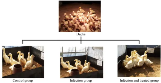
Fig. 1. Three different groups of ducks for in vivo antibacterial assay. Control group, infected group, infected and treated group.
2.4. In vivo antibacterial assay
Ducks were assigned to three groups, including a control group (group 1), infected group (group 2), and infected and treated group (group 3) (Fig. 1). In the control group, nine ducks were not infected with bacteria. In the infection group, 45 ducks were
infected with the Salmonella spp. and E. coli. In the infection and injection group, 45 ducks were infected with the Salmonella spp. and E. coli like the injection group, but these ducks were fed with plant extracts. The bacteria were administered to the group 2 and group 3 ducks orally. Ducks in group 3 were fed Table 1. Inhibition zone (mm) of bacterial growth by plant extracts
Plant extract E. coli S. aureus S. typhi- murium
S. typhi- murium
(NAR)
S. enter- itidis
S. galli- narum
S. pull- orum
S. chole-
rasuis S. dublin Nelumbo
nucifera gaertner
- - - - - - - - -
Asparagi
tuber - - - - - - - - -
Dalbergiae odoriferae
lignum
- - - - - - - - -
Galla rhois 24 22 21 26 24 22 27 23 22
Coptidis
rhizome - - - - - - - - -
Scutellaria baicalensis
georgi
- - - - - - - - -
Schizandrae
fructus - - - - - - - - -
Phellodendri
cortex - - - - - - - - -
Geranii
herba - - - - - - - - -
Thujae semen 22 16 18 19 21 18 20 18 22
Sophorae
radix - - - - - - - - -
Arecae semen - - - - - - - - -
Sophorae flos - - - - - - - - -
Flos
carthami - - - - - - - - -
Zizyphi spinosae
semen
- - - - - - - - -
Armeniacae
semen - - - - - - 22 - -
Lycii fructus - - - - - - - - -
Lycii radicis
cortex - - - - - - - - -
Cassiae
semen - - - - - - - - -
Paeonia
japonica 22 15 13 - 12 21 - 15 21
Epimedii
herba - - - - - - - - -
Acanthopana
cis cortex - - - - - - - - -
Astragali
radix - - - - - - - - -
Theae folium - - - - - - - - -
with the plant aqueous extracts after the bacterial administration.
The antibacterial effects of the plant extracts were determined by analyzing fecal and blood samples taken from all three groups of ducks.
2.5. Gene expression analysis of animal infection by rev- erse transcription-polymerase chain reaction
Total RNA was extracted from blood and fecal samples of the ducks after the treatment with the natural antibiotics (NABs) present in the plant extracts as above for 24 and 48 h. RNA samples were reverse-transcribed to cDNA for 30 min at 42°C using the High Capacity cDNA reverse transcription (RT) kit (Applied Biosystems). Quantitative polymerase chain reaction
(PCR) was performed using the following conditions: 2 min at 50°C, 10 min at 95°C, and 40 cycles for 15 s at 95°C and 1 min at 60°C using 1 μL of cDNA, 2× SYBR Green PCR master mix (Applied Biosystems), and 200 nM forward and reverse primers.
3. RESULTS
3.1. In vitro antibacterial assay
Twenty-four different plants were screened for their antibacter- ial potential (Table 1). The antibacterial activities of the differ- ent plant extracts were determined by disc diffusion method in vitro. Clear zones of bacterial growth were measured for each Table 2. Inhibition zone (mm) of bacterial growth treated with plant extracts
Bacteria
Concentrations (mg/mL) of plant extracts*
1.25 2.5 3.75 5
A B A B A B A B
E. coli 12 11 20 19 21 22 25 24
S. aureus 15 16 19 20 23 21 27 24
S. typhimurium 11 13 14 16 17 19 24 23
S. typhimurium (NAR) 16 12 18 20 20 23 21 25
S. enteritidis 11 14 14 16 19 19 20 22
S. gallinarum 14 16 19 18 21 22 24 25
S. pullorum 11 11 15 14 20 21 25 26
S. cholerasuis 12 13 16 17 19 21 21 23
S. dublin 13 11 15 14 20 19 24 21
*Plant extracts: A, Galla rhois; B, Thujae semen.
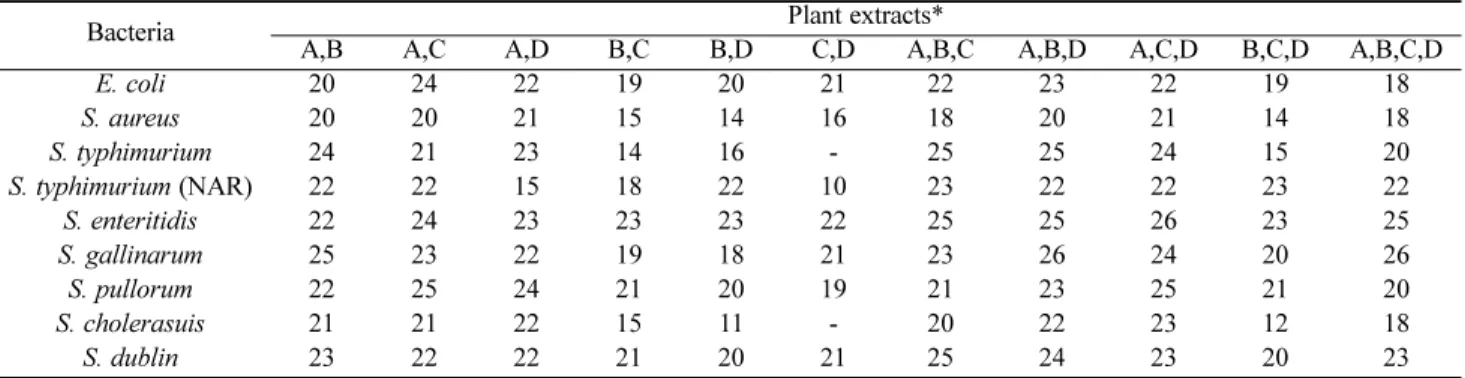
Table 3. Inhibition zone (mm) of bacterial growth by plant extracts in combination
Bacteria Plant extracts*
A,B A,C A,D B,C B,D C,D A,B,C A,B,D A,C,D B,C,D A,B,C,D
E. coli 20 24 22 19 20 21 22 23 22 19 18
S. aureus 20 20 21 15 14 16 18 20 21 14 18
S. typhimurium 24 21 23 14 16 - 25 25 24 15 20
S. typhimurium (NAR) 22 22 15 18 22 10 23 22 22 23 22
S. enteritidis 22 24 23 23 23 22 25 25 26 23 25
S. gallinarum 25 23 22 19 18 21 23 26 24 20 26
S. pullorum 22 25 24 21 20 19 21 23 25 21 20
S. cholerasuis 21 21 22 15 11 - 20 22 23 12 18
S. dublin 23 22 22 21 20 21 25 24 23 20 23
*Plant extracts: A, Galla rhois; B, Thujae semen; C, Paeonia japonica; D, Armeniacae semen.
Table 4. Inhibition zone (mm) of bacterial growth by plant extracts in combination in different ratio
Bacteria Plant extract ratio*
1:1:1 2:1:1 2:2:1 2:1:2 3:1:1 3:2:1 3:3:1 3:1:3 1:3:3 3:3:2 1:3:1
E. coli 21 18 16 23 27 23 25 23 27 23 20
S. typhimurium 19 21 22 23 25 23 27 25 21 18 19
S. typhimurium (NAR) 21 24 24 25 23 20 18 21 25 20 25
S. enteritidis 20 21 19 20 21 27 22 22 21 24 22
S. gallinarum 18 20 21 20 22 23 20 22 20 21 18
S. pullorum 16 21 20 20 25 27 22 26 24 20 20
S. cholerasuis 19 21 23 22 23 23 20 21 20 27 20
S. dublin 19 21 19 20 27 22 25 23 25 18 19
*Plant extracts: Galla rhois, Thujae semen, and Paeonia japonica (1:1:1 = 50:50:50 mg/mL).
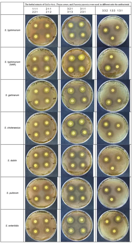
Fig. 2. The antibacterial effects of the natural antibiotics present in the different plant extract against E. coli, S. aureus and serovars of Salmonella enterica subsp by disc diffusion method.
extract and compared with those of the standard antibiotic am- picillin used in the experiments. Among the extracts, only four plant, i.e., Galla rhois, Thujae semen, P. japonica, and Armen- iacae semen, showed antibacterial activities. These four plant extracts were further tested for antibacterial activities against
the Salmonella spp. and E. coli individually and in combination at different ratios. When tested individually, the Galla rhois and Thujae semen extracts showed the highest antibacterial effects at different concentrations against the used bacterial strains (Table 2). When the four plant extracts were used in combination, the antibacterial effects against the Salmonella spp. and E. coli inc- reased compared with those of the individual extracts (Table 3).
When the plant extracts of Galla rhois, Thujae semen, and P.
japonica were then used in combination at different ratios, the antibacterial activities further increased (Table 4, Fig. 2).
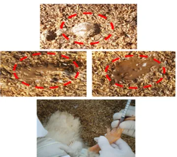
3.2. In vivo antibacterial assay
The blood and fecal samples taken from the ducks of the three groups (Fig. 3) were analyzed for the presence of the bacteria.
mRNA was extracted from the samples and analyzed by RT- PCR. The results showed the presence of the bacterial sequ- ences in the samples taken from the ducks of group 2, while no bacteria was detected in the blood and feces samples from groups 1 and 3. The result indicated that the plant extracts inhi- bited bacterial growth in the ducks of group 3 (Fig. 4 and 5).
4. DISCUSSION
Salmonellae are gram-negative, non-spore-forming pathogenic bacteria causing infections and diseases in animals and hum- ans, and the infections are zoonotic. Salmonella spp. are also the major cause of bacterial food-borne illnesses. These bacteria develop resistance to commonly used antibiotics [7]. There is a strong need for economical and effective antibacterial agents against Salmonella and other gram-negative bacteria that would suppress their resistance and have broad mode of action. The drug resistance properties of bacteria, high prices of synthetic antimicrobial drugs, and compromising safety have forced microbiologists to explore new sources of antibiotics. Natural sources now attract researchers’ attention for the presence of cheap, safe, and potent antimicrobial agents. Medicinal plants
Fig. 3. Sampling from blood and feces of ducks of the three differ-ent groups.
Fig. 4. Gene expression level of the bacteria in the blood samples from all groups of ducks.
Fig. 5. Gene expression level of the bacteria in the feces samples from all groups of ducks.
Table 5. Primers sequences used for anti-bacterial assay
Strains Sequence
E. coli-F CCG ATA CGC TGC CAA TCA GT E. coli-R ACG CAG ACC GTA GGC CAG AT S. enteritidis-F GCA GCG GTT ACT ATT GCA GC S. enteritidis-R TGT GAC AGG GAC ATT TAG CG S. choleraesuis-F AAG GAA AAG ATC ATG GCA CAA S. choleraesuis-R GAA CCC ACC ATC AAT AAC TTT G S. typhimurium-F CCT TTC TCC ATC GTC CTG AA S. typhimurium-R TGG TGT TAT CTG CCT GAC CA S. gallinarum-F GTA TGG TTA TTA GAC GTT GTT S. gallinarum-R TAT TCA CGA ATT GAT ATA TCC
present rich sources of natural antibiotics that can potentially be used as an alternative to chemicals synthetic drugs [8]. Rec- ently, the interest in medicinal plant extracts and pure com- pounds has increased as an alternative method to control patho- genic microorganisms [9]. Plant products have been shown to specifically target resistant pathogenic bacteria [10]. Drug res- istance is one of the major and serious problems that make che- motherapy more difficult. Moreover, in developing countries, the costs of most chemotherapeutic agents are not bearable [11].
The necessity to develop alternative antimicrobial drugs has been widely addressed by microbiologists [12,13]. Therefore, it is essential to search for and develop effective natural, non- toxic antimicrobial agents. Studies have shown that medicinal plants exhibit antiviral, antifungal, antibacterial, antimolluscal, antihelmintic, and anti-inflammatory activities [2]. Plant anti- microbials would thus appear to be an excellent choice [14].
Natural antibiotics can replace synthetic antibiotics to prevent overuse of synthetic antibiotics. Poultry dense breeding weak- ens the immune system, but the effect of natural products imp- roves immunity, then it is possible to raise healthy poultry. The present study was designed to screen medicinal plants for the presence and effectiveness of natural antibiotics. Certain plants and their formulations, such as Galla rhois, Thujae semen, and P. japonica were found to exhibit antimicrobial activities against Salmonella spp. and E. coli. The antibacterial activities increa- sed when the plant extracts were used in combinations and at different ratios both in vitro and in vivo. However, the antibac- terial effects of the plant extract compositions were different against different strains of the bacteria. Bacterial growth was inhibited in the ducks of group 3 by feeding them natural anti- biotic supplements present in the plant extracts. Bacterial gene expression was only found in the group 2 ducks but not in the group 3 ducks, although bacteria were administered equally to the ducks in both groups. When natural antibiotics are used, quality is improved because there is no antibiotic residue in the poultry body. It is simple and efficient because it supplies natu- ral antibiotics through existing water supply devices.
5. CONCLUSION
The plants extracts of Galla rhois, Thujae semen and Paeonia japonica showed its potential against various zoonotic Salmo- nella species and E. coli. The plant extracts successfully inhibit the growth of the bacteria individually and in combination with different extent. Our results confirmed that the above mentioned plants possess antibacterial activities, the results were confirmed by RT-PCR analysis. Hence these natural products are recom- mended to use in the production and formulation of antibiotics
either in pure form or in combination with the pharmaceuticals.
Further studies are suggested to determine and isolate the com- pounds responsible for the antibacterial activities.
Acknowledgements
The research reported in this manuscript was funded by Chon- nam Nat’l University Research Fund.
REFERENCES
1. Diallo, D., B. Hveem, M. A. Mahmoud, G. Betge, B. S. Paulsen, and A. Maiga (1999) An ethnobotanical survey of herbal drugs of Gourma district. Mali. Pharm Biol. 37: 80-91.
2. Mahesh, B. and S. Satish (2008) Antimicrobial activity of some important medicinal plant against plant and human pathogens. Wor.
J. Agr. Sci. 4: 839-843.
3. Sivastava, J., J. Lambart, and Vietmeyer. (1996) Medicinal plants, an expanding role in development. Wor. Ban. T. 1: No. 320.
4. Odikamnoro, O. O. and C. A. Uhuo (2014) Antibacterial activities of two medicinal herbs on Salmonella typhi isolates in Abakaliki, Ebonyi State, Nigeria: Improvement to herbal medicine. J Bacte- riol. 7: 14-18.
5. Krittika, N. and G. H. Ryu (2012) Inhibitory effect of plant extracts on Salmonella spp. InTech. DOI:10.5772/30782.
6. Lewis, W. H. and M. P. Elvin-Lewis (1995) Medicinal plants as sources of new therapeutics. Ann. Missouri Bot. Gard. 82: 16-24.
7. Neu, H. C. (1992) The crisis in antibiotic resistance. Science 257:
1064-1073.
8. Nimri, L. F., M. M. Meqdam, and A. Alkofahi (1999) Antibacte- rial activity of Jordanian medicinal plants. Pharm. Biol. 37: 196- 201.
9. Aqil, F., M. S. Khan, M. Owais, and I. Ahmad (2005) Effects of certain bioactive plant extracts on clinical isolates of beta-lactam- ase producing methicillin. J. Basic Microbiol. 45: 106-114.
10. Nostro, A., L. Cellini, and S. D. Bartolomeo (2006) Effects of com- bining extracts (from propolis or Zingiber officinale) with clarith- romycin on Helicobacter pylori. Phytother Res. 20: 187-190.
11. Gopalkrishnan S., G. Shibumon, and P. J. Benny (2010) Antimi- crobial effect of Punica grantum on pyogenic bacteria. J. Pharm.
Biomed. Sci. 3: 1-3.
12. Poole, K. (2002) Mechanisms of bacterial biocide and antibiotic resistance. Symp. Ser. Soc. Appl. Microbiol. 92: 55-64.
13. Sibanda, T. and A. I. Okoh (2007) The challenges of overcoming antibiotic resistance; Plant extracts as potential sources of antimi- crobial and resistance modifying agents. Afr. J. Biotechnol. 6: 2886- 2896.
14. Mahady, G. B., S. L. Pendland, A. Stoia, F. A. Hamill, D. Fabri- cant, B. M. Dietz, and L. R. Chadwick (2005) In vitro susceptibility of Helicobacter pylori to botanical extracts used traditionally for the treatment of gastrointestinal disorders. Phytother Res. 19: 988- 991.