Received: 31 January, 2017 Revised: 22 February, 2017 Accepted: 22 February, 2017
Ⓒ The Korean Society of Mycology
This is an Open Access article distributed under the terms of the Creative Commons Attrib- ution Non-Commercial License (http://creative- commons.org/licenses/by-nc/4.0/) which permits unrestricted non-commercial use, distribution, and reproduction in any medium, provided the original work is properly cited.
Kor. J. Mycol. 2017 March, 45(1): 54-62 https://doi.org/10.4489/KJM.20170006
pISSN : 0253-651X eISSN : 2383-5249
OPEN ACCESS
RESEARCH NOTE
First Report of Fruit Rot Caused by
Fusarium decemcellulare in Apples in Korea
Seung-Yeol Lee
1, Su-Jin Park
1, Jae-Jin Lee
1, Chang-Gi Back
1,2, Leonid N. Ten
1, In-Kyu Kang
1, Hee-Young Jung
1*1
College of Agriculture and Life Sciences, Kyungpook National University, Daegu 41566, Korea
2
National Institute of Horticultural and Herbal Science, Wanju 55365, Korea
*Corresponding author: [email protected]
Abstract
In 2014, abnormal brown spots were observed on Hongro apples in fields in Gyeongsangbuk-do Province and during low-temperature storage. The spots were round, blight brown, and different from the symptoms of previously reported apple diseases. A fungal pathogen was isolated and cultured on potato dextrose agar, and it was morphologically similar to Fusarium decemcellulare. A pathogenicity test showed the same brown spots on both wounded and unwounded Hongro and Fuji apple cultivars. RPB1 and RPB2 sequences of F. decemcellulare KNU-GC01 matched with those of F. decemcellulare NRRL 13412 (98.3% and 97.6%
similarities, respectively); both strains clustered together in the phylogenetic tree, indicating their close relationship at the species level. Therefore, F. decemcellulare is a newly reported pathogen that causes brown spots on apples in Korea.
Keywords: Apple fruit, Brown spot, Cultivar Fuji, Cultivar Hongro, Fusarium decemcellulare
Apple (Malus pumila Mill.) is cultivated worldwide, and its production was approximately 84 million tons in 2014 (FAOSTAT, Food and Agriculture Organization of the United Nations, http://faostat.fao.org/). In Korea, the cultivation area for apple is approximately 31,620 ha, of which cultivars Fuji and Hongro occupy about 70% and 15%, respectively;
the total cultivation area of the two cultivars has gradually increased (KOSTAT, Statistics Korea, http://kostat.go.kr/portal/eng/). To date, about 40 pathogens have been reported for apple[1], among which, the causal agents of apple blotch, anthracnose, white rot, Alternaria leaf spot, and bacterial shoot blight are of economic concern for apple cultivation[2].
Because of the economic losses caused by the major apple diseases, rapid and accurate
identification of the causal agent of the disease is an essential prerequisite for the fungicide
spray program[3]. Apple anthracnose and white rot caused by Colletotrichum spp. and
Botryosphaeria dothidea, respectively, are clearly distinct from each other on the basis of
typical signs and symptoms[3], rendering their identification easier. Apple blotch caused by
Marssonina coronaria can be observed on the fruit, but it is distinguished using its unique
symptoms and signs[4]. Recently, abnormal brown spots were observed on Hongro fruits
apples in low-temperature storage (Fig. 1C, 1D). To isolate the causal agent from the abnormal brown spots, the surface of an apple was wiped with 70% EtOH and the diseased peel was removed using a sterilized blade. Then, the surface of the collected diseased tissue was sterilized in 70% ethanol and 1% sodium hypochlorite and washed 3 times with double-distilled water. The surface-sterilized tissues were transferred onto potato dextrose agar (PDA) plates and maintained in an incubator at 25°C. After 3 days, small white colonies were observed, and they were transferred onto a new PDA plate. The colonies formed abundant white to pink mycelia (Fig. 2A), and dark carmine-red pigment was produced under the PDA after 7 days (Fig. 2B). In addition, creamy yellow sporodochia were observed on the 20-day-old mycelia cultured on PDA (Fig. 2C). Two morphologically
Fig. 1. Photographs of showing abnormal brown spot on cultivar Hongro diseased apple fruits
collected from Gimcheon in Gyeongsangbuk-do Province in 2014. A, Collected from field; B,
Close-up view of an infected apple of A; C, Collected from low-temperature storage; D,
Close-up of an infected apple of C. Red arrowhead indicate lenticels on the surface.
Fig. 2. Morphological characteristics of the isolated Fusarium decemcellulare KNU-GC01 on potato dextrose agar (PDA). A, Feature of the colony after 7 days of growth on PDA; B, Reverse side of the 7 days growth colony showed dark carmine-red pigment; C, Creamy yellow sporodochia on the after 20 days cultured mycelium (scale bar = 1 mm).
Fig. 3. The observed macroconidia and microconidia of Fusarium decemcellulare KNU-GC01.
A, Monophialides and immature and mature macroconidia; B, Macroconidia, predominantly
5~9 septate depends on the length; C, Microconidia produced in long chains on potato dextrose
agar; D, Microconidia, 0~1 septate and oval (scale bars: A~D = 10 µm).
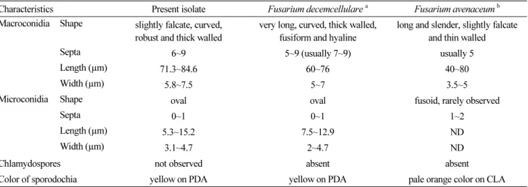
cultural and morphological characteristics of previously reported Fusarium spp. in fruit [5-7], the isolated fungal strains were found to be the closest to Fusarium decemcellulare (Table 1). In addition, KNU-GC01 and KNU-GC02 showed identical morphological and cultural characteristics (data not shown).
To confirm pathogenicity, F. decemcellulare KNU-GC01 isolated in this study was inoculated into 5 healthy Hongro and Fuji fruits. For the inoculum, the spore suspension was prepared using F. decemcellulare cultured for 20 days, and the conidial concentration was determined with a hemocytometer and adjusted to approximately 1.75 × 10 5 conidia/mL. The surface of a healthy apple was wiped with 70% EtOH and then air-dried.
Two points of the lenticel were wounded using a sterilized needle, and paper disks containing 20 µL of the spore suspension were attached and sealed using foil. Identical paper disks were also attached to unwounded lenticels and sealed using foil. Fruits inoculated with sterilized water were used as the control. All the inoculated fruits were incubated at 25°C, and after 3 days, the disks were removed. After 15 days, brown spots could be observed on both wounded and unwounded Hongro fruits (Fig. 4A~4D), although the size and shape of the spots were somewhat smaller on the Hongro fruits. However, pathogenicity was confirmed for Fuji (Fig. 4E~4H). From each of the inoculated fruits, F.
Table 1. Comparison of morphological characteristics of isolated fungi from abnormal brown spot with the previous descriptions of Fusarium spp. on fruit disease
Characteristics Present isolate Fusarium decemcellulare
aFusarium avenaceum
bMacroconidia Shape slightly falcate, curved,
robust and thick walled
very long, curved, thick walled, fusiform and hyaline
long and slender, slightly falcate and thin walled
Septa 6~9 5~9 (usually 7~9) usually 5
Length (µm) 71.3~84.6 60~76 40~80
Width (µm) 5.8~7.5 5~7 3.5~5
Microconidia Shape oval oval fusoid, rarely observed
Septa 0~1 0~1 1~2
Length (µm) 5.3~15.2 7.5~12.9 ND
Width (µm) 3.1~4.7 2~4.7 ND
Chlamydospores not observed absent absent
Color of sporodochia yellow on PDA yellow on PDA pale orange color on CLA
PDA, potato dextrose agar; ND, not described; CLA, carnation leaf agar.
a
Description by Leslie and Summerell[6]; Serrato-Diaz et al.[5].
b