http://dx.doi.org/10.11620/IJOB.2013.38.4.169
Fast temporal detection of intracellular hydrogen peroxide by HyPer
Yu-Mi Yang, Sung Jun Lee, and Dong Min Shin*
Department of Oral Biology, BK21 PLUS Project, Yonsei University College of Dentistry, Seoul 120-752, Korea (received November 26, 2013; revised December 11, 2013; accepted December 16, 2013)
*Correspondence to: Dong Min Shin, DDS, PhD, Department of Oral Biology, Yonsei University College of Dentistry, 50 Yonsei-ro, Seodaemun-gu, Seoul 120-752, Korea
Tel: +82-2-2228-3051, Fax: +82-2-364-1085 E-mail: [email protected]
This is an Open-Access article distributed under the terms of the Creative Commons Attribution Non-Commercial License(http://creati- vecommons.org/licenses/by-nc/3.0) which permits unrestricted non- commercialuse, distribution, and reproduction in any medium, pro- vided the original work is properly cited.
HyPer is the genetically encoded biosensor of intracellular hydrogen peroxide (H2O2), the most stable of the reactive oxygen species (ROS) generated by living cells. HyPer has a high sensitivity and specificity for detecting intracellular H2O2 by confocal laser microscopy. However, it was not known whether high speed ratiometric imaging of H2O2 by HyPer is possible. We thus investigated the sensitivity of HyPer in detecting changes to the intracellular H2O2 levels in HEK293 and PC12 cells using a microfluorometer imaging system. Increase in the HyPer ratio were clearly evident on stimulations of more than 100 μM H2O2 and fast changes in the HyPer ratio were observed on ratiometric fluorescent images after H2O2 treatment. These results suggest that HyPer is a potent biosensor of the fast temporal production of intracellular H2O2.
Key words: HyPer, H2O2, ROS, high speed detection
서 론
활성산소종 (reactive oxygen species, ROS)은 정상적인 산소 대사반응을 통해 자연적으로 생성되나 자외선이나 열과 같은 환경적 스트레스에 지속적으로 노출될 경우, 세포 내 활성산소종이 급격히 증가 되어 결국 심각한 세 포의 손상을 초래한다. 이러한 세포 내 산화적 손상 (oxidative damage)을 유발시키는 인자 중 과산화수소 (H2O2)는 산화 용량 (oxidizing capacity)이 높아 활성산소 종에 의한 세포 손상과 매우 밀접한 관련이 있는 것으로 여겨지고 있다 [1-3]. 이렇게 과산화수소는 세포독성 효과 를 가지고 있지만, 최근 동물세포에서 신호전달 물질로써 세포의 성장, 분화, 그리고 물질 대사와 같은 작용들에 중 요한 역할을 담당하는 것으로 보고되고 있다 [4-6].
활성산소종의 생체 외 (in vitro) 생성을 측정하고자 여 러 가지 측정방법들이 개발되어졌다. 이중 DCF (dichloro- fluorescein) 유도체는 활성산소종에 의한 산화반응 후 형 광을 나타내기 때문에 세포 내 활성산소종을 감지하기 위 해 가장 많이 사용되어져 왔다. 그러나, DCF 유도체는 일 부 활성 산소 및 산화 질소와 같은 다양한 활성산소종에 반응을 하며, 특정 세포 내 구역의 활성산소종은 감지하 지 못한다는 문제점을 가지고 있다. 가장 큰 문제점은 빛 에 노출이 되면 DCF가 자체적으로 활성산소종을 증가시 킬 수 있어 DCF 유도체에 의한 활성산소종의 정량이 부 정확해 질 수 있다는 것이다 [7-9]. 이러한 문제점들을 개 선하여 살아있는 세포 내 (in vivo) 활성산소종의 하나인 과산화수소를 감지할 수 있게 개발된 형광 센서가 HyPer 이다 [10]. HyPer는 원핵생물의 과산화수소 감지 단백질 (H2O2-sensing protein)인 OxyR의 조절 도메인 (regulatory
169
170 Yu-Mi Yang, Sung Jun Lee, and Dong Min Shin
domain, OxyR-RD) 안에 cpYFP (circularly permuted yellow fluorescent protein)를 넣어서 만든 것으로 μM 수준의 과산 화수소를 감지할 수 있고, 세포질이나 미토콘드리아에서 의 과산화수소 생성을 모니터링 할 수 있다. 또한, HyPer 의 장점은 DCF와는 달리 과산화수소 이외의 활성산소종 에는 반응을 하지 않으며, 세포 내 pH 변화에도 민감하지 않다. HyPer의 개발 이후 여러 논문들에서 다양한 세포들 과 약물들을 이용하여 HyPer의 과산화수소 민감도와 특 이성이 높다는 것을 증명하였다. 그러나, HyPer의 효율에 대한 이전까지의 연구들은 대부분이 공초점 현미경 (confocal microscope)을 사용하여 얻은 결과들로 형광 이 미징 시스템의 카메라 셔터 속도가 느려 단시간 동안의 세포 내 과산화수소의 변화를 관찰할 수 없었다 [11-13].
본 연구는 이러한 카메라 셔터의 속도를 개선한 이미징 시스템을 이용하여 HyPer의 과산화수소에 대한 민감도를 확인하고, HyPer의 활용 가능성에 대하여 알아보았다.
재료 및 방법
세포 배양 및 실험 재료
실험에 사용된 HEK293 (human embryonic kidney cell line; Korean Cell Line Bank, Korea) 세포는 Dulbecco’s modified eagle media (DMEM; Invitrogen, Carlsbad, CA, USA)에 10% fetal bovine serum (FBS; Invitrogen)과 1% 항 생제 (Invitrogen)를 첨가하여 배양 후 사용하였고, PC12 (neuron-like rat pheochromocytoma cell line; American Type Culture Collection, Manassas, VA, USA) 세포는 7일 동안 collagen (10 μg/ml; Upstate Biotechnology, Lake Pacid, NY, USA) 코팅한 plate에서 DMEM에 1% FBS와 2%
heat-inactivated horse serum (HS; Invitrogen), 1% 항생제, 그리고 50 ng/ml NGF (Alomone Laboratory, Jerusalem, Israel)를 첨가하여 배양 후 사용 하였다. HyPer (pHyPer -Cyto) 벡터는 이서구 박사 (Yonsei Univerisity College of Medicine, Seoul, Korea)에게 제공받았다. 과산화수소는 Sigma-Aldrich (St Louis, MO, USA)에서 구입하였다.
Transient transfection
HEK293과 PC12 세포를 2 × 10⁵ cells/60-mm dish가 되도 록 분주하고 세포가 80%정도 찼을 때 HyPer 벡터를 Lipofectamine 2000 (Invitrogen)을 이용하여 transfection을 시 행하였다. 24-48 시간 후 세포의 활성 변화를 측정하였다.
세포 내 과산화수소량 측정
세포 내 과산화수소량의 변화를 측정하기 위하여
HyPer 벡터를 transfection하여 준비된 HEK293 세포는 생 리식염수 (140 mM NaCl, 5 mM KCl, 1 mM MgCl2, 1 mM CaCl2, 10 mM HEPES, 10 mM glucose, 310 mOsm, pH 7.4 with NaOH)에 담근 후 중력을 이용한 관류장치를 통해 실험 진행 동안 용액을 교환하였다. HyPer에 의한 형광 발색은 이전 실험방법 등을 참조하여 측정하였다 [10,14,15]. HyPer의 형광은 Molecular Devices (Universal Imaging Co., Downingtown, PA, USA) 이미징 시스템을 이 용하였으며, 이 때 파장은 excitation 파장 (420 nm와 500 nm)과 emission 파장 (530 nm)을 사용하여 형광의 변화를 측정하였다 (Ratio = F500/F420). HyPer의 형광 변화 이미지 들은 도립 현미경 (inverted microscope, Nikon Instruments Inc., Tokyo, Japan)에 부착된 CCD 카메라 (Photometrics, Tucson, AZ, USA)를 통하여 컴퓨터에서 0.26-2초 간격으 로 기록하였다.
실험자료의 분석
모든 실험자료의 분석은 MetaFlour software (Molecular Devices)를 이용하였다. 과산화수소 자극에 대한 HyPer 의 형광반응 수치 값은 평균값 ± 표준오차 (mean ± S.E.)로 표시하였다.
결과
HyPer는 μM 수준의 과산화수소를 감지하는 것으로 알려져 있다 [10,16]. HyPer를 transfection시킨 HEK293 세포와 PC12 세포에서 다양한 농도의 과산화수소의 처 리에 따른 HyPer의 형광 변화를 관찰하였다. 그 결과, HEK293 세포에서는 30 μM 이상의 과산화수소 농도에 서 HyPer의 형광 변화를 관찰할 수 있었고 (Fig. 1A와 1B), PC12 세포에서는 100 μM 이상의 과산화수소 농도 에서 HyPer의 활성을 감지할 수 있었다 (Fig. 1C와 1D).
비록 이전 연구들에서 보고 되어진 HyPer의 세포 내 과 산화수소 감지 농도인 20 μM에 비하여 좀 더 높은 농 도에서 형광이 나타났지만, 과산화수소에 대한 형광 반 응의 크기는 이전 연구들과 비슷하게 나타나는 것을 확 인하였다 [10].
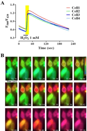
이전 연구들에서 HyPer 활성을 측정하기 위해 사용한 공초점 현미경에서의 이미징들은 주로 분 단위 수준에 서 얻어진 결과들이었다. 따라서 본 연구에서는 형광 이 미지 시스템의 이미징 획득 시간을 최소한으로 조절하 여 과산화수소에 대한 HyPer 형광 반응을 관찰하였다. 1 mM 과산화수소를 처리한 후 0.26초의 빠른 간격으로 HyPer의 형광 변화를 확인하였다 (Fig. 2A). 세포 내 과산
Fig. 1. Concentration-dependent H2O2 sensitivity of HyPer in HEK293 and PC12 cells. Application of H2O2 produced an increase of HyPer fluorescent and the changes of HyPer fluorescent (Ratio = F500/F420) were traced as a function of H2O2 concentration (A and C).
Concentration-dependent bar graph for H2O2-induced increase of HyPer fluorescent (B and D, n = 4). HyPer generated a fluorescent for intracellular H2O2 in a concentration-dependent manner in HEK293 (A and B) and PC12 cells (C and D).
Fig. 2. Fast temporal changes of HyPer responses to added H2O2 in HEK293 cells. Ratio changes of HyPer by 1 mM H2O2
traced in A. Also, fast interval (interval time: 0.26 sec) images represented the production of intracellular H2O2 after application of 1 mM H2O2 (gray region in A) in B.
화수소량의 변화는 실험 시작 20초 정도에서 과산화수 소를 처리한 후 약 33.5초 정도에서 감지되기 시작하여 2초 후에 최대 증가량을 보이는 것을 확인할 수 있었다 (Fig. 2B). 이는 HyPer를 이용하여 보고되어진 이전 연구 들의 세포 내 과산화수소량의 변화 시간 보다 빠른 시 간 동안의 HyPer의 형광 변화를 보여준 것으로, HyPer 가 세포 내 과산화수소량의 변화에 대한 반응 민감도가 높으며 [10,12,13,16-18], 시간적 반응도도 높다는 것을 의미한다. 이러한 결과들은 HyPer를 이용하여 세포 내 활성산소종 조절과 관련이 있는 연구를 할 때 활성산소 종과 관련 신호전달 단백들 간의 상관관계를 실시간 모 니터링이 가능할 수 있음을 시사한다.
고 찰
본 연구는 형광 이미징 시스템을 이용하여 HyPer가 세포 내 과산화수소의 증가를 얼마나 빠르고 정확하게 감지하는지를 조사하였다. HyPer는 초 단위의 짧은 시 간 동안에도 세포 내 과산화수소의 변화를 감지할 수 있음을 확인하였다. 이전의 연구들에서 HyPer의 형광 발현의 변화는 주로 분 단위 수준에서 보여주었으며, 과 산화수소에 대한 HyPer의 반응 변화를 실시간으로 모니
172 Yu-Mi Yang, Sung Jun Lee, and Dong Min Shin
터링 한 결과를 보여준 연구 결과는 없었다.
세포 내 활성산소종의 감지를 위한 노력은 여러 형광 유도체들의 개발을 이끌어 내었다. 그러나, 다른 활성산 소종과의 간섭 현상과 세포 소기관 별 특이성을 가지지 못했다는 여러 문제점들이 나타났다. Belousov 등은 이 러한 문제점을 해결한 과산화수소 특이적인 형광 벡터 인 HyPer를 개발하여, 활성산소종에 대한 연구를 다양 한 세포와 약물들을 이용하여 생리활성 자극 수준의 저 농도 과산화수소도 감지할 수 있다고 보고 하였다. 특 히, 이들이 사용한 HyPer는 과산화수소에 대하여 생체 외 실험 (in vitro)에서 250 nM, 박테리아 세포 (E. coli) 에서 5 μM, 동물 세포 (HeLa cells)에서 50 μM부터 감 지를 할 수 있었다 [10]. 본 연구에서도 HEK293 세포를 사용하여 과산화수소 농도가 30 μM 수준부터 HyPer 벡 터에 의해 감지가 가능하다는 것을 보여주었다. 그러나, 명확한 반응을 감지하기 위해서는 100 μM 이상의 과산 화수소를 사용하여 실험을 하였을 때 대부분의 세포에 서 감지를 하는 것으로 나타나 세포를 이용한 실험에서 각 세포에 알맞은 과산화수소의 농도를 조절하는 것이 중요할 것으로 여겨진다. 또한, HyPer의 반응을 감지하 기 위해 공초점 현미경을 사용하는 이미징 시스템은 레 이저를 이용하여 파장을 변화시키기 때문에 이미징 획 득 속도가 빠르지 못하다는 단점이 있었으며, 이 때문에 그 동안의 HyPer 반응들은 분 단위 수준에서 대부분 나 타났다 [12,13,16-18]. 최근 Mishina 등은 다양한 세포주 에서 여러 이미징 시스템의 이미징 획득 간격을 고속으 로 얻은 결과들을 통해 HyPer의 민감도와 과산화수소에 대한 특이성이 높다는 것을 보여주었다 [18]. 본 연구 역시 형광현미경을 사용하는 고속 이미징 시스템에서 빠른 파장 변화를 위한 셔터의 개폐 속도 개선을 통하 여 얻은 결과들이 Mishina 등의 결과와 견주어 HyPer 의 민감도와 특이성이 뒤떨어지지 않는 것으로 나타났 다. HyPer의 민감도와 과산화수소에 대한 높은 특이성 은 향후 활성산소종에 대한 연구를 빠르고 간편하게 수 행할 수 있을 것으로 사료된다. Woo 등은 국지적 과산 화수소의 농도 변화가 세포막 주변에 분포한 perioxiredoxin 효소와 같은 과산화수소의 독성을 제거하 는 효소들의 활성을 인산화를 통하여 조절할 수 있다는 연구 결과를 발표한 바 있다 [19]. 또한, Wu 등은 소포 체 (endoplasmic reticulum, ER)에서 국지적으로 생성되는 과산화수소는 Nox4에 의존적이며 ERK의 활성을 유도하 는 Ras의 지속적인 활성을 통해서 분비되어진다는 보고 를 하였다 [20]. 이러한 보고들은 과산화수소의 국지적 생성이 주변 신호전달 단백들의 변화와 밀접한 상관관 계가 있음을 보여주며, 과산화수소의 변화와 주변 신호
전달 단백들의 상관관계를 연구하는데 있어서 HyPer의 역할이 중요하다는 것을 의미한다. HyPer를 이용하여 이러한 신호전달 단백들의 변화를 관찰하고자 한 대부 분의 실험들이 분 단위 수준의 관찰이었다는 것을 감안 하면 본 연구의 결과는 보다 빠르고 정확하게 국지적 과산화수소의 변화 관찰이 가능하게 되었음을 보여준다. HyPer를 이용하여 주변 신호전달 단백들의 변화뿐만 아 니라 세포막에 존재하는 활성산소종 유발과 관련한 채 널들의 변화도 실시간으로 관찰 할 수 있게 되어 활성 산소종에 대한 실험 수행시 세포 내 칼슘 변화와 과산 화수소의 변화 등 세포 내부의 다양한 이온의 유입/농 도 변화도 함께 측정할 수 있을 것으로 기대된다. 본 결 과들을 통하여 HyPer는 세포 내 과산화수소의 증가와 관련한 활성산소종에 대한 실험을 수행할 때 형광 이미 징 시스템을 이용하여 빠르고 정확한 신호작용기전에 대한 연구 수행이 가능할 것으로 여겨진다.
감사의 글
이 논문은 연세대학교 치과대학 2012년도 연구비 (6-2012-0060)에 의하여 이루어진 것임.
References
1. Rhee SG. Redox signaling: hydrogen peroxide as intracellular messenger. Exp Mol Med. 1999;31:53-9 2. Simon HU, Haj-Yehia A, Levi-Schaffer F. Role of reactive
oxygen species (ROS) in apoptosis induction. Apoptosis.
2000;5:415-8
3. Cai H. Hydrogen peroxide regulation of endothelial function: origins, mechanisms, and consequences.
Cardiovasc Res. 2005;68:26-36
4. Rhee SG, Bae YS, Lee SR, Kwon J. Hydrogen peroxide: a key messenger that modulates protein phosphorylation through cysteine oxidation. Sci STKE. 2000;2000:pe1 5. Sundaresan M, Yu ZX, Ferrans VJ, Irani K, Finkel T.
Requirement for generation of H2O2 for platelet-derived growth factor signal transduction. Science. 1995;270:296-9 6. Rhee SG. Cell signaling. H2O2, a necessary evil for cell
signaling. Science. 2006;312:1882-3
7. LeBel CP, Ischiropoulos H, Bondy SC. Evaluation of the probe 2',7'-dichlorofluorescin as an indicator of reactive oxygen species formation and oxidative stress. Chem Res Toxicol. 1992;5:227-31
8. Marchesi E, Rota C, Fann YC, Chignell CF, Mason RP.
Photoreduction of the fluorescent dye 2'-7'-dichloro- fluorescein: a spin trapping and direct electron spin resonance study with implications for oxidative stress measurements.
Free Radic Biol Med. 1999;26:148-61
9. Rota C, Fann YC, Mason RP. Phenoxyl free radical formation during the oxidation of the fluorescent dye 2',7'-dichlorofluorescein by horseradish peroxidase.
Possible consequences for oxidative stress measurements.
J Biol Chem. 1999;274:28161-8
10. Belousov VV, Fradkov AF, Lukyanov KA, Staroverov DB, Shakhbazov KS, Terskikh AV, Lukyanov S. Genetically encoded fluorescent indicator for intracellular hydrogen peroxide. Nat Methods. 2006;3:281-6
11. Rhee SG, Chang TS, Jeong W, Kang D. Methods for detection and measurement of hydrogen peroxide inside and outside of cells. Mol Cells. 2010;29:539-49
12. Markvicheva KN, Bogdanova EA, Staroverov DB, Lukyanov S, Belousov VV. Imaging of intracellular hydrogen peroxide production with HyPer upon stimulation of HeLa cells with epidermal growth factor.
Methods Mol Biol. 2008;476:79-86
13. Markvicheva KN, Bilan DS, Mishina NM, Gorokhovatsky AY, Vinokurov LM, Lukyanov S, Belousov VV. A genetically encoded sensor for H2O2 with expanded dynamic range. Bioorg Med Chem. 2011;19:1079-84 14. Hong JH, Lee SY, Shin DM. Real-time imaging of inositol
1,4,5-triphosphate movement in mouse salivary gland cells. Int J Oral Biol. 2008;33:124-29
15. Jin MH, Koo NY, Jin MH, Hwang SM, Park KP. A role of sodium bicarbonate cotransporter (NBC) in formation in human salivary gland acinar cells Int J Oral Biol.
2008;33:143-47
16. Malinouski M, Zhou Y, Belousov VV, Hatfield DL, Gladyshev VN. Hydrogen peroxide probes directed to different cellular compartments. PLoS One. 2011;6:e14564 17. Jin BY, Sartoretto JL, Gladyshev VN, Michel T.
Endothelial nitric oxide synthase negatively regulates hydrogen peroxide-stimulated AMP-activated protein kinase in endothelial cells. Proc Natl Acad Sci U S A.
2009;106:17343-8
18. Mishina NM, Markvicheva KN, Bilan DS, Matlashov ME, Shirmanova MV, Liebl D, Schultz C, Lukyanov S, Belousov VV. Visualization of intracellular hydrogen peroxide with HyPer, a genetically encoded fluorescent probe. Methods Enzymol. 2013;526:45-59
19. Woo HA, Yim SH, Shin DH, Kang D, Yu DY, Rhee SG.
Inactivation of peroxiredoxin I by phosphorylation allows localized H2O2 accumulation for cell signaling. Cell.
2010;140:517-28
20. Wu RF, Ma Z, Liu Z, Terada LS. Nox4-derived H2O2
mediates endoplasmic reticulum signaling through local Ras activation. Mol Cell Biol. 2010;30:3553-68