Gangneung-Wonju National University
This report described a technique utilizing a computer-aided design (CAD) /computer-aided machining (CAM) - guided surgical implant placement and prefabricated fixed complete denture for an immediately loaded restoration. A patient with an edentulous maxilla and mandible received 6 implants in maxilla and 6 implants in the mandible using CAD/CAM surgical templates. Prefabricated provisional maxillary and mandibular implant supported fixed prostheses were connected immediately after implant installation. Provisional prostheses were evaluated for aesthetics, function during 6 months.
Definitive prostheses were fabricated.
Key words: CAD/CAM technology, flapless surgery, immediate loading, prefabricated prosthesis
(J.K.A.of Stomatognathic Function and Occlusion 2009:25(3):243~253)
INTRODUCTION
The original Brånemark implant protocol required submerged healing time of 3-4 months in mandible and 5-6 months in maxilla to obtain predictable osseointegration.1,2 However, with the trend of shortening treatment time and reducing patient discomfort, immediate loading of implants has emerged. With superior initial stability, immediate implant loading has achieved a similar success rates as those reported in the delayed 2-stage approach in edentulous patients.3-10
Correspondence to : Professor Chan-Jin Park
Department of Prosthodontics, College of Dentistry, Gangneung-Wonju National University, 120 Gangneung Daehangno, Gangneung city, Gangwon-do, South Korea, 210-702
Tel : +82-33-640-3153, Fax : +82-33-640-3113, E-mail : [email protected]
Received :April 03, 2009, Last Revision :May 23, 2009, Accepted: September 25, 2009
With the use of computed tomography (CT), computer - aided design / computer - assisted machining (CAD/CAM) technology, and internet, the implant dentistry has been evolved.11-13 CAD/CAM systems such as stereolithographic rapid prototyping have been developed to fabricate precision surgical templates.11-13 The surgical templates made by CAD/CAM technology and precise installation of implants, permit restorations to be inserted immediately after implants have been placed. From these, the surgical and prosthetic treatment times are minimized. The advantages of
CAD/CAM guided implant procedures are flapless, minimally invasive surgery and shorter surgery duration. With this technique, less postoperative morbidity and delivery of prosthesis for immediate function would be possible.14-18
This report describes about the technique utilizing a computer-assisted surgical design, CAD/CAM surgical template, a flapless surgical procedure, and a prefabricated fixed complete denture for immediately loaded restoration.
CLINICAL REPORT
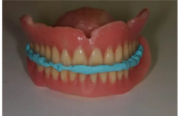
A 61-year-old man was referred for an edentulous maxilla and mandible (Fig. 1). The patient complained of unstable mandibular and uncomfortable maxillary complete denture. Clinical examinations and radiographic assessments were conducted. CT examinations on maxilla and mandible revealed that alveolar bone volume was sufficient for the implants placement. Various treatment alternatives were discussed, the patient consented to have dental implant placed simultaneously in maxilla and mandible with CAD/CAM guided surgery and immediately loaded
Fig. 1. Frontal view of edentulous arches at an approximated occlusal vertical dimension.
restoration using Nobel Guide system.
New temporary complete dentures were fabricated to determine and confirm tooth position for esthetics, phonetics, and occlusal vertical dimension. (Fig. 2) An interocclusal record was taken with a rigid vinyl polysiloxane (Regisil Rigid; Densply Intl, Milford, USA) at the patient's appropriate centric position and occlusal vertical dimension. Nine 2 mm-diameter gutta percha (Temporary stopping; GC corporation Tokyo, Japan) markers were placed into the denture base of the maxillary and mandibular dentures to serve as radiographic markers. The CT scan of maxilla and mandible were generated using a double-scan technique.19 First CT scan was made while patient was wearing the radiographic guide together with an interocclusal record. Using the same CT settings, the scanning was repeated with the radiographic guide alone. Dicom data were converted into a file format compatible with implant planning soft ware (Procera Software; Nobel Biocare, Gothenburg, Sweden). The scanning data were superimposed according to radiographic makers. Then, the images were uploaded for processing. With the zoom, rotate, and translate functions of the three dimensional viewer, any detail
Fig. 2. New temporary dentures and inter- occlusal record.
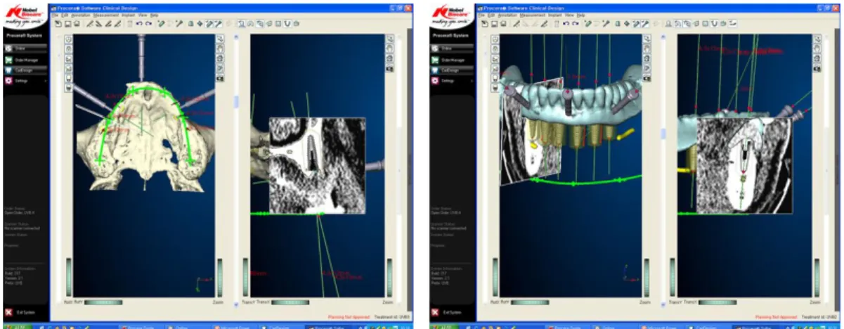
Fig. 3. Three dimensional image-based treatment planning software(Procera software; Nobel Biocare AB). Using the program, 6 regular platform implants and 3 horizontal stabilization pins were planned for each arch.
Fig. 4. Stereolithographic surgical templates
Fig. 5. Prefabricated provisional fixed prostheses
can be inspected. From this information, the sizes and positions of the dental implants were digitally evaluated and placed (Fig. 3). Using implant planning software (Procera Software; Nobel Biocare), 6 regular platform implants (Nobel Replace Tapered Groovy; Nobel Biocare) and 3 horizontal stabilization pins were planned for each arch. The data was transferred to the milling center (Procera;
Nobel Biocare) to fabricate the stereolithographic surgical templates(Fig. 4) with the preplanned surgical sites for the dental implants. The provisional
Fig. 6. Maxillary and mandibular surgical templates in situ. The surgical templates were positioned with the centric relation interocclusal record.
Fig. 7. Three stabilizing transalveolar pins fix surgical guide
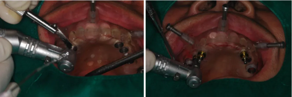
Fig. 8. A, Preparation of alveolus using metal sleeves B, Fixture placement.
fixed prostheses with metal framework were fabricated on the working cast prior to the implant placement (Fig. 5).
The flapless implant surgery was performed under the local anesthesia. Surgical template was inserted and positioned with the centric relation interocclusal record (Fig. 6) Three guided anchor pins were used to maintain the accurate position of the mandibular surgical template during the surgical procedure (Fig. 7). The mandibular implants were placed using the surgical template following the predetermined direction and depth, based on the computer model planning. All implants were place with 45 Ncm insertion torque. Surgical template was removed. And then, preplanned multi-unit abutments (Nobel Replace; Nobel Biocare) were inserted. Upon completion of the mandibular implant placement, the same procedure was repeated to place the implants in the maxilla (Fig.
8, 9).
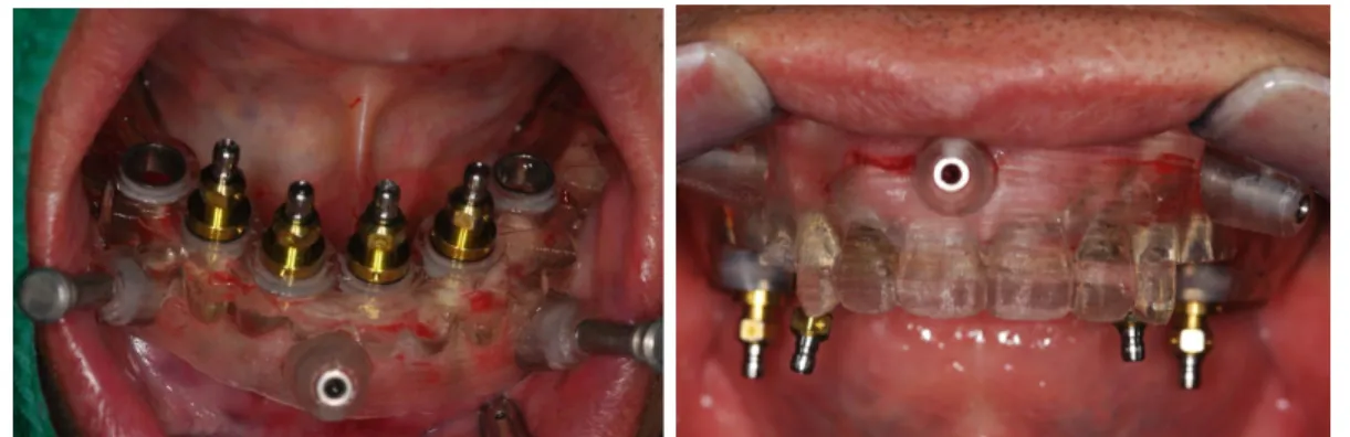
Prefabricated customized metal reinforced acrylic bridge was fixed by anterior two abutments. And four temporary cylinders (Nobel Replace; Nobel Biocare) were connected with auto polymerized resin (Jet, Lang dental, Wheeling, USA) in the mouth. The occlusion was adjusted to have
Fig. 9. Implant installation using surgical templates.
simultaneous centric relation contact and a canine protected occlusal scheme. Direct relining material (Tokuyama rebase II, Tokuyama Dental Corporation, Tokyo, Japan) was used for relining of the soft tissue defect. In frontal view of temporary prosthesis, midline discrepancy was seen (Fig. 10).
Simultaneous treatment of both maxilla and mandible was more technique sensitive in terms of surgical template positioning, precise implant placement location, and maintenance of an accurate occlusion relationship. Another possibility of the discrepancy was an interocclusal recording error.
Once the prosthesis was inserted, the postoperative radiographs were made to confirm the fit of the prostheses. The patient was instructed to take a soft diet for 6 to 8 weeks.
At one month recall check, there was seen marginal bone loss and radiolucent band around left lower most distal fixture (#36i) (Fig. 11). This was diagnosed as an osseointegration failure. After surgery, this fixture showed high primary initial stability (ISQ 79). This failure might been caused by early loading and distal cantilever effect. The fixture was removed and regular platform, 10 mm another fixture was re-installed on the posterior site.
The provisional restoration was repaired and
Fig. 10. Frontal view of temporary prostheses.
immediately loaded. From the failure of the fixture, the patient's masticatory force was suspected to be strong. Therefore, the patient's incidence of repair would be much frequent. We decided to treat the patient with a screw type, metal and resin fixed complete denture.
At 6 months after surgery (Fig. 12), the impression taking was performed on abutment level with transfer type impression coping with polyvinyl siloxane (Examixfine, GC corporation, Tokyo, Japan). It was for transferring the position of the abutment to a working model. Master casts (Die keen, Heraeus Kulzer Inc. Lafayette Blvd., America) were fabricated (Fig. 13). Occlusal rims
Fig. 11. Osseointegration failure in the lower left most distal fixture. Marginal bone loss and radiolucent band around the fixture. A, Panoramic radiograph B, Periapical radiograph
Fig. 12. Frontal view at 6 months after surgery
were made with the plastic temporary cylinder. The patient complained of anterior protuberance of the provisional restoration. After adjustment of the
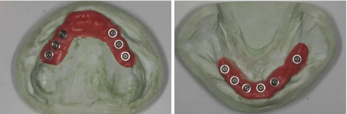
Fig. 13. Master casts were fabricated
anterior wax rim, the jaw relation was recorded (Fig. 14). Anterior artificial teeth were re-selected and set up palatally in wax rim. Wax dentures were inserted and evaluated the esthetic, phonetic and occlusal vertical dimension (Fig. 15). The master cast and the wax denture were sent to the milling center where they were scanned. The frameworks were obtained by CAD/CAM milling of the titanium block using the Procera implant bridge system.20 The wax dentures with titanium framework were fabricated for metal-acrylic resin prosthesis (Fig. 16).21 After obtaining the patient's consent, definitive prosthesis were fabricated. The occlusion was evaluated and adjusted to have simultaneous centric relation contact and a canine
Fig. 14. After adjustment of wax rim, the interocclusal recording was taken.
Fig. 15. Wax denture without titanium framework
Fig. 16. Wax denture with titanium framework
Fig. 17. Frontal view of definitive metal and acrylic fixed complete dentures.
Fig. 18. Panoramic view after definitive prostheses inserted.
protected occlusal scheme.22 After final prostheses were inserted (Fig. 17), panoramic radiography (Fig.
18) and periapical radiographs were taken for the examination. One week later, the occlusion was evaluated, screw access holes were filled with the flowable resin (Elite-flo, Bisco Inc., Schaumburg, USA).
DISCUSSION
CAD/CAM guided implant procedures are seemed to be accurate and predictable. With the aid of the
CT scans and planning software, a custom-made precision drill guide and a prefabricated prosthesis can be made before the implant surgery. Therefore, the patient was elated with the short surgical time and minimal discomfort.
However, complications might be encountered.
23,24 Yong and Moy reported23 that early
complications of this technique are immediate implant failure, bony interference of prosthesis seating, prosthesis loosening, speech problems, and bilateral cheek biting. Moreover, late complications including implant failure, persistent pain, buccal soft tissue defect, screw loosening, acrylic fracture, and aesthetic dissatisfaction could be presented.
In this case, using the provisional fixed restorations could provide the opportunity to overcome the complications. With a provisional restoration, it is possible to have a chance of asking the patient's satisfaction. Osseointegration from immediate loading is also not yet predictable. From the researchs, it is known that the majority of implant failures would occur in the 3-6 months following implant placement.25-27 If an implant would fail to integrate, the implant must be removed and the provisional prosthesis might be modified to splint the remaining integrated implants. 25-27 If the prefabricated fixed definitive prosthesis was used, the loss of implant would require complete re-fabrication. Moreover, esthetic trial evaluation of prosthesis could not be made. In possible fracture of a acrylic resin, we could remove the definitive prosthesis and replace it with the provisional prosthesis, while laboratory work is being completed to repair the definitive restoration.15
SUMMARY
In this case, fixed provisional prostheses using CAD/CAM technology were fabricated prior to the
surgical procedure so that prosthesis may be inserted immediately after the implants are placed. A double scan technique was used to acquire the CT data. An implant planning software program allowed the clinicians to study the structure of the alveolar bone in relation to the position of the artificial teeth. The surgical template contains all the necessary information for making the master cast, prosthesis, and implant placement. In treatment procedure, an accurate positioning of the surgical template is crucial for the placement of the implants. The flapless surgical procedure was performed according to the protocol. Provisional prostheses were inserted immediately and evaluated for aesthetics and function during 6 months. Finally, the definitive prostheses were fabricated. Patient was highly satisfied with the treatment result.
REFERENCES
1. Brånemark PI. Osseointegration and its experimental background. J Prosthet Dent 1983;50:399-410.
2. Adell R, Lekholm U, Rockler B, Brånemark PI. A 15-year study of osseointegrated implants in the treatment of the edentulous jaw. Int J Oral Surg 1981;10:387-416.
3. Gapski R, Wang HL, Mascarenhas P, Lang NP.
Critical review of immediate implant loading. Clin Oral Implants Res 2003;14:515-27.
4. Östman PO. Immediate/early loading of dental implants. Clinical documentation and presentation of a treatment concept. Periodontol 2000 2008:47;
90-112.
5. Degidi M, Piattelli A, Felice P, Carinci F. Immediate functional loading of edentulous maxilla; a 5-year restrospective study of 388 titanium implants. J Periodontol 2005;76:1016-24.
6. Horiuchi K, Uchida H, Yamamoto K, Sugimura M.
Immediate loading of Brånemark system implants following placement in edentulous patients: a clinical report. Int J Oral Maxillofac Implants 2000;15:
9. Wolfinger GJ, Balshi TJ, Rangert B. Immediate functional loading of Brånemark system implants in edentulous mandibles: clinical report of the results of developmental and simplified protocols. Int J Oral Maxillofac Implants 2003;18:250-7.
10. Ioannidou E, Doufexi A. Does loading time affect implant survival? A meta-analysis of 1,266 implants.
J Periodontol 2005;76:1252-8.
11. Fuster-Torres MA, Albalat-Estela S, Alcañiz-Raya M, Peñarrocha-Diago M. CAD / CAM dental systems in implant dentistry: update. Med Oral Patol Oral Cir Bucal 2009;14:E141-5.
12. Strub JR, Rekow ED, Witkowski S. Computer-aided design and fabrication of dental restorations: current systems and future possibilities. J Am Dent Assoc 2006;137:1289-96.
13. Verstreken K, Van Cleynenbreugel J, Marchal G, Naert I, Suetens P, van Steenberghe D. Computer- assisted planning of oral implant surgery: a three- dimensional approach. Int J Oral Maxillofac Implants 1996;11:806-10.
14. Tee-Khin N, Cheng AC, Lee H, Wee AG, Leong EW. The management of a completely edentulous patient using simultaneous maxillary and mandibular CAD/CAM-guided immediately loaded definitive implant-supported prostheses: a clinical report. J Prosthet Dent 2008;99:416-20.
15. Balshi SF, Wolfinger GJ, Balshi TJ. Surgical planning and prosthesis construction using computed tomography, CAD/CAM technology, and the Internet for immediate loading of dental implants. J Esthet Restor Dent 2006;18:312-23.
16. Kupeyan HK, Shaffner M, Armstrong J. Definitive
18. Marchack CB. An immediately loaded CAD/
CAM-guided definitive prosthesis: a clinical report. J Prosthet Dent 2005;93:8-12.
19. van Steenberghe D, Naert I, Andersson M, Brajnovic I, Van Cleynenbreugel J, Suetens P. A custom template and definitive prosthesis allowing immediate implant loading in the maxilla: a clinical report. Int J Oral Maxillofac Implants 2002;17:663-70.
20. Takahashi T, Gunne J. Fit of implant frameworks: an in vitro comparison between two fabrication techniques. J Prosthet Dent 2003;89:256-60.
21. Torsello F, di Torresanto VM, Ercoli C, Cordaro L.
Evaluation of the marginal precision of one-piece complete arch titanium frameworks fabricated using five different methods for implant-supported restorations. Clin Oral Implants Res 2008;19:772-9.
22. Kim Y, Oh TJ, Misch CE, Wang HL. Occlusal considerations in implant therapy: clinical guidelines with biomechanical rationale. Clin Oral Implants Res 2005;16:26-35.
23. Yong LT, Moy PK. Complications of computer- aided-design/computer-aided-machining-guided (NobelGuide) surgical implant placement: an evaluation of early clinical results. Clin Implant Dent Relat Res 2008;10:123-7.
24. Komiyama A, Klinge B, Hultin M. Treatment outcome of immediately loaded implants installed in edentulous jaws following computer-assisted virtual treatment planning and flapless surgery. Clin Oral Implants Res 2008;19:677-85.
25. Rocci A, Martignoni M, Gottlow J. Immediate loading of Brånemark System TiUnite and machined-surface implants in the posterior mandible:
a randomized open-ended clinical trial. Clin Implant Dent Relat Res 2003;5 Suppl 1:57-63.
26. Grunder U. Immediate functional loading of immediate implants in edentulous arches: two-year results. Int J Periodontics Restorative Dent 2001;21:545-51.
27. Schnitman PA, Wöhrle PS, Rubenstein JE, DaSilva JD, Wang NH. Ten-year results for Brånemark implants immediately loaded with fixed prostheses at implant placement. Int J Oral Maxillofac Implants 1997;12:495-503
임플란트 술식에서 방사선 영상 기술, 컴퓨터 소프트웨어의 발전으로 정확한 진단 및 surgical guide의 제작이 가 능해졌다. 본 증례는 양악 무치악 환자에서 고정성 임플란트 보철을 위해 CAD/CAM technique을 이용하여 수술을 하고 즉시 하중을 가한 증례이다. Planning software program을 이용하여 해부학적 구조물과 단면상을 고려하여 상 하악에 각각 6개씩의 임플란트를 최적의 위치에 계획하였다. 정밀한 surgical guide 이용하여 미리 계획된 위치와 방향으로 무절개 임플란트 식립 수술을 시행하였다. 즉시 사용 가능한 고정성 임시 보철물을 미리 제작해 수술 직 후 장착하여 환자의 만족도를 높였으며, 이를 6개월간 평가하여 심미적이며, 기능적으로 안정적인 최종 보철물을 제작할 수 있었다.
주요어: CAD/CAM technology, 무절개 수술, 즉시 하중, 임플란트 보철
교신저자 : 박찬진
강원도 강릉시 강릉대학로120, 강릉원주대학교 치과대학, 210-702
Tel : +82-33-640-3153, Fax : +82-33-640-3113, E-mail : [email protected] 원고접수일 :2009년 04월 03일, 원고수정일 :2009년 05월 23일, 원고채택일 :2009년 09월 25일