Antiangiogenic Activity of Coptis chinensis Franch. Water Extract in in vitro and ex vivo Angiogenesis Models
Eok-Cheon Kim, Seo Ho Kim, Jin-Ho Lee and Tack-Joong Kim*
Division of Biological Science and Technology, College of Science and Technology, Yonsei University, Wonju, Gangwon-do 220-710, Korea Received August 5, 2016 /Revised December 20, 2016 /Accepted January 16, 2017
Angiogenesis, the formation of new blood vessels, plays an important role in tumor growth and meta- stasis; therefore, it has become an important target in cancer therapy. Novel anticancer pharmaceutical products that have relatively few side effects or are non-cytotoxic must be developed, and such prod- ucts may be obtained from traditional herbal medicines. Coptis chinensis Franch. is an herb used in traditional medicine for the treatment of inflammatory diseases and diabetes. However, potential anti- angiogenic effects of C. chinensis water extract (CCFWE) have not yet been studied. The purpose of this study was to determine the antiangiogenic effect of CCFWE in order to evaluate its potential for an anticancer drug. We found that the treatment with CCFWE inhibited the major steps of the angio- genesis process, such as the endothelial cell proliferation, migration, invasion, and capillary-like tube formation in response to vascular endothelial growth factor (VEGF), and also resulted in the growth inhibition of new blood vessels in an ex vivo rat aortic ring assay. We also observed that CCFWE treat- ment arrested the cell cycle at the G0/G1 phase, preventing the G0/G1 to S phase cell cycle pro- gression in response to VEGF. In addition, the treatment reduced the VEGF-induced activation of ma- trix metalloproteinases 2 and 9. Taken together, these findings indicate that CCFWE should be consid- ered a potential anticancer therapy against pathological conditions where angiogenesis is stimulated during tumor development.
Key words : Angiogenesis, anticancer drug, Coptis chinensis Franch. water extract, endothelial cell,
vascular endothelial growth factor
*Corresponding author
*Tel : +82-33-760-2242, Fax : +82-33-760-2183
*E-mail : [email protected]
This is an Open-Access article distributed under the terms of the Creative Commons Attribution Non-Commercial License (http://creativecommons.org/licenses/by-nc/3.0) which permits unrestricted non-commercial use, distribution, and reproduction in any medium, provided the original work is properly cited.
Journal of Life Science 2017 Vol. 27. No. 1. 78~88 DOI : https://doi.org/10.5352/JLS.2017.27.1.78
Introduction
Angiogenesis has been heavily researched recently as a promising field in anticancer drug development [4, 39].
Novel anticancer pharmaceutical products that have rela- tively few side effects or are non-cytotoxic must be devel- oped, and such products may be obtained from traditional herbal medicines [17, 52]. Several natural herbal products have been found to have antitumor activity, and they have become the main sources of anticancer drugs [23, 61].
Coptis chinensis Franch. is an herb used in traditional med- icine for the treatment of inflammatory diseases, diabetes, and cancer. Plant-derived alkaloids have traditionally gained interest due to their pronounced physiological activities [27, 42, 45]. C. chinensis contains several compounds thought to
enhance health, including the isoquinoline alkaloids berber- ine, palmatine, hydrastine, and coptisine [25, 26]. Recent re- ports have shown a variety of activities for C. chinensis alka- loids, such as antibacterial, neuroregenerative, antidiabetic, antioxidative, anti-atherosclerotic, and anti-inflammatory ef- fects [13, 50, 53, 58, 59, 60, 62]. A variety of pharmacological effects have been reported for C. chinensis extracts or in- gredients, but the potential antiangiogenic effects of C. chi- nensis water extract (CCFWE) have not yet been studied.
Angiogenesis is a complex process, which is tightly con-
trolled through a balance between proangiogenic and anti-
angiogenic regulatory factors and is triggered by vascular
endothelial growth factor (VEGF), a major proangiogenic
mediator in cancer [51, 38]. This complex multistep process
is associated with basal membrane injury, endothelial cell
proliferation, migration, invasion, and capillary-like struc-
ture formation, ending up in the recruitment and adhesion
of pericytes or smooth muscle cells [18]. The aim of the cur-
rent research was to examine the antiangiogenic effects of
CCFWE on the VEGF-induced angiogenic process both in
vitro and ex vivo.
Materials and Methods
Preparation of C. chinensis water extract
C. chinensis was harvested in Eumseong, Korea, and ex- tracted three times with 2 l of water (20~25°C) within one day. The extracts were filtered through Whatman No. 1 pa- per, combined, and the supernatant was concentrated using a rotary evaporator. The extract was then freeze-dried to ob- tain a powder extract. The powder extract was suspended in sterilized distilled water at appropriate concentrations and stored at -70°C until use.
Animals
Sprague–Dawley rats (7-week-old, male) were purchased from Orient Co. (Sungnam, Korea) and maintained on a standard chow and water available ad libitum. All animal experiments were conducted in accordance with the Guide for the Care and Use of Laboratory Animals adopted by the United States National Institutes of Health and approved by the Ethics Committee, Institutional Animal Care and Use Committee (IACUC no. YWC-131127-1) of Yonsei University (Wonju, Korea).
Cell culture
Human umbilical vein endothelial cells (HUVECs) were purchased from the American Type Culture Collection (Manassas, VA, USA) and incubated in M199 medium (Gibco, Grand Island, NY, USA) supplemented with 20%
(v/v) fetal bovine serum (FBS; Gibco), 100 U/ml penicillin, 100 μg/ml streptomycin (Gibco), 3 ng/ml basic fibroblast growth factor (Komabiotech, Seoul, Korea), and 5 U/ml hep- arin (Sigma, St. Louis, MO, USA) at 5% CO
2in a 37°C incubator. HUVECs were used between passages 4 and 8 for all experiments.
Cell viability assay
Cell viability was evaluated using a 3-(4,5-dimethylthia- zol-2-yl)-2,5-diphenyltetrazolium bromide (MTT; USB Co- rporation, Cleveland, OH, USA) assay. One day before the extract treatment, HUVECs (1×10
5cells) were seeded into each well of a gelatin (Sigma)-coated 24-well tissue culture plate. The cells were treated with 1-100 μg/ml CCFWE for 48 hr, then the medium was replaced with a fresh medium containing 0.5 mg/ml MTT, and the plate was incubated for an additional 4 hr. After the incubation, the medium was carefully removed from the plate, and dimethyl sulfoxide
was added to solubilize the formazan produced from MTT.
Cell viability was determined by measuring the absorbance at 560 nm using a microplate reader (Molecular Devices, Sunnyvale, CA, USA).
Cell proliferation assay
HUVECs were seeded at a density of 4×10
4cells per well in gelatin-coated 12-well plates. On the next day, the cells were treated with various concentrations of CCFWE in the absence or presence of 20 ng/ml VEGF (PEPRO TECH, Rocky Hill, NJ, USA). After 48 hr of incubation, proliferation and normal growth were determined using a Luna
TMauto- mated cell counter (Logos Biosystems, Anyang, Korea).
Cell cycle analysis
HUVECs were seeded at a density of 1×10
6cells into 100-mm plates. After 24 hr, the cells were washed with M199 medium and incubated for 6 hr in M199 medium containing 1% FBS. The cells were precultured with CCFWE for 40 min and then stimulated by the addition of VEGF to 20 ng/ml.
After 20 hr of incubation, the cells were harvested and then fixed in 70% ice-cold ethanol. The fixed cells were dehy- drated at 4°C for 30 min in phosphate-buffered saline con- taining 2% FBS and 0.1% Tween 20 (USB Corporation), then centrifuged at 1,500 rpm for 5 min, and resuspended in 0.5 ml of the same buffer. RNase digestion (5 μg/ml, Sigma) was carried out at 37°C for 1 hr, followed by staining with propidium iodide (5 μg/ml, Sigma). The cells were analyzed using a Laser BD FACSCalibur flow cytometer (BD Biosci- ence, San Jose, CA, USA).
Migration assay
Chemotactic motility of HUVECs was assayed using a
Transwell apparatus (Corning Costar, Cambridge, MA,
USA). Briefly, cell culture inserts with membrane filters (3-μ
m pore size) were coated with 10 μg of gelatin on the lower
surface. Cell suspensions were incubated for 30 min at 37°C
in M199 medium (1% FBS) containing various concentrations
of CCFWE and then seeded into the inserts at 5×10
4cells/insert. A chamber containing VEGF alone served as a
positive control. After an incubation for 4 hr, migrated cells
were fixed and stained with hematoxylin and eosin stain
and quantified using an optical microscope (200×). Each
treatment was performed in triplicate, and eight fields of
view were counted for each assay.
Invasion assay
The invasive behavior of endothelial cell in the presence of CCFWE was quantified using a double-chamber assay kit (Trevigen, Gaithersburg, MD, USA) by counting the number of cells that invaded Matrigel (BD Biosciences, Bedford, MA, USA)-coated inserts, according to the manufacturer's in- structions. The membrane of the upper chamber was coated with Matrigel to prevent the migration of noninvasive cells [1]. VEGF was used as a positive chemoattractant.
Gelatin zymography
Activity of matrix metalloproteinases (MMPs) 2 and 9 was determined using a gelatin zymogram. In brief, to remove cellular debris, cultured media were centrifuged at 1,500 rpm for 5 min at 4°C, and the supernatants containing 20 μg of secreted protein were mixed with sodium dodecyl sulfate polyacrylamide gel electrophoresis (SDS–PAGE) loading buffer without a reducing agent. The protein samples were separated on a 10% SDS–PAGE gel copolymerized with 2 mg/ml gelatin. The gels were washed with 2.5% (v/v) Triton X-100 (USB Corporation) for 30 min to remove SDS, then rinsed with incubation buffer (50 mM Tris–HCl, pH 7.5, containing 10 mM CaCl
2plus 1 μM ZnCl
2), and in- cubated at 37°C for 3 hr or overnight. The gels were stained with a 0.25% Coomassie brilliant blue R250 (Sigma) solution and then destained until clear bands appeared.
Tube formation assay
To assess the inhibitory potential of CCFWE for tube for- mation by HUVECs, Matrigel was used. In brief, 2×10
5cells were preincubated with CCFWE for 40 min and then seeded to each well of 24-well plates coated with 250 μl of Matrigel, followed by the addition of VEGF (20 ng/ml). After 20 hr of incubation, tube-like structures were visualized and pho- tographed under light microscopy at 40× magnification. The average branch areas were measured using Image-Pro Plus (Media Cybernetics, Bethesda, MD, USA).
Rat aortic ring sprouting assay
Rat aortic ring angiogenesis was investigated using the three-dimensional rat aortic ring model as described pre- viously [3]. Basically, aortic rings of about 1-mm thickness, excised from thoracic aortas, were placed into a 48-well plate coated with 120 μl of Matrigel and sealed in place with an overlay of 50 μl of Matrigel. After an incubation for 30 min at 37°C, 600 μl of M199 medium containing VEGF or CCFWE
was added into each well. On day 7, the outgrowth of the sprouting microvessels was measured using an inverted light microscope (Olympus, Center Valley, PA, USA) at 100×
magnification. Sprouting was estimated using the following scale: 0 = no sprouting; 1 = migrated cells without sprouting;
2 = isolated sprouting; 3 = sprouting in 25-50% of the arterial ring circumference; 4 = sprouting in 50-75% of the circum- ference; and 5 = sprouting in 75-100% of the circumference.
The results were scored from 0 to 5 in a double-blinded manner. Each data point was quantified in sextuplicate.
Results
CCFWE inhibits VEGF-induced proliferation of HUVECs
To determine non-toxic concentrations of CCFWE against HUVECs, an MTT cytotoxicity assay was performed using cells exposed to various concentrations of CCFWE for 24 hr.
CCFWE did not show any cytotoxicity up to 25 μg/ml as shown in Fig. 1A. After having established that CCFWE does not affect cell viability, all experiments were performed at non-toxic concentrations of CCFWE. Since angiogenesis be- gins with local proliferation of endothelial cells in response to a proangiogenic stimulus from VEGF, we first determined the antiproliferative effect of CCFWE on human endothelial cells in vitro. The CCFWE treatment significantly inhibited the proliferation of VEGF-stimulated HUVECs in a concen- tration-dependent manner (Fig. 1B).
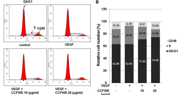
CCFWE induces arrest of endothelial cells in G0/
G1 phase
We then examined the effect of CCFWE on cell cycle pro- gression using fluorescence-activated cell sorting (FACS) analysis. After the treatment of HUVECs with 20 ng/ml VEGF, with or without CCFWE, for 20 hr, the percentage of cells in the G0/G1, S, and G2/M phases was monitored.
VEGF induced HUVECs to enter into the S phase, whereas
the treatment with CCFWE significantly increased the pro-
portion of cells at G0/G1 (Fig. 2), which indicated that
CCFWE induced a cell cycle arrest. Thus, these results in-
dicate that CCFWE affects the transition of cells from G0/G1
to the S phase. Taken together, the inhibitory effect on the
growth of endothelial cells should be attributed to the cell
cycle-arresting pharmacological properties of CCFWE.
A B
Fig. 1. Effect of CCFWE on viability and proliferation of HUVECs. (A) HUVECs were exposed to CCFWE at concentrations of 1, 5, 10, 25, 50, and 100 μg/ml. After 24 hr, the viability (%) was determined by the MTT assay. Vehicle-treated cells were used as the control (100%). (B) HUVECs were treated with the indicated concentrations of CCFWE for 40 min before the exposure to VEGF (20 ng/ml). After 24 hr, the number of proliferating cells was counted. Data are expressed as the mean
± standard deviation (SD) of three independent experiments. *p<0.01 versus VEGF alone.
A B
Fig. 2. CCFWE causes a G0/G1 arrest in HUVECs. HUVECs were preincubated with increasing concentrations of CCFWE for 40 min and treated with VEGF (20 ng/ml) for up to 24 hr. After the incubation, the representative images of cell cycle analysis shown in (A) were acquired, and the percentage of cells in each phase after the treatment is presented as a histogram (B).
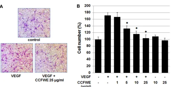
CCFWE inhibits VEGF-induced migration and invasion of HUVECs
We additionally examined the influence of CCFWE on cell motility using an in vitro cell migration assay. Endothelial cell migration is one of the crucial events in neovessel formation. As shown in Fig. 3A and B, after the incubation with various concentrations of CCFWE for 4 hr, the migra- tion of HUVECs in response to VEGF was dose-dependently
inhibited. At 25 μg/ml CCFWE, the number of migrating
cells was reduced to basal levels. However, CCFWE alone
had no significant effect on the basal migration of endothe-
lial cells. We next tested whether CCFWE could inhibit the
invasion ability of endothelial cells. The invasion property
of endothelial cells was analyzed in a Matrigel-coated
Boyden chamber in the presence of various concentrations
of CCFWE. As shown in Fig. 4A, the number of the cells
A B
Fig. 4. Effect of CCFWE on invasion and MMP expression in HUVECs. (A) HUVEC invasion was studied using a Transwell plate.
HUVECs were pretreated for 40 min with 10 and 25 μg/ml CCFWE before the exposure to VEGF (20 ng/ml). After the incubation with VEGF for 16 hr, invaded endothelial cells were evaluated. (B) MMP (MMP-2 and MMP-9) activities were measured by a zymogram assay. After the incubation with 10 or 25 μg/ml CCFWE for 40 min, the cells were treated with VEGF (20 ng/ml) for 12 hr. The cultured media were electrophoresed, incubated at 37°C for 3 hr (upper panel) or 12 hr (lower panel), and stained with Coomassie brilliant blue R250. The culture medium from HUVECs treated with phorbol myristate acetate (PMA, 40 ng/ml) for 12 hr was used to distinguish between the different types of MMPs. Experiments were repeated three times, and the results are shown as the mean ± SD of triplicate determinations. *p<0.01 versus VEGF alone.
A B
Fig. 3. Effect of CCFWE on migration of HUVECs. HUVECs were pretreated for 40 min with various concentrations (1, 5, 10, and 25 μg/ml) of CCFWE before the exposure to VEGF (20 ng/ml). After incubation for 4 hr, chemotactic migration was evaluated. (A) Images of migration were captured under a phase-contrast microscope. The cells successfully migrated to the lower surface of the insert. (B) The cell number (%) decreased in a dose-dependent manner following the treatment with CCFWE. In the histogram, the data are expressed as the mean ± SD from triplicate experiments. *p<0.01 versus VEGF alone.
that invaded through the Matrigel-coated filter was dose-de- pendently reduced by CCFWE, indicating that CCFWE can inhibit the VEGF-induced invasion ability of endothelial cells. This inhibition of migration and invasion is not due to a cytotoxic effect of CCFWE because the viability of these
cells was not affected by CCFWE in the concentration range tested (Fig. 1A).
CCFWE suppresses MMP-2 and MMP-9 secretion
by HUVECs
A B
Fig. 5. Inhibition of VEGF-induced tube formation on a Matrigel matrix. HUVECs were incubated for 40 min with 10 or 25 μg/ml CCFWE, then plated on Matrigel-coated plates, and treated with 20 ng/ml VEGF. After 20 hr, microphotographic images were obtained (×40). (A) Representative images of endothelial tube formation. (B) The area covered by the capillary-like tubes was measured using the Image-Pro Plus software. Data shown are the mean ± SD of triplicate determinations. *p<0.01 versus VEGF alone.
A vital step in cell migration and invasion includes the degradation of the extracellular matrix (ECM), and MMPs play a central role in the process [41]. Therefore, we further performed a zymogram assay to examine the effect of CCFWE on the VEGF-stimulated secretion of MMP-2 and MMP-9 into the supernatant of cultured media. Gelatin zy- mography of the serum-free cultured medium supernatants revealed that the activities of MMP-2 and MMP-9 in VEGF- treated cells were substantially higher than those in un- treated cells. However, their activities were significantly suppressed by the treatment with CCFWE (Fig. 4B).
CCFWE inhibits tube formation by endothelial cells To characterize the antiangiogenic activity of CCFWE, a tube formation assay, which mimics the neovascularization process, was conducted using endothelial cells. HUVECs were cultured on Matrigel-formed, tube-like networks (Fig.
5A) for 20 hr. The inhibitory effect of CCFWE on the tube formation induced by VEGF was quantitated by measuring the tube area. It is noteworthy that the VEGF-treated HUVECs formed organized capillary tubes; however, 25 μ g/ml CCFWE completely abrogated the integrity of the en- dothelial tube network, reducing both the width and length of the tube-like structures (Fig. 5B).
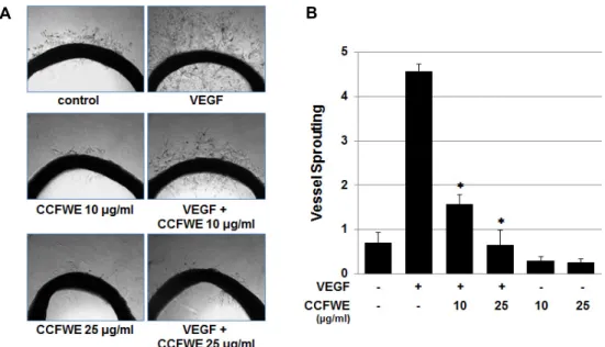
CCFWE inhibits sprouting of microvessels from rat aortic rings
Finally, to verify the antiangiogenic effect of CCFWE ex
vivo, a rat aortic ring assay was conducted. The rat aortic ring sprouting assay is a widely used antiangiogenic model that mimics several stages of angiogenesis, including pro- liferation, migration, and invasion of vascular endothelial cells, as well as tube formation. Fig. 6 shows that in the ab- sence of CCFWE, sprouts emerged from the aortic ring and grew outward after seven days in culture with VEGF. In contrast, the treatment with CCFWE exhibited a dramatic dose-dependent inhibitory effect on microvessel formation.
This inhibition of vessel sprout formation by CCFWE was unlikely due to cytotoxicity.
Discussion
C. chinensis has been used in China to treat inflammatory diseases and diabetes for centuries. In recent years, it has been reported that C. chinensis exhibits various pharmacobio- logical effects, such as antidiabetic [14, 55], neuroprotective [13], antipyretic [29], antibacterial [11], immunomodulatory [37, 43], antioxidant [22], and anticancer activities [48].
Active ingredients of CCFWE include alkaloids, such as ber-
berine, jatrorrhizine, coptisine, and palmatine [34]. Berberine
shows strong antibacterial bioactivity against Shigella dysen-
teriae [54], induces apoptosis via a mitochondrial pathway
in liver cancer cells [56], provides neuroprotective effects in
mice [8], prevents the development of left ventricular hyper-
trophy induced by pressure overload in rats [21], and in-
hibits angiogenesis through the suppression of various
A B
Fig. 6. CCFWE inhibits vessel sprouting from rat aorta. Rat aortic rings were placed in Matrigel and treated with 10 or 25 μg/ml CCFWE before the exposure to VEGF (20 ng/ml). The effect of CCFWE on sprout formation by the aorta samples was examined on day 7. (A) Representative images were photographed. (B) CCFWE inhibits the VEGF-induced vessel sprouting.
The results were scored from 0(least positive) to 5(most positive), and the data are shown as the mean ± SD (n=6). *p<0.01 versus VEGF alone.
proinflammatory and pro-angiogenic factors in melanoma cells and C57BL/6 mice [19]. Jatrorrhizine inhibits pro- liferation of metastatic melanoma cell and neovasculariza- tion in mice [35] and Coptisine inhibits aggressive osteo- sarcoma cell proliferation [57]. Palmatine exhibits an ex- tensive range of pharmacological actions, including anti- bacterial activity against Escherichia coli [53], as well as anti- viral [24], anti-inflammatory [30], and anticancer effects [2].
Although various beneficial effects of CCFWE have been re- ported, the exact mechanisms of action of the extract and its active components, associated with the diverse biological functions, are still unclear. Until now, there have been no reports on antiangiogenic activity of CCFWE. To validate CCFWE as a potential antiangiogenic material, we explored well-established angiogenesis assays [47]. Consequently, we demonstrated that the treatment with CCFWE suppressed the VEGF-induced angiogenesis progression in vitro and ex vivo.
Angiogenesis plays a key role in tumor growth and meta- stasis [15], and it is a multi-step process requiring coordi- nated endothelial functions, such as the activation of endo- thelial cells, degradation of the basement membrane, migra- tion and proliferation of endothelial cells, elongation and branching of vessels, acquisition of pericytes, and ECM re- modeling [49]. VEGF is a key factor in angiogenesis, and it is highly expressed in a wide variety of human cancers
[12]. It has been demonstrated that antiangiogenic drugs that target molecules within the angiogenic signaling pathway inhibit the tumor growth [46]. Recently, more efforts have been focused on the discovery of novel, non-cytotoxic, anti- angiogenic compounds or natural product extracts, some of which have been used clinically for thousands of years as important alternative remedies for a wide variety of dis- eases, including cancer [16, 32, 33, 36].
To investigate the antiangiogenic activity of CCFWE, we
first performed a test of endothelial cell proliferation in re-
sponse to VEGF. Cell growth is determined by the balance
between cell proliferation and death. As shown in Fig. 1,
the treatment with CCFWE dramatically reduced the
VEGF-induced proliferation of endothelial cells in a concen-
tration-dependent manner (Fig. 1B), but cell viability did not
significantly change at concentrations as high as 25 μg/ml,
as determined by the MTT assay (Fig. 1A). These results in-
dicate that the antiangiogenic potential of CCFWE is not due
to cytotoxicity. In order to gain deeper insights into the
mechanism of the antiangiogenic activity of the extract, we
investigated the effect of CCFWE on the phosphorylation
of extracellular signal-regulated kinases (ERKs) 1/2 and p38
mitogen-activated protein kinase (MAPK) in endothelial
cells. MAPK pathways are involved in the VEGF signaling
cascades and regulate a variety of physiological processes,
including endothelial cell growth in angiogenesis [6, 31]. In
these experiments, we observed that the CCFWE treatment did not inhibit the VEGF-stimulated phosphorylation of ERK1/2 and p38 MAPK (data not shown). Using FACS anal- ysis, we found that CCFWE induces a cell cycle arrest in the G0/G1 phase (Fig. 2). The G0/G1 to S phase transition is a major checkpoint responsible for DNA replication, and it is strongly regulated by the combined activity of the cyclin D1/cyclin-dependent kinase (CDK) 4 complex [40]. The p21 protein, a proliferation inhibitor, plays a role in the G0/G1 arrest by binding to and inhibiting the activity of cy- clin/CDK complexes [20]. In our previous work, we showed that a hot water extract of Coptis japonica Makino inhibited the proliferation of HUVECs in response to VEGF through the G0/G1 arrest [28].
The migratory and invasive behavior of endothelial cells is another mainstay in angiogenesis. Cell migration and in- vasion requires ECM degradation and involves the activa- tion of many intracellular signaling pathways, such as those regulating MMPs. Our findings show that the number of migrated and invaded endothelial cells decreased very effec- tively among VEGF-stimulated, CCFWE-treated HUVECs (Fig. 3, Fig. 4A). Therefore, these data suggest that CCFWE significantly reduced the endothelial cell migration and in- vasion and reveal the potential of this extract as an anti- angiogenic agent. The MMPs, MMP-2 and MMP-9, are key enzymes involved in the migratory and invasive progress of angiogenesis [7, 44]. We investigated the MMP-2 and MMP-9 enzyme activities in response to VEGF in CCFWE- treated HUVECs. The activities of MMP-2 and MMP-9, in- creased by VEGF, decreased in CCFWE-treated cells in a dose-dependent manner (Fig. 4B). However, we did not in- vestigate the mechanism by which the CCFWE treatment downregulates the MMP-2 and MMP-9 expression. Taken together, our results suggest that CCFWE exerts its anti- angiogenic effects on HUVECs by inhibiting the activity of the proangiogenic proteins, MMP-2 and MMP-9. After endo- thelial cells invade and migrate into the perivascular space, the cells differentiate and form a capillary network [9, 10].
To further study the differentiation of endothelial cells, we measured the tube formation on a Matrigel matrix. Matrigel is a tumor-derived matrix that contains all the components necessary to start the formation of blood vessel-like struc- tures by endothelial cells [5]. As a result, CCFWE showed the potent inhibition of tube formation in a dose dependent manner (Fig. 5). Finally, to verify the antiangiogenic effect of CCFWE, we carried out an ex vivo rat aortic ring sprouting
assay. The results showed that the VEGF-stimulated out- growth and dendritic branching of new blood vessels were potently and dose-dependently inhibited in the rat aortic rings treated with CCFWE (Fig. 6). Collectively, the in- hibition of microvessel outgrowth from rat aortic rings, as well as the inhibition of HUVEC proliferation, migration, in- vasion, and tube formation on a Matrigel matrix, provides strong evidence of the antiangiogenic effect of CCFWE.
In conclusion, we demonstrated that a water extract of C. chinensis could be an interesting antiangiogenic candidate that targets the VEGF signaling pathway and may be a po- tential novel therapeutic agent for the treatment of cancer and other angiogenesis-related diseases.
Acknowledgment
This research was supported by the Basic Science Research Program through the National Research Foundation of Korea (NRF-2014R1A1A2059027).
References
1. Albini, A., Iwamoto, Y., Kleinman, H. K., Martin, G. R., Aaronson, S. A., Kozlowski, J. M. and McEwan, R. N. 1987.
A rapid in vitro assay for quantitating the invasive potential of tumor cells. Cancer Res. 47, 3239-3245.
2. Ali, H. and Dixit, S. 2013. Extraction optimization of Tinospora cordifolia and assessment of the anticancer activity of its alkaloid palmatine. ScientificWorldJournal 2013, 376216.
3. Aplin, A. C., Fogel, E., Zorzi, P. and Nicosia, R. F. 2008.
The aortic ring model of angiogenesis. Methods Enzymol. 443, 119-136.
4. Atanasov, A. G., Waltenberger, B., Pferschy-Wenzig, E. M., Linder, T., Wawrosch, C., Uhrin, P., Temml, V., Wang, L., Schwaiger, S., Heiss, E. H., Rollinger, J. M., Schuster, D., Breuss, J. M., Bochkov, V., Mihovilovic, M. D., Kopp, B., Bauer, R., Dirsch, V. M. and Stuppner, H. 2015. Discovery and resupply of pharmacologically active plant-derived nat- ural products: A review. Biotechnol. Adv. 33, 1582-1614.
5. Benton, G., Kleinman, H. K., George, J. and Arnaoutova, I. 2011. Multiple uses of basement membrane-like matrix (BME/Matrigel) in vitro and in vivo with cancer cells. Int.
J. Cancer 128, 1751-1757.
6. Chrzanowska-Wodnicka, M., Kraus, A. E., Gale, D., White, G. C. 2nd. and Vansluys, J. 2008. Defective angiogenesis, en- dothelial migration, proliferation, and MAPK signaling in Rap1b-deficient mice. Blood 111, 2647-2656.
7. Doyle, J. L. and Haas, T. L. 2009. Differential role of beta-cat- enin in VEGF and histamine-induced MMP-2 production in microvascular endothelial cells. J. Cell Biochem. 107, 272-283.
8. Durairajan, S. S., Liu, L. F., Lu, J. H., Chen, L. L., Yuan,
Q., Chung, S. K., Huang, L., Li, X. S., Huang, J. D. and Li, M. 2012. Berberine ameliorates β-amyloid pathology, gliosis, and cognitive impairment in an Alzheimer's disease trans- genic mouse model. Neurobiol. Aging 33, 2903-2919.
9. Eccles, S. A., Court, W., Patterson, L. and Sanderson, S. 2009.
In vitro assays for endothelial cell functions related to angio- genesis: proliferation, motility, tubular differentiation, and proteolysis. Methods Mol. Biol. 467, 159-181.
10. Fan, T. P., Yeh, J. C., Leung, K. W., Yue, P. Y. and Wong, R. N. 2006. Angiogenesis: from plants to blood vessels.
Trends Pharmacol. Sci. 27, 297-309.
11. Feng, X., Yan, D., Zhao, K. J., Luo, J. Y., Ren, Y. S., Kong, W. J., Han, Y. M. and Xiao, X. H. 2011. Applications of mi- crocalorimetry in the antibacterial activity evaluation of var- ious Rhizoma Coptidis. Pharm. Biol. 49, 348-353.
12. Ferrara, N., Gerber, H. P. and LeCouter, J. 2003. The biology of VEGF and its receptors. Nat. Med. 9, 669-676.
13. Friedemann, T., Otto, B., Klätschke, K., Schumacher, U., Tao, Y., Leung, A. K., Efferth, T. and Schröder, S. 2014. Coptis chinensis Franch. exhibits neuroprotective properties against oxidative stress in human neuroblastoma cells. J. Ethnophar- macol. 155, 607-615.
14. Ge, A. H., Bai, Y., Li, J., Liu, J., He, J., Liu, E. W., Zhang, P., Zhang, B. L., Gao, X. M. and Chang, Y. X. 2014. An activ- ity-integrated strategy involving ultra-high-performance liq- uid chromatography/quadrupole-time-of-flight mass spec- trometry and fraction collector for rapid screening and char- acterization of the α-glucosidase inhibitors in Coptis chinensis Franch. (Huanglian). J. Pharm. Biomed. Anal. 100, 79-87.
15. Gensicka, M., Głowacka, A., Dzierzbicka, K. and Cholewin- ski, G. 2015. Inhibitors of Angiogenesis in Cancer Therapy - Synthesis and Biological Activity. Curr. Med. Chem. 22, 3830-3847.
16. Goey, A. K., Chau, C. H., Sissung, T. M., Cook, K. M., Venzon, D. J., Castro, A., Ransom, T. R., Henrich, C. J., McKee, T.
C., McMahon, J. B., Grkovic, T., Cadelis, M. M., Copp, B.
R., Gustafson, K. R. and Figg, W. D. 2016. Screening and Biological Effects of Marine Pyrroloiminoquinone Alkaloids:
Potential Inhibitors of the HIF-1α/p300 Interaction. J. Nat.
Prod. 79, 1267-1275.
17. Gordaliza, M. 2007. Natural products as leads to anticancer drugs. Clin. Transl. Oncol. 9, 767-776.
18. Greenberg, J. I., Shields, D. J., Barillas, S. G., Acevedo, L.
M., Murphy, E., Huang, J., Scheppke, L., Stockmann, C., Johnson, R. S., Angle, N. and Cheresh, D. A. 2008. A role for VEGF as a negative regulator of pericyte function and vessel maturation. Nature 456, 809-813.
19. Hamsa, T. P. and Kuttan, G. 2012. Antiangiogenic activity of berberine is mediated through the downregulation of hy- poxia-inducible factor-1, VEGF, and proinflammatory medi- ators. Drug Chem. Toxicol. 35, 57-70.
20. Harper, J. W., Adami, G. R., Wei, N., Keyomarsi, K. and Elledge, S. J. 1993. The p21 Cdk-interacting protein Cip1 is a potent inhibitor of G1 cyclin-dependent kinases. Cell 75, 805-816.
21. Hong, Y., Hui, S. C., Chan, T. Y. and Hou, J. Y. 2002. Effect
of berberine on regression of pressure-overload induced car- diac hypertrophy in rats. Am. J. Chin. Med. 30, 589-599.
22. Jang, M. H., Kim, H. Y., Kang, K. S., Yokozawa T. and Park, J. H. 2009. Hydroxyl radical scavenging activities of iso- quinoline alkaloids isolated from Coptis chinensis. Arch. Pharm.
Res. 32, 341-345.
23. Ji, H. F., Li, X. J. and Zhang, H. Y. 2009. Natural products and drug discovery. Can thousands of years of ancient med- ical knowledge lead us to new and powerful drug combina- tions in the fight against cancer and dementia? EMBO Rep.
10, 194-200.
24. Jia, F., Zou, G., Fan, J. and Yuan, Z. 2010. Identification of palmatine as an inhibitor of West Nile virus. Arch. Virol.
155, 1325-1329.
25. Jung, H. A., Yoon, N. Y., Bae, H. J., Min, B. S. and Choi, J. S. 2008. Inhibitory activities of the alkaloids from Coptidis Rhizoma against aldose reductase. Arch. Pharm. Res. 31, 1405- 1412.
26. Kamath, S., Skeels, M. and Pai, A. 2009. Significant differ- ences in alkaloid content of Coptis chinensis (Huanglian) from its related American species. Chin. Med. 4, 17.
27. Kingston, D. G. and Newman, D. J. 2007. Taxoids: can- cer-fighting compounds from nature. Curr. Opin. Drug Discov.
Devel. 10, 130-144.
28. Kim, S. H., Kim, E. C., Kim, W. J., Lee, M. H., Kim, S. Y.
and Kim, T. J. 2015. Coptis japonica Makino extract sup- presses angiogenesis through regulation of cell cycle-related protein. Biosci. Biotechnol. Biochem. 80, 1095-1106.
29. Kong, X., Wan, H., Su, X., Zhang, C., Yang, Y., Li, X., Yao, L. and Lin, N. 2014. Rheum palmatum L. and Coptis chinensis Franch. exert antipyretic effect on yeast-induced pyrexia rats involving regulation of TRPV1 and TRPM8 expression. J.
Ethnopharmacol. 153, 160-168.
30. Kwon, O. J., Kim, M. Y., Shin, S. H., Lee, A. R., Lee, J. Y., Seo, B. I., Shin, M, R., Choi, H. G., Kim, J. A., Min, B. S., Kim, G. N., Noh, J. S., Rhee, M. H. and Roh, S. S. 2016.
Antioxidant and anti-inflammatory effects of Rhei Rhizoma and Coptidis Rhizoma mixture on reflux esophagitis in rats.
Evid. Based Complement. Alternat. Med. 2016, 2052180.
31. Lal, B. K., Varma, S., Pappas, P. J., Hobson, R. W. 2nd. and Durán, W. N. 2001. VEGF increases permeability of the en- dothelial cell monolayer by activation of PKB/akt, endothe- lial nitric-oxide synthase, and MAP kinase pathways.
Microvasc. Res. 62, 252-262.
32. Lee, B., Kim, K. H., Jung, H. J. and Kwon, H. J. 2012.
Matairesinol inhibits angiogenesis via suppression of mi- tochondrial reactive oxygen species. Biochem. Biophys. Res.
Commun. 421, 76-80.
33. Lee, S. H., Lee, J., Jung, M. H. and Lee, Y. M. 2013. Glyceol- lins, a novel class of soy phytoalexins, inhibit angiogenesis by blocking the VEGF and bFGF signaling pathways. Mol.
Nutr. Food Res. 57, 225-234.
34. Liu, L. H. and Chen, Z. L. 2012. Analysis of four alkaloids of Coptis chinensis in rat plasma by high performance liquid chromatography with electrochemical detection. Anal. Chim.
Acta 737, 99-104.
35. Liu, R., Cao, Z., Pan, Y., Zhang, G., Yang, P., Guo, P. and Zhou, Q. 2013. Jatrorrhizine hydrochloride inhibits the pro- liferation and neovascularization of C8161 metastatic mela- noma cells. Anticancer Drugs 24, 667-676.
36. Lu, J., Zhang, K., Nam, S., Anderson, R. A., Jove, R. and Wen, W. 2010. Novel angiogenesis inhibitory activity in cin- namon extract blocks VEGFR2 kinase and downstream signaling. Carcinogenesis 31, 481-488.
37. Luo, Y., Zhao, H., Liu, Z., Ju, D., He, X., Xiao, C., Zhong, G., Chen, S., Yang, D., Chan, A. S. and Lu, A. 2010.
Comparison of the enteric mucosal immunomodulatory ac- tivity of combinations of Coptis chinensis Franch. Rhizomes and Evodia rutaecarpa (Juss.) Benth. Fruits in mice with dex- tran sulphate sodium-induced ulcerative colitis. Planta Med.
76, 766-772.
38. Majeti, B. K., Lee, J. H., Simmons, B. H. and Shojaei, F. 2013.
VEGF is an important mediator of tumor angiogenesis in malignant lesions in a genetically engineered mouse model of lung adenocarcinoma. BMC Cancer 13, 213.
39. Mikstacka, R., Stefański, T. and Różański, J. 2013. Tubulin- interactive stilbene derivatives as anticancer agents. Cell Mol. Biol. Lett. 18, 368-397.
40. Morgan, D. O. 1995. Principles of CDK regulation. Nature 374, 131-134.
41. Nagase, H. and Woessner, J. F. Jr. 1999. Matrix metal- loproteinases. J. Biol. Chem. 274, 21491-21494.
42. Oberlies, N. H. and Kroll, D. J. 2004. Camptothecin and tax- ol: historic achievements in natural products research. J. Nat.
Prod. 67, 129-135.
43. Park, E. K., Rhee, H. I., Jung, H. S., Ju, S. M., Lee, Y. A., Lee, S. H., Hong, S. J., Yang, H. I., Yoo, M. C. and Kim, K. S. 2007. Antiinflammatory effects of a combined herbal preparation (RAH13) of Phellodendron amurense and Coptis chinensis in animal models of inflammation. Phytother. Res.
21, 746-750.
44. Puyraimond, A., Weitzman, J. B., Babiole, E. and Menashi, S. 1999. Examining the relationship between the gelati- nolytic balance and the invasive capacity of endothelial cells. J. Cell. Sci. 112, 1283-1290.
45. Ram, V. J. and Kumari, S. 2001. Natural products of plant origin as anticancer agents. Drug News Perspect. 4, 465-482.
46. Scott, L. J. 2007. Bevacizumab: in first-line treatment of metastatic breast cancer. Drugs 67, 1793-1799.
47. Staton, C. A., Stribbling, S. M., Tazzyman, S., Hughes, R., Brown, N. J. and Lewis, C. E. 2004. Current methods for assaying angiogenesis in vitro and in vivo. Int. J. Exp. Pathol.
85, 233-248.
48. Wang, X. N., Xu, L. N., Peng, J. Y., Liu, K. X., Zhang, L.
H. and Zhang, Y. K. 2009. In vivo inhibition of S180 tumors by the synergistic effect of the Chinese medicinal herbs Coptis chinensis and Evodia rutaecarpa. Planta Med. 75, 1215- 1220.
49. West, D. C. and Burbridge, M. F. 2009. Three-dimensional in vitro angiogenesis in the rat aortic ring model. Methods
Mol. Biol. 467, 189-210.
50. Wu, M., Wang, J. and Liu, L. T. 2010. Advance of studies on anti-atherosclerosis mechanism of berberine. Chin. J.
Integr. Med. 16, 188-192.
51. Wu, X., Yang, T., Liu, X., Guo, J. N., Xie, T., Ding, Y., Lin, M. and Yang, H. 2015. IL-17 promotes tumor angiogenesis through Stat3 pathway mediated upregulation of VEGF in gastric cancer. Tumour Biol. 37, 5493-5501.
52. Xu, W., Towers, A. D., Li, P. and Collet, J. P. 2006. Traditio- nal Chinese medicine in cancer care: perspectives and expe- riences of patients and professionals in China. Eur. J. Cancer Care 15, 397-403.
53. Yan, D., Jin, C., Xiao, X. H. and Dong, X. P. 2008. Antimicro- bial properties of berberines alkaloids in Coptis chinensis Franch by microcalorimetry. J. Biochem. Biophys. Methods 70, 845-849.
54. Yan, D., Li, J., Xiong, Y., Zhang, C., Luo, J., Han, Y., Wang, R., Jin, C., Qian, H., Li, J., Qiu, L., Peng, C., Lin, Y., Song, X. and Xiao, X. 2014. Promotion of quality standard of herb- al medicine by constituent removing and adding. Sci. Rep.
4, 3668.
55. Yang, T. C., Chao, H. F., Shi, L. S., Chang, T. C., Lin, H.
C. and Chang, W. L. 2014. Alkaloids from Coptis chinensis root promote glucose uptake in C2C12 myotubes. Fitoterapia 93, 239-244.
56. Yip, N. K. and Ho, W. S. 2013. Berberine induces apoptosis via the mitochondrial pathway in liver cancer cells. Oncol.
Rep. 30, 1107-1112.
57. Yu, D., Fu, S., Cao, Z., Bao, M., Zhang, G., Pan, Y., Liu, W. and Zhou, Q. 2014. Unraveling the novel anti-osteosarco- ma function of coptisine and its mechanisms. Toxicol. Lett.
226, 328-336.
58. Yu, H. H., Kim, K. J., Cha, J. D., Kim, H. K., Lee, Y. E., Choi, N. Y. and You, Y. O. 2005. Antimicrobial activity of berberine alone and in combination with ampicillin or ox- acillin against methicillin-resistant Staphylococcus aureus. J.
Med. Food 8, 454-461.
59. Zhang, J., Yang, J. Q., He, B. C., Zhou, Q. X., Yu, H. R., Tang, Y. and Liu, B. Z. 2009. Berberine and total base from Rhizoma Coptis chinensis attenuate brain injury in an alumi- num-induced rat model of neurodegenerative disease. Saudi Med. J. 30, 760-766.
60. Zhang, Q., Piao, X. L., Piao, X. S., Lu, T., Wang, D. and Kim, S. W. 2011. Preventive effect of Coptis chinensis and berberine on intestinal injury in rats challenged with lipopolysaccharides.
Food Chem. Toxicol. 49, 61-69.
61. Zhao, J., Jiang, P. and Zhang, W. D. 2009. Molecular net- works for the study of TCM pharmacology. Brief. Bioinform.
11, 417-430.
62. Zhou, H., Jiang, T., Wang, Z., Ren, S., Zhao, X., Wu, W., Jiang, L., Liu, Z. and Teng, L. 2014. Screening for potential α-glucosidase inhibitors in Coptis chinensis Franch extract us- ing ultrafiltration LC–ESI–MS. Pak. J. Pharm. Sci. 27, 2007- 2012.
초록: In vitro 와 ex vivo 혈관신생 모델에서 황련 냉수추출물의 신생혈관 억제효과
김억천․김서호․이진호․김택중*
(연세대학교 과학기술대학 생명과학기술학부)