T 세포 및 대식세포 기농에 대한 Silybin의 조절효과
조 재 열*
강원대 학교 BT학부대 학 생 물소재 공학전공 (Received May 28, 2007; Revised July 19, 2007)
Immunomodulatory Effect of Silybin on T Cell- and Macrophage-mediated Functions
Jae Youl Che/
School of Bioscience and Biotechnology, Kangwon National University, Chucheon 200-701, Korea
Abstract — Silybin is known to be a major active flavonoid component isolated from Silybum marianum, a hepatoprotective medicinal plant. In this study, we examined the immunomodulatory role of silybin on T cell and macrophage-mediated immune responses. To do this, the proliferation of splenic lymphocytes and CD8+ CTLL-2 cells under mitogenic stim
ulation with lipopolysaccharide (LPS), concanavalin (Con) A and interleukin (IL)-2 and the production of TNF-a and NO from LPS- and IFN-y-activated macrophages was evaluated under silybin treatment. The mitogenic proliferation of splenic lym
phocytes induced by LPS and Con A was strongly diminished by silybin in a dose-dependent manner. Moreover, the pro
liferation of CD8+ CTLL-2 cells was also negatively modulated by the compound. In contrast, silybin did not strongly suppress the proliferation of normal splenocytes and T cell line Sup-Tl cells, indicating that the inhibitory effect of silybin may be due to blocking only mitogenic responses of splenic lymphocytes. In addition, silybin inhibited TNF-a production in LPS-stimulated RAW264.7 cells. Effect of silybin however was distinct, according to NO-inducing stimuli. Thus, silybin only blocked NO production induced by IFN-y but not LPS and the inhibition was increased when PMA was co-treated with IFN-y. Unlike NO inhibition, however, this compound protected the cytotoxic damage of RAW264.7 cells induced by both LPS and IFN-y. Therefore, our data suggest that silybin may participate in host immune responses mediated by T cells and macrophages via regulating mitogenic proliferation, and the production of TNF-a and NO, depending on cellular stimuli.
Keywords □ silybin, T cell proliferation, NO release, TNF-a production
여러 간장질환과 염증과정과의 관계는 매우 밀접한 것으로 알 려져 있다. 이와같은 사실은 그람 옴성균으로 부터 생성된 lipopolysaccharide(LPS)와 같온 내 득소(endotoxin)와 D-galactos- amine(D-GalN) 및 CCl^와 같은 실험적 간장해 유발물질에 의한 여러 간염모델에서 증명되 어졌 다. * 특 히 endotoxin-mediated liver injury는 nitric oxide(NO)나 tumor necrosis fector(TNF)- a에 의해 매개되는 가장 대표적인 간장질환의 하나로 알려져 있 다_2) 이들 질환의 발생은 감염균으로 부터 방출된 LPS에 의해 간 대식세포인 Kupffer cell이 자극을 받옴으로써 분비되는 NO 및 TNF-a에 의해 주로 매개되는 것으로 보고되어 있는데, 이때 분비된 NO 및 TNF-a는 간세포의 직 • 간접적인 파괴를 유도하 는 것으로 알려져 있다.^^ 이는, 특벌히 이들 물질의 저해제가 간
*본 논문에 관한 문의는 저자에게로 (전화) 033-250-6562 (팩스) 033-253-6560 (E-mail) [email protected]
손상에 의한 높은 사망율을 저하시킨다는 시설로 볼 때 그 관련 성을 더욱 확인할 수 있다. 게다가 일부 대식세포나 수지상세포 들은 여러가지 감염원으로부터 얻어진 항원들을 기공하고 MHC class n 분자 위에 제시함으로써 CD4+ T cell의 증식, 분화 및 활성을 유도하게 된다."*^ 이때 활성화된 T 세포들은 interferon (IFN)-y와 같은 대식세포 활성 A뻐토카민을 분비하여 간 대식세 포의 기능을 더욱 항진시킴으로써, 더욱 악화된 간염발생과정을 유도하는 것으로 알려져 있다.^>
Silybin은 여러 간장질환에 효과가 증명된 Silybum marianum 에서 분리된 주요 폴라보노이드류 화합물이다.®> 간장질환과 염 증과의 상관성이 밝혀지면서 최근 silybin이 가지는 염증면역 과 정에 관한 직접적인 조절기전에 관한 다양한 연구돌이 진행되고 었다. 실제로 수지상세포의 기능억제, T cell 분열억제 등과 같 은 여러 증거듬로 볼 때 silybin의 간질환 치료효능온 본 약물이 가지는 염증면역의 조절효고H1 서 기인된 것으로 간주되고 있다/'®
특별히 silybin이 염증매개 단백질의 발현에 될수적인 NF-kB의
활성을 억제한다는 사실이 보고되면서,®> 항염증 효과에 관한 분 자수준에서의 약리기전이 이해되어져 가고 있다. 그럼에도 불구 하고, silybin이 가지는 대식세포 기눙 및 T cell 활성에 관한 조 절 작용은 여견히 많은 부분이 이해되어져 있지 않으므로, 본 연 구에서는 T cell 중식과정 및 대식세포 유래 NO/TNF-a 분비 과 정에 관한 silybin의 조절 작용을 심도있게 조사하였다.
실험 방법
실험재료
Pentoxifylline, prednisolone, silybin, E. co/j(0111:B4) 유래 LPS, concanavalin A 및 MTT(3-[4,5-dimethylthiazol-2-yl]-2,5- diphenyltetrazolium bromide)는 Sigma(USA)로부터 구매하여 사 용하였으며, leflunomide 유도체인 A77,1726은 대응제약 합성연 구팀에서 합성된 것을 이용하였다. Murine 대식세포주인 RAW264.7 cell 및 CD4+ T cell line인 Sup-Tl cell은 ATCC (USA)S■부터 구입하여 실험하였다. 또한 세포배양시 용된 RPMI 1640 및 fetal bovine senim(FBS)은 Gibco사(USA)로부터 구입 하였다. 그외 사용된 모든 시약은 Sigma제품을 이용하였다. 초 자의 경우 24 well plate는 Falcon사(USA) 제품을, TNF-a enzyme-linked immnunosorbent assay(ELISA) kit는 Amersham Life Science Co.(Arlington Heights, U.K.) 사로부터 구입하여 정량에 이용하였으며 ELISA reader로는 Spectramax 250 microplate reader(Molecular Devices, U.S.A.)를 사용하였다.
Lymphocyte 증식 정량
Silybin 처리에 의한 splenocyte 증식 조절 능 평가 시험은 다 옴의 방법으로 실시하였다.고® BALB/C 생쥐로 부터 무균조작으 로 비장을 적출하고 주사기을 이용하식 차가운 RPMI 1640 배 지로 비장세포를 분리하였다. 분리된 비장세포를 원심분리로 모 은 후 0.83% ammonium chloride-20 mM Tris 완중 액 (pH 7.4) 을 이용하여 적혈구를 용해시켰다. 다시 Hanks' balanced salts solution 및 RPMI 1640 배지로 3회 세척 후 10% FBS 함유 RPMI 1640 배지를 이용하여 세포를 5x10®cell/m/ 농도로 96 well plates에 접종하였다. 여러 농도의 LPS를 처리하고 48시간
Silybin Fig. 1 - Chemical structure of silybin.
동안 배양하였다. Lymphocyte 분열증식은 MTT법으로 확인하 였다.
MTT 법에 의한 세포 증식 평가
Silybin(0~50 |ig/m/)과 LPS를 48시간 동안 처리 후 , MTT 용 액(5 mg/m/) 20 m/룰 첨가하고 다시 4시간 동안 동일 조건에서 배양하였다. 발색은 30% SDS용액을 첨가하식 유도하였으며 흡 광도는 540nm에서 Spectramax microplate reader(Molecular Devices)를 이용하여 측정하였다.
TNF-a 정량
Murine 대식세포주인 RAW264.7 세포를 penicillin(100IU/
m/) 및 streptomycin(100 과 10%의 FBS를 함유하는 RPMI1640 배지률 이용해서 I X 10® cell/mi로 조절한 후, 24 well plate에 접중하고, 5% C02 및 37T 에서 18시간 동안 견배양 하 였다. 이후 배지를 제거하고 10배 농도로 조제된 시험물질 50 |i/
와 450 n/의 LPS(최중농도 1 ng/m /)! 함유한 새로운 배지률 동 시에 처리하여 전배양과 동일 조건에서 배양하였다. 6시간 후 배양 배지률 원심분리 (12,000 rpm, 3분 간 ) 상 층 액 을 얻고 효 소면역 측정법(ELISA)으로 정량 전까지 -20°C 이하게서 보관하 였다.
Nitric Oxide의 정량
배양 배지에서의 nitric oxide 정량은 Cho 등의 방법으로 RAW264.7 세포(2xl0®cell/m/)l- 이용하여 실시하였다.^®^ 배양 배지 100 |o/와 Griess시약[5%(v/v) phoshophoric acid 용액 내 l%(w/v) sulfanilamide 및 0.1%(w/v) naphthylethylenediamide]
100 |o/를 흔합한 후 상온에서 10분 동안 방치한 후에 발색된 정 도틀 540 nm에서 microplate reader틀 이용하여 즉정하였다. 표 준 정량곡선은 sodium nitrite를 적정한 능도로 희석하여 동일 방법으로 흡광도를 측정한 후 완성하였다.
통계처리
각 data는 득럽적으로 3회(각 실험시 n=3) 실시하여 얻어진 결과률 평균±SEM으로 나타낸 것이며, AS package# 이용하여 Duncan의 다중비교법 이용하여 P<0.05 이상 수준에서 유의성 있다고 관정하였다.
결과 및 고찰
비장유래 임파구의 증식과정에 미처는 Silybin의 효과 Silybin은 T cell의 증식과정 및 분열시 유래되는 사의토카인 들의 분비를 효과적으로 억제한다고 보고되어짐에 따 라 , 본 연구에서는 silybin의 T cell 증식조절 작용에 관해 심도 있게 조
(C)
RAW264.7 - O - Sup-T1
Concentration (ug/ml)
(B)
3.2 6.2512.5 25 50
Concentration (ug/ml)
2.5 5 10 20 40 100
Concentration (jig/ml)
0.51 5 10 20
Concentration (ug/ml)
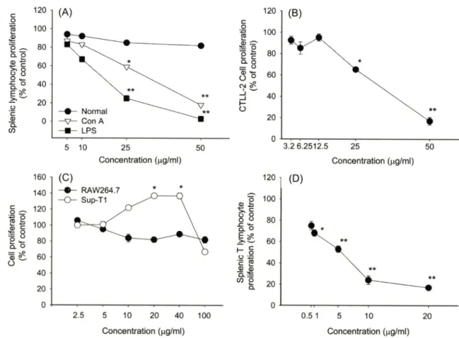
Fig. 2 - Effect of silybin on splenic lymphocyte and CD8+ CTLL-2 cell proliferation. (A) Splenocytes (5xl0®cells/m/) were incubated with various concentrations of silybin in the presence or absence of mitogens such as Con A (1 |ag/m/) and LPS (10 |ig/m/) for 48 h. (B) CTLL-2 cells (5x10® cells/m/) were treated with various concentrations of silybin in the presence or absence of IL-2 (50 U/ra/). (C) RAW264.7 or Sup-Tl cells (5x10® cells/m/) were treated with various concentrations of silybin for 48 h. (D) Splenocytes (5x10® cells/
m/) were incubated with various concentrations of A77.1726 in the presence or absence of Con A ( 1 ng/m/) for 48 h. Cell proliferation was evaluated by conventional MTT assay as described in Materials and Methods. *p<0.05 and **p<0.01 represent significant difference compared to control (A, B and D) or normal (C).
사하였다. 이률 위해, 다 중 식 유 도 mitogen(LPS 및 Con A) 및 CD4+ 흑은 CD8+ T cell의 증식모델을 도입하여 실험에 이 용하였다. Fig. 2에서 제시되어져 었듯이, silybin은 정상 splenocyte의 세포생존율은 억제하지 않는 것으로 확인되었다.
또한 기존 보고처럼, silybin은 강력한 T cell mitogen인 Con A 유도 T cell증식현상을 농도의존적으로 저해하였다(K:5o=42 ^g/
ml). 하지만 홍머롭게도 silybin은 LPS에 의해 유도된 비장세포 의 분열을 Con A 자극시 보다 더 강하게 억제한 것으로 나타났 다 (IC5o=16 ng/m/).
특별히 Con A는 CD4+ T cell의 분열을 유도하는 것으로 알 려져 있으므로, CD8+ T cell의 증식눙에 미치는 효과를 확인하 기 위해 대표적인 CD8+ T cell인 CTLL-2를 이용하여 조사하 였다. 이들 세포는 IL-2 의존성 증식과정을 보이므로, IL-2 처리 시 유도되는 증식과정에 대한 silybin의 효과를 검증해 보았다.
Fig. 2B에서 되 듯이, silybin은 Con A처리와 유사한 CD8+
T cell 증식 억제능(IC5o=38ng/m/)을 갖는 것으로 나타났다.
한편 실험의 대조약물로 사용한 A77,1726은 Con A 유도에 의 한 T cell 중식과정을 매우 강력하게 억제(1€50=6.4 ^1보)하였다 (Fig. 2D).
LPS는 강력한 B cell mitogen^.?. 알려져 었기 때문에, 본 결 과(Fig. 2A)는 silybin이 T cell 특이적인 증식과정을 억제한다는 기존의 결과와는 상반된 것으로 판단되어 , CD4+ T cell 암세포 주인 Sup-Tl cell을 이용e H 분열과정에서의 silybin 작용을 확 인해 보았다. 에상대로, silybin은 Sup-Tl의 분열과정을 억제하 지 못하였으며, 오히려 일부 농도(20 및 에서는 유의적 으로 분열과정을 더 촉진하는 것으로 나타났다. 따라서 본 결과 는 그 동안 보고된 T cell의 증식 에 관한 silybin의 효과는 mitogenic effect에서 기인되는 증식과정에 대해서 T 및 B lymphocyte에 상관없이 비특이적으로 작용하는 것으로 관단된다.
하지만 어떻게 LPS유도에 의한 비장 임파구 분화과정이 더 민 감하게 억제되는지는 추가적인 연구률 통해서 설명할 수 있을 것 으로 사료된다. 다만 이들 기견은 epidermal growth factor에 의
oooooo (D)
2
0
8
6
4
2
{loj}uoo
jo
%>uo!Jal®j!IOJd
이jaoocicjlua
i 1ulu^ldw oooooo
2
0
8
6
4
2
0OJJU8
uolaild10 Oo/J 타 -11& 3
ooooooo
2
0
8
6
4
2 0OJHJ8 iO
uo!}nJ^jlloJc
i 아
AUOLICiuJA;
l olu^ld^
o o o o o o o o 6 4 2 0 8 6 4 2 {laau8;0%>
® = 0
(B)
6 13 25
C o n ce n tra tio n (|ig /m l) 50 해 분열되는 epidermal cell의 분열 억제시 확 인 되 었 듯 이 / 세 포의 생존을 조절하는 여러가지 신호견달 과정[에: NF-kB, Akt (protein kinase B) 촉은 mitogen activated protein kinases (MAPK)]의 네 억제에서 기인된 것으로 관단된다.
대식세포 활성에 미처는 Silybin의 효과
최근 대식세포의 염중과정에 관한 silybin의 조절작용이 밝혀 지면서 대식세 포 매개성 면역반§ •에 관한 길항 흑은 항진작용 에 관한 다양한 연구둘이 진행되고 있다. 특별히 강 등(2002)은 silybin을 다량 함유한 silymarin의 경우, 전사인자인 NF-kB의 활 성을 농도의존적으로 억제함으로써 대식세포 유래 iNOS 발현 및 그에 따른 NO 생성이 억제된다는 결과률 제시한 적이 있다.^®
따라서 본 연구진 역시 silybin이 가지는 대식세포 기능 조절 능
1 1 0 5 0 1 0 0 5 0 0 1 0 0 0
C o n c e n tra tio n OaM)
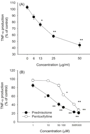
Fig. 3 - Effect of silybin on the production of TNF-a in LPS- activated RAW264.7 cells. (A and B) RAW264.7 cells
( 2 X1 0^ cells/m/) were incubated with various concentrations of silybin (A) or other standard drugs (pentoxifylline and prodnosolone) (B) in the presence or absence of LPS (1 |ig/
m/) for 6 h. Culture supernatants were assayed for TNF-a determination by ELISA. *p<0.05 and **p<0.01 represent significant difference compared to control.
을 대표적인 염증유발 물질로 알려져 있는 NO 및 TNF-a 분비 정도를 이용하여 평가해 보았다. Fig. 3 및 Fig. 4는 silybin이 대 식세포의 기능을 효파적으로 조절할 수 있다는 가능성을 제시한 다고 하겠다. 죽 Fig. 3A에서 보여지듯, silybin은 농도의존적으로 TNF-a의 생성을 억제하는 것으로 나타났다. 하지만 그 효과는 기존에 알려져 있는 prednisolone(IC5o=21.4 |jM), pentoxifyllin (IC50=236.2 mM) 및 cynaropicrin(IC5o=7.3 나M)베 등과 같은 여 러가지 항염증 물질이 나타내는 억제능 보다는 약한 것으로 보 여졌다(Fig. 3B).
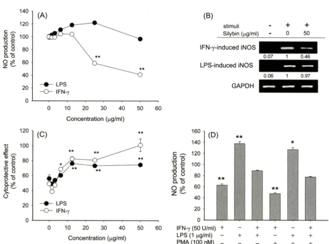
NO에 관한 silybin의 조절 효과는 특별히 기존 보고와는 다른 양상을 나타내었다. 즉 기존 보고돌은 silybin이 LPS 조건에서 농도의존적 억제경향을 보인다고 제시하였지만,®^ 본 연구진의 조 건에서는 전혀 억제효과률 확인할 수 없었다. 오히려 10 및 25 Ug/m/ 농도조건에서는 NO 생성을 더 유도하는 것으로 나타났다.
하지만 흥미롭게도 silybin은 IFN-y의 자국에 의해 유도된 NO생 성은 농도의존적으로 저해하는 것으로 나타났다. 비록 iNOS의 발현을 촉진하는 지국원은 다르지만 기존의 보고에서 처럼,®> 이 들 억제 작용은 증가된 iNOS 발현이 유의적으로 감소된다는 결 과로 볼 때 견사수준에서 진행될 것으로 관단된다. 한편 NO 생 성을 유도하는 LPS나 IFN-y와 같은 대식세포 활성 자국원돌은 24시간이나 48시간 처리시 이돌 물질에 의해 분비된 NO에 기인 된 세포득성 현상(세포사멸>을 유도하게 된다.^® 실제로 Fig. 4C 에서도 정상군(100%) 대비 40%에서 50% 정도의 세포가■ 사멸 된 결괴를 확인할 수 었었으며 , 본 연구자는 이들 현상<>1 NO 억 제제인 N-MMA처리시 완벽히 저해될 수 있다는 것을 보고한 바 있다. Fig. 4C는 확실히 silybin이 이들 대식세포 활성 자국원 에 의한 세포득성 과정을 매우 효과적으로 저해할 수 있다는 것 을 보여주고 있는데, 특별히 IFN-y 자극하에서는 50ng/mZ에서 거의 100%까지 세포사멸로부터 RAW264.7 cell을 보호하는 것 로 나타났다. LPS 자극 하에서도 silybin^ 유의적으로 세포사 멸을 억제한 것으로 확인되었다. 하지만, LPS 조건에서 silybin 은 전혀 NO 분비를 억제하지 앉은 것으로 보아, 이는 NO에 의 해 유도되는 세포사멸 유도용 라디칼 생성을 silybin이 직접 억 제하였거나, 흑은 세포사멸 유도시 발생되는 내부적인 신호전달 과정을 억제하쉬 간접적으로 보호작용을 나타낸 것으로 사료된 다. 하지만 MTT assay는 단지 미토콘드리아의 효소활성을 측정 한 방법이므로, silybin에 의한 세포보호작■용서 apoptosis 과정 억 제에서 유래되는 것인지 혹은 세포 분열유도에서 기인되는 것인 지에 관한 추가적인 연구가 apoptosis관련 지표 및 유세포분석기 등을 이용하여 진행될 예정이다.
특별히 IFN-y 처러시에도 NO 억제는 60% 정도에서 그쳤지 만 득성현상으로부터 세포는 거의 정상세포군 수준으로 보호작 용을 보여준 것>2로 보아 자국원에 상관없이 40% 정도는 silybin 약물 자체의 약러작용게서 기인된 것으로 생각된다. 실제로 silybin
0OJHI8J0%
}
uolpnF>OJC
i bIj
zH
0 (A)
0 0
0 0
0 0
c
0 9
8 7
6 5
4 0
{I04U8 JO %
}uollonpojcl
**
LPS
— 0 ~ IFN-y
(A)
— 0 ~ IFN-y
0 10 20 30 40
Concentration (나g/ml)
50 60
(C)
(B)
Stimuli
Silybin (|ig/ml) IFN-Y-inckJced iNOS
LPS-inchJced iNOS
GAPDH
+ +
0 50
160 140 (D)
0 10 20 30 40
Concentration (|ig/ml)
50 60 IFN-y (50 U/ml) LPS {1 배/ml) PMA (100 nM)
Fig. 4 - Effect of silybin on the production of NO in LPS-activated RAW264.7 cells. (A) RAW264.7 cells (2 x 10^ cells/m/) were incubated with various concentrations of silybin in the presence or absence of LPS (1 나g/m/) for 24 h. Culture supernatants were assayed for NO determination by Griess assay. (B) RAW264.7 cells (5 x 10^ cells/m/) were incubated with silybin (50 \ig/rr\l) in the presence or absence of LPS (1 fig/m/) or IFN-y (50 U/m/) for 6 h. After preparing total RNA and cDNA, mRNA levels of iNOS and GAPDH were determined by RT-PCR as described in Materials and Methods. (C) RAW264.7 cells (1x10찐 cells/m/) were treated with various concentrations of silybin in the presence of absence of LPS (1 p.g/m[) for 24 h. The protective effect of silybin on LPS-mediated cytotoxicity in RAW264.7 cells was evaluated by conventional MTT assay. (D) RAW264.7 cells (1x10^ cells/m/) were pre-treated with PMA 100 nM in the presence or absence of silybin (25 [ig/wl) or LPS (1 [xg/ml) for 24 h. Supernatants were collected and NO level from the supernatants was determined by Griess reagent, as described in Materials and Methods. *p<0.05 and **p<0.01 represent significant difference compared to stimuli also (A and D) or LPS alone (C).
은 발생되는 라디칼에 대한 소거작용이 매우 우수한 항산화제로 알려져 있으며, 여러가지 간득성 유발물질에 의해 발생되는 활 성산소중이나 다른 라디칼의 중화작용이 주요 간보호 약리기전
^ 알려져 있 다 . 그 럼 에 도 불구하고, 현재까지 몇 가지 증 게 은 s ily b i n 자체가 세포 내에서 신호전달과정의 조절에 참여 할 가능성들을 제시하고 있다. 죽 s ily b i n은 L P S 자국에 의해 활 성화된 NF-kB 활성 신호전달 과정을 조절하는 것으로 확인되었 으며 ,^®^ 이를 통해 여러 염증유전자들의 전사과정을 억제하는 것 으로 알려져 었다.^^* 현재 구체적으로 어떤 단백질이 s i l y b i n의 표적단백질인지는 확인되어 있지 않은 실정이다. 강 등( 2 0 0 3 )의 결과에서도 s ily m a r i n은 L P S에 의해 증가된 M A P K ( p 3 8 , E R K
및 J N K )의 활성은 견혀 억제하지 않았으며/ 5> N F - i c B를 제외한
A P - 1 이나 O C T와 같은 다른 중류의 전사인자 활성도 저해하지
않는 것으로 나타났다.^® 그러나, 흥미롭게도 F ig . 4 D는 s ily b i n
의 표적 단백질로 특벌히 P K C를 고려할 수 있다는 것을 시사한 다. 즉 s ily b i n의 I F N - y 매개성 N O 생성의 억제효과는 P K C 활 성화인자인 P M A와 병용처리시 그 억제 효과가 더욱 증가되었 다. 반면에 L P S의 경우는 P M A 병용처리에 의한 번화가 관찰되 지 않았으며, 오히려 L P S 및 I F N -y와의 병용 처리시에는 억제 효능이 감소되었다. 이는 두 자국원에 의한 N O 생성 유도 신호 가 증폭됨으로써, s ily b i n의 상대적인 활성감소에서 기인된 것으 로 관단된다. P M A는 a t y p ic a l P K C is o f o r m을 제외하고 대부분의
c o n v e n t i o n a l f o r m ( P K C a / p / V )및 n o v e l f o r m ( P K C 5 / E )들의 활성 을 유도하므로,2기 S i l y b i n에 의한 P K C 조절 가능성에 관한 이전 보 고 처 럼 , 어 떤 P K C is o f o r m의 활성이 s ily b i n의 억제활성에 관련되어 있는지는 is o f o r m 특이 저해제 및 W e s t e r n b lo t을 이
용한 m e m b r a n e t r a n s lo c a li z a t io n 정도의 분석을 통해 확인가능
할 것으로 사료되므로, 관련 연구들을 추 가 적 로 진행할 에정이다.
♦ V HJ광
1
벌LI
o o o o o o 2 0 8 6 4 2
0OJJU8%>
uollonpojc
l o
z
o o o o o o 4 o 2 0 8 6 4 2
{10JJU8i0%}
uolpnpojdoz
20
00
결 론
S i l y b i n의 T c e l l 및 대식세포의 활성에 관한 조절현상 연구를
통해 다옴과 같은 결론을 도출할 수 있었다. 1. S i l y b i n은 비장유 래 임파구둘의 증식 및 I L - 2 -의존성 C D8+ C T L L - 2 세포 분열 과정을 매우 효과적으로 억제하였다. 2 . S i l y b i n은 L P S 자극에 의한 대식세포 활성시 분비되는 T N F - a의 생성을 매우 유의적으 로 억제하였다. 3 . S ily b in - ^ 대식세포에서 L P S자극에 의해 생성 된 N O분비는 미약하게 억제하였으나, I F N - y 자국시 발생되는
N O 생성은 매우 효과적으로 억제하였다. 4 . S i l y b i n은 L P S 및
I F N - y에 의해 유도되는 세포득성으로부터 세포사멸을 억제하였
다. 5 . S il y b in의 N O 생성 저해작용은 P K C a c t iv a t o r인 P M A와 병용처리시 더 증가된 것^ 나타났다.
감사의 글
본 논문은 학술진흥재단 2 0 0 6년 기초과화면구지원사업(과제번
호: C 0 0 4 5 5 )의 지원에 의해 일부 수행되어졌다.
참고문헌
1) J a e s c h k e , H . : R o le o f in f la m m a t io n in t h e m e c h a n is m o f a c e t a m in o p h e n - in d u c e d h e p a t o t o x ic it y . Expert Opin. Drug Metab. Toxicol 1 , 3 8 9 (2 0 0 5 ) .
2 ) S c h w a b e , R . E a n d B r e n n e r , D . A , : M e c h a n is m s o f l i v e r in ju r y . I. T N F - a lp h a - in d u c e d l i v e r in j u r y : r o le o f I K K , J N K , a n d R O S p a t h w a y s . Am. J. Physiol. Gastrointest Liver Physiol. 2 9 0 , G 5 8 3 (2 0 0 6 ).
3 ) T ilg , H ., K a s e r , A . a n d M o s c h e n , A . R . : H o w t o m o d u la t e in f la m m a t o r y c y t o k in e s in li v e r d is e a s e s . Liver h it 26, 1 0 2 9 (2 0 0 6 ).
4 ) L i, Z . a n d D i e h l, A . M . : I n n a t e im m u n it y in t h e liv e r . Curr.
Opin. Gastroenterol 1 9 , 5 6 5 (2 0 0 3 ).
5 ) R o b e r t s , R . A ., G a n e y , P E . , J u , C ., K a m e n d u lis , L . M . , R u s y n , I. a n d K la u n ig , J , E . : R o le o f t h e K u p f f e r c e ll in m e d ia t in g h e p a t ic t o x ic it y a n d c a r c in o g e n e s is . Toxicol. Sci 9 6 , 2 (2 0 0 7 ).
6) S c h u m a n n , J ., P r o c k l, J., K ie m e r , A . K . , V o llm a r , A . M . , B a n g , R . a n d T ie g s , G . : S il ib i n in p r o t e c t s m ic e f r o m T c e ll- d e p e n d e n t l i v e r in ju r y . J. Hepatol 3 9 , 3 3 3 (2 0 0 3 ) .
7) G u p t a , 0 . R , S in g , S ., B a n i, S ., S h a r m a , N ., M a lh o t r a , S ., G u p t a , B . D ., B a n e r j e e , S . K . a n d H a n d a , S . S . : A n t i- in f la m m a t o r y a n d a n t i- a r t h r it ic a c t iv it ie s o f s ily m a r in a c t in g t h r o u g h in h ib it io n o f 5 - lip o x y g e n a s e . Phytomedicine. 7 , 2 1 (2 0 0 0 ).
8) G u , M . , S in g h , R . R , D h a n a la k s h m i, S., A g a r w a l, C . a n d A g a r w a l, R . : S il ib i n in in h ib it s in f la m m a t o r y a n d a n g io g e n ic a t t r ib u t e s in p h o t o c a r c in o g e n e s is in S K H - 1 h a ir le s s m ic e .
Cancer Res, 67, 3483 (2007).
9) Kang, J. S., Jeon, Y. J., Kim, H. M., Han, S. H. and Yang, K . H. : Inhibition of inducible nitric-oxide synthase expression by silymarin in lipopolysaccharide-stimulated macrophages. J.
Pharmacol Exp. Ther. 302, 138 (2002),
10) Cho, J. Y., Baik, K. U., Jung, J. H. and Park, M. H .: In vitroanti
inflammatory effects of cynaropicrin, a sesquiterpene lactone, from Saussurea lappa. Eur. J. Pharmacol. 398, 399 (2000).
11) Lee, J. S.,Kim, S. G., Kim, H. K., Lee, T. H., Jeong, Y. L, Lee, C. M,, Yoon, M. S., Na, Y. J., Suh, D . S., Park, N. C., Choi, I. H., Kim, G . Y., Choi, Y. H., Chung, H. Y. and Park, Y. M. : Silibinin polarizes Thl/Th2 immune responses through the inhibition of immunostimulatory function of dendritic cells. /.
Cell Physiol. 210, 385 (2007).
12) Polyak, S. J., Morishima, C., Shuhart, M. C., Wang, C. C., Liu, Y. and Lee, D. Y. : Inhibition of T-cell inflammatory cytokines, hepatocyte NF-kappaB signaling, and HCV infection by standardized silymarin. Gastroenterology132, 1925 (2007).
13) Singh, R , R , Dhanalakshmi, S., Mohan, S., Agarwal, C. and Agarwal, R . : Silibinin inhibits UVB- and epidermal growth factor-induced mitogenic and cell survival signaling involving activator protein-1 and nuclear factor-kappaB in mouse epidermal JB6 cells. Mol Cancer Ther. 5, 1145 (2006).
14) Beere, H. M. : Death versus survival: functional interaction between the apoptotic and stress-inducible heat shock protein pathways. J. Clin. Invest 115, 2633 (2005).
15) Kang, J. S., Park, S. K., Yang, K. H. and Kim, H. M. : Silymarin inhibits TNF-alpha-induced expression of adhesion molecules in human umbilical vein endothelial cells. FEBS Lett.550, 89 (2003).
16) Lee, S. K., Kim, H. S., Lee, H. J., Lee, J., Jeon, B. H., Jun, C. D., Lee, S. K. and Kim, E. C. : Dual effect of nitric oxide in immortalized and malignant human oral keratinocytes:
induction of apoptosis and differentiation. J. Oral Pathol. Med.
35, 352 (2006).
17) Kim, B. H. and Cho, J. Y. : Honokiol-mediated cytoprotective effects. /. Pharm. Pharmacol Submitted (2007).
18) Das, S. K. and Vasudevan, D. M. : Protective effects of silymarin, a milk thistle (Silybium marianum) derivative on ethanol-induced oxidative stress in liver. Indian J. Biochem.
Biophys. 43, 306 (2006).
19) Moulisova, V, Srbova, M., Jedlickova, O., Sebestian, J. and Jegorov, A. : Silybin reduces lipid peroxidation of rat hepatocyte membrane caused by cyclosporin A. Biochemistry (Mosc). 71, 1110 (2006).
20) Manna, S. K., Mukhopadhyay, A., Van, N. T. and Aggarwal, B. B. : Silymarin suppresses TNF-induced activation of NF- kappa B, c-Jun N-terminal kinase, and apoptosis. J. Immunol 163, 6800 (1999).
21) Yoo, H. G., Jung, S. N., Hwang, Y, S., Park, J. S„ Kim, M.
Jeong, M., Ahn, S. J., Ahn, B. W, Shin, B. A., Park, R. K. and Jung, Y. D. : Involvement of NF-kappaB and caspases in silibinin-induced apoptosis of endothelial cells. Int. J. Mol Med.
13, 81 (2004).
22) Cho, J. Y., Skubitz, K. M., Katz, D. R. and Chain, B. M. : CD98- dependent homotypic aggregation is associated with trans
location of protein kinase Cdelta and activation of mitogen- activated protein kinases. Exp. Cell Res. 286, 1 (2003).
23) Varga, Z,, Ujhelyi, L , Kiss, A., Balia, J,, Czompa, A. and Antus, S. : Effect of silybin on phorbol myristate actetate-induced protein kinase C translocation, NADPH oxidase activity and apoptosis in human neutrophils. Phytomedicine. 11,206 (2004).