Abstract. Background: Chemo-resistance to cisplatin- centered cancer therapy is a major obstacle to effective disease treatment. Recently, salinomycin was proven to be highly- effective for the elimination of cancer stem cells both in vitro and in vivo. The objective of the present study was to evaluate the anticancer properties of salinomycin in cisplatin-resistant ovarian cancer cells (A2780cis). Materials and Methods: The tetrazolium dye (MTT) assay was used to determine cell viability. Flow cytometric analysis was performed to analyze the effect on cell cycle and apoptosis.
The expression of apoptosis-related proteins was evaluated by western blot analysis. Results: Cell viability was significantly reduced by salinomycin treatment in a dose- dependent manner. Flow cytometry showed an increase in sub-G1phase cells. Salinomycin increased the expression of death receptor-5 (DR5), caspase-8 and Fas-associated protein with death domain (FADD). A decline in the expression of FLICE-like inhibitory protein (FLIP), activation of caspase-3 and increased poly ADP-ribose polymerase (PARP) cleavage, triggered apoptosis.
Furthermore, annexin-V staining also revealed the apoptotic induction. Conclusion: These findings provide important insights regarding the activation of caspase-8 and DR5, to our knowledge, for the first time in salinomycin-treated cisplatin-resistant ovarian cancer and demonstrate that salinomycin could be a prominent anticancer agent.
Despite attempts to implement effective early detection, the long term survival of ovarian cancer remains poor.
Maximal surgical cytoreduction with platinum-based chemotherapy is the standard treatment for patients with ovarian cancer. However, the recurrence of ovarian cancer due to chemoresistance takes place in up to 75% of the patients (1). Oncogene expression, increased drug efflux, reduced accumulation of drug, enhanced DNA repair and defective apoptotic program are believed to be major causes of chemoresistance. Currently, no specific treatment has been reported to reduce chemoresistance in ovarian cancer.
Several antitumor drugs, or cytokines, have been developed for the prevention of ovarian cancer but resistance to apoptosis is a major obstacle to chemotherapeutic treatment of cancer. The ability to induce apoptosis makes salinomycin a potentially effective therapeutic agent to combat malignancy. Recently, in a high-throughput screening test, salinomycin was found to be the most effective agent against breast cancer stem cells. Among the 16,000 natural and commercial chemical compounds studied, salinomycin was 100-times more effective than paclitaxel (2). Salinomycin was originally used as antimicrobial agent to kill bacteria and fungi (3-5). It is a monocarboxylic polyether antibiotic derived from Streptomyces albus, which is extensively used as coccidiostat in poultry and commonly fed to ruminant animals to improve feeding efficacy (6). As an ionophore with strict selection for alkali ions and a strong preference for potassium, it interferes with the transmembrane potassium potential. It has been reported that salinomycin can overcome drug resistance in human cancer cells (7). The combination treatment of paclitaxel and salinomycin resulted in strong antitumor efficacy for the eradication of breast cancer and cancer stem cells (8). The anticancer mechanism of salinomycin has been recognized based on its ability to induce apoptosis and cause growth inhibition in diverse types of chemo-resistant cancer cells (9-11).
The apoptotic signals induced by salinomycin are transduced by its binding to death receptors (DRs). The DRs are members of the tumor necrosis factor receptor Correspondence to: Chi-Heum Cho, MD, Ph.D., Department of
Obstetrics and Gynecology, Keimyung University School of Medicine, 56 Dalseong-ro, Jung-gu, Daegu 700-712, Republic of Korea. Tel: +82 532507518, Fax: +82 532507599, e-mail:
Key Words: Salinomycin, DR4, DR5, apoptosis, ovarian cancer, A2780cis cells.
Salinomycin Induces Apoptosis via Death Receptor-5 Up-regulation in Cisplatin-resistant Ovarian Cancer Cells
BIDUR PARAJULI1, SO-JIN SHIN1, SANG-HOON KWON1, SOON-DO CHA1, ROSA CHUNG1, WON-JIN PARK1, HYUN-GYO LEE2and CHI-HEUM CHO1,2
1Department of Obstetrics and Gynecology, 2Institute for Cancer Research, Keimyung University School of Medicine, Daegu, Republic of Korea
superfamily and comprise a subfamily that is characterized by an intracellular death domain. DR4 and DR5 are capable of engaging the cell suicide apparatus. Among all the DRs, tumor necrosis factor-related apoptosis-inducing DR4 and DR5 are selectively expressed in cancer cells and, thus, offer an advantage for targeted therapy and prevention (12, 13).
The present study examined the effect of salinomycin in cell growth-inhibition, cell-cycle progression and regulation of apoptosis on cisplatin-resistant ovarian cancer cells. To the best of our knowledge our results provide the first evidence that salinomycin induces DR5-mediated apoptosis. This may offer a promising therapeutic approach to overcome cisplatin-resistant ovarian cancer.
Materials and Methods
Reagents and cell lines. Dulbecco’s modified Eagle’s medium was obtained from GIBCO BRL (Grand Island, NY, USA).
Salinomycin was purchased from Sigma-Aldrich (Sigma-Aldrich, St. Louis, MO, USA) and dissolved in dimethylsulfoxide (DMSO).
The ovarian cancer cells (A2780) and cisplatin-resistant ovarian cancer cells (A2780cis) were obtained from the European Collection of Animal Cell Cultures (Salisbury, UK). The cells were cultured at 37˚C in 5% CO2.
Cell viability assay. The number of viable cells was evaluated by a colorimetric 3-(4,5-dimethylthiazol-2,5-diphenyl tetrazolium bromide (MTT) assay. Initially, cells were seeded in 96-well plates, and then cultured for 24 h to allow their adhesion to the plates. After this pre-incubation, the culture medium was changed to experimental medium supplemented with salinomycin (0.1, 0.5, 1, 5 and 10 μM) or DMSO (control) for 48 h. The MTT reagent was then added and cells incubated for an additional 4 h at 37˚C.
The intensity of the purple color formed in this assay is proportional to the number of viable cells. The optical density was measured at 495 nm. The mean value and their standard deviation were calculated from triplicate experiments.
Determination of cell-cycle distribution. To determine the cell distribution, cisplatin-resistant ovarian cancer cells were treated with various doses of salinomycin or DMSO (control). After 48 h treatment of salinomycin, cells were harvested by trypsinization and centrifugation, washed with cold phosphate-buffered saline (PBS), and were fixed in ice-cold 70% ethanol at 4˚C for 24 h.
Ethanol-fixed cells were then washed and treated with RNaseA for 30 min at 37˚C, stained with propidium iodide (PI) and then incubated for 30 min at room temperature. DNA fluorescence was measured by flow cytometry (FACS Calibur™; Becton Dickison, Franklin Lakes, NJ, USA). The percentage of cells in each cell- cycle phase was determined using the ModFit LT™ software (Becton-Dickinson) based on the DNA histogram.
Protein isolation and immunoblotting. Extracts of cells (2×10⁵/ml) were prepared in lysis buffer [10 mM Tris (7.4) 5 mM EDTA, 130 mM NaCl, 1% Triton X-100, serine protease inhibitor phenylmethylsulphonyl fluoride (10 μg/ml), leupeptin (10 μg/ml), aprotinin (10 μg/ml), 5 mM phenanthroline, and 28 mM benzamidine-HCl]. Protein concentrations were measured using
the Bio-Rad Protein Assay Reagent (Bio-Rad, Hercules, CA, USA), following the manufacturer’s protocol. Aliquots of protein were separated by 8% to 15% sodium dodecyl sulfate polyacrylamide gel electrophoresis (SDS-PAGE) and transferred to polyvinylidine difluoride membrane (Millipore, Bedford, MA, USA). The membrane was blocked with Tris-buffered saline containing 5% skimmed milk (Becton Dickinson) and 0.2% Tween 20 (Amresco, Solon, OH, USA). After washing, the membranes were incubated with primary antibodies to DR4 (Santa Cruz Biotechnology, Santa Cruz, CA, USA), DR5 (Koma Biotech, Seoul, Korea), p53, cleaved caspase-3, cleaved caspase-8, Fas- associated protein with death domain (FADD) and poly ADP- ribose polymerase (PARP; Cell Signaling, Beverly, MA, USA), FLICE-like inhibitory protein (FLIP; Enzo Life Science International, Farmingdale, NY, USA) and β-actin (Santa Cruz Biotechnology). After reaction with horseradish peroxidase- conjugated secondary antibodies (Santa Cruz Biotechnology), bands on the membranes were visualized by an enhanced chemiluminescence system (Thermo Scientific, Rockford, IL, USA), following the manufacturer’s suggested procedure.
Apoptotic assessment by annexin-V/PI staining. Cells were incubated for 48 h at 37˚C with different concentrations of salinomycin or DMSO (control). After washing with cold PBS, cells were re-suspended in 1× binding buffer. Staining of apoptotic cells was performed using the annexin V-fluorescein isothiocynate (FITC)/PI apoptosis assay kit (Becton Dickinson, Pharmingen, Heidelberg, Germany) according to the manufacturer’s instructions.
Determination of apoptotic cells was performed by flow cytometry using a FACScan flow cytometer and Cellquest software (Becton Dickinson).
Statistical analysis. The data are presented as the mean±SD.
Statistical analysis was conducted using one-way analysis of variance (ANOVA), followed by Duncan’s multiple range test for a post-hoc comparison by SPSS 17.0 (SPSS Inc., Chicago, IL, USA).
Statistical significance was set at p<0.05.
Results
Growth-inhibitory effect of salinnomycin on A2780cis cells.
To evaluate the effect of salinomycin on ovarian cancer cells, A2780 parental and cisplatin-resistant ovarian cancer cells were grown in the presence of different doses of salinomycin. In comparison with control, salinomycin reduced the viability of A2780 and A2780cis cells in a dose- dependent manner (Figure 1).
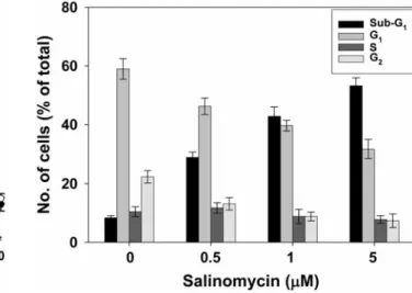
Effect of salinomycin on the cell cycle. To assess whether salinomycin-induced growth inhibition is mediated via alternations in the cell cycle, we examined the effect of salinomycin on the cell-cycle distribution. PI staining was carried out so that the DNA content of untreated and treated cisplatin-resistant ovarian cancer cells could be measured by flow cytometric analysis. DNA histograms showed an increased percentage of cells in sub-G1phase compared to untreated cells (Figure 2).
Induction of apoptotic cell death by salinomycin. Apoptosis assay was performed using A2780cis cells to evaluate the mechanism of the inhibition of cell growth using annexin-V FITC/PI kit. Flow cytometric analysis showed a significant increase of apoptotic cells upon 48 h exposure to salinomycin (Figure 3).
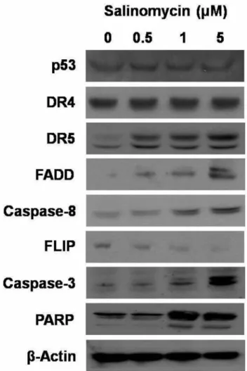
Role of salinomycin on apoptotic signaling pathway. The apoptotic cell death is related to the activation of regulatory genes. Therefore, the expression level of its related proteins was measured. The expression of DR5, caspase-8 and FADD proteins were significantly increased. In addition, we observed a down-regulation of FLIP expression. There was no change in the expression of DR4 and p53. Caspase-3 and PARP protein were increased after treatment (Figure 4).
These results suggest that salinomycin-induced cell growth- inhibition and induction of apoptotic cell death involve the activation of DR-mediated pathway.
Discussion
The present study demonstrated that salinomycin inhibited the growth of cisplatin-resistant human ovarian cancer cells.
The growth-inhibition effects of salinomycin and salinomycin-induced apoptosis in A2780cis cell was associated with DR5-mediated cell death pathway.
In our study, salinomycin decreased the viability of cisplatin-resistant cancer cells in a dose-dependent manner.
The growth-inhibitory effect of salinomycin demonstrated in the cell viability assay may corroborate the increased number of cells in the sub-G1phase of the cell cycle. Salinomycin significantly increased the proportion of cells in the sub-G1 phase upon treatment while the S phase remained almost unchanged and those of G2/M phase decreased with treatment of increasing concentrations of salinomycin. This shows that salinomycin causes apoptosis without causing G1/G2 arrest, which confirms previous findings that salinomycin activates an apoptotic pathway not accompanied by cell-cycle arrest (14).
Apoptosis, a form of programmed cell death, is an important homeostatic mechanism that balances cell division and cell death, maintaining the appropriate cell numbers in the body (15). It requires the cascaded activation and the execution of a series of regulatory molecules and cysteine- aspartic proteases known as caspases (16). Activation of pro- caspases stimulates the down-stream signaling cascades transmitted through two major pathways, which are mitochondria-dependent and mitochondria-independent. The mitochondria-independent signaling pathway directly activates caspase-3 by activating caspase-8. After the activation of caspase-3, PARP cleavage is induced, a classical marker in the apoptosis cascade. Early apoptosis was consistently marked by the annexin-V apoptotic assay in the present study.
Previous studies have also shown that salinomycin can induce apoptosis of human lymphoma and leukemia cells Figure 1. Antiproliferative effect of salinomycin on cisplatin-sensitive
and -resistant ovarian cancer cells (A2780 and A2780cis). Cells were treated with dimethyl sulfoxide (DMSO) control or salinomycin (0.5, 1, 5 and 10 μM) for 48 h. Cell viability was measured by the tetrazolium dye (MTT) assay and results were expressed as the percentage of viable cells. Values are means±SD of three measurements.
Figure 2. Effect of salinomycin in the cell cycle. A2780cis cells were treated with dimethyl sulfoxide (DMSO) control or salinomycin (0.5, 1 and 5 μM) for 48 h. After treatment with salinomycin, cells were harvested, fixed, stained with propidium iodide and analyzed by flow cytometric analysis. The values shown represent the number of cells in different phases of the cell cycle as a percentage of total cells. Values are means±SD of three measurements.
(17). Moreover, cancer cell-specific apoptotic effects of salinomycin were documented in neuronal, colorectal and prostate cancer cells (18-20). These findings show that the cell growth inhibition by salinomycin was performed through an apoptosis-dependent mechanism. To elucidate the signaling pathways activated during salinomycin treatment, we evaluated the key receptor proteins known to participate in the apoptotic pathways. Treatment with salinomycin
resulted in an increase in the expression of DR5 in a dose- dependent manner, however, the DR4 levels were unchanged.
DR4 and DR5 associate through their functional cytoplasmic death domain motifs with FADD upon activation by the extrinsic DR pathway, since FADD is known to be involved in the activation of caspase-8 (21). Several studies have also reported that DRs, such as tumor necrosis factor receptor (TNF-R) can induce apoptosis in a ligand-independent Figure 3. Induction of apoptosis after salinomycin treatment of A2780cis cells. As described in Materials and Methods, cells were exposed to dimethyl sulfoxide (DMSO) control or salinomycin (0.5, 1 and 5 μM) for 48 h. Apoptosis was quantified by flow cytometric analysis using annexin-V- fluorescein isothiocynate (FITC)/propidium iodide (PI) staining. Values are means±SD of three measurements.
manner (22, 23). We therefore raise the possibility that salinomycin might induce apoptosis through a DR5-mediated extrinsic pathway. However, the dependency on DRs and apoptosis, with respect to salinomycin, merits further investigation.
In conclusion, the results of this research demonstrated that salinomycin-suppressed cell proliferation by inducing apoptosis. Apoptosis induction was associated with the activation of caspase-8 and FADD along with the elevated expression of DR5. Our results may contribute to the development of salinomycin-based therapy for patients with cisplatin-resistant ovarian cancer.
References
1 Agarwal R and Kaye SB: Ovarian cancer: Strategies for overcoming resistance to chemotherapy. Nat Rev Cancer 3: 502- 516, 2003.
2 Gupta PB, Onder TT, Jiang G, Kuper Wasser C, Weinberg RA and Lander ES: Identification of selective inhibitors of cancer stem cells by highthroughput screening. Cell 138: 645-659, 2009.
3 Miyazaki Y, Shibuya M, Sugawara H, Kawaguchi O and Hirsoe C: Salinomycin, a new polyether antibiotic. J Antibiot 27: 814- 821, 1974.
4 Mahmoudi N, De Julian-Ortiz JV, Ciceron L, Galvez J, Mazier D, Danis M, Derouin F and Garcia-Domench F: Identification of new antimalarial drugs by linear discriminant analysis and topological virtual screening. J Antimicrob Chemother 57: 489- 497, 2006.
5 Naujokat C, Fuch D and Opelz G: Salinomycin in cancer: A new mission for an old agent. Mol Med Report 3: 555-559, 2010.
6 Mitani M, Yamanishi T and Miyazaki Y: Salinomycin: A new monovalent cation ionophore. Biochem Biophys Res Commun 27: 1231-1236, 1975.
7 Fuchs D, Heinold A, OPelz G, Daniel V and Naujokat C:
Salinomicin induces apoptosis and overcomes apoptosis resistance in human cancer cells. Biochem Biophys Res Commun 390: 734-749, 2009.
8 Zhang Y, Zhang H, Wang X, Wang J, Zhang X and Zhang Q: The eradication of breast cancer and cancer stem cells using octreotide- modified paclitaxel active targeting micelles and salinomycin passive targeting micelles. Biomaterials 33: 679-691, 2012.
9 Lu D, Choi MY, Yu J, Castro JE, Kipps TJ and Carson DA:
Salinomycin inhibits WNT signaling and selectively induces apoptosis in chronic lymphocytic leukemia cells. Proc Natl Acad Sci USA 108: 13253-13257, 2011.
10 Kim WK, Kim JH, Yoon K, Kim S, Ro J, Kang HS and Yoon S:
Salinomycin, a P-glycoprotein inhibitor, sensitizes radiation treated cancer cells by increasing DNA damage and inducing G2 arrest. Invest New Drugs 30: 1311-1318, 2013.
11 Riccioni R, Dupuis ML, Bernabei M, Petrucci E, Pasquini L, Mariani G, Cianfriglia M and Testa U: The cancer stem cell selective inhibitor salinomycin is a P-glycoprotein inhibitor.
Blood Cells Mol Dis 45: 86-92, 2010.
12 Srivastava RK: TRAIL/APO 2L: mechanisms and clinical applications in cancer. Neoplasia 3: 535-46, 2001.
13 Shankar S, Chen X and Srivastava RK: Effects of sequential treatments with chemotherapeutic drugs followed by TRAIL on prostate cancer in vitro and in vivo. Prostate 62: 165-86, 2005.
14 Zhang B, Wang X, Cai F, Chen W, Loesch U, Bitzer J and Zhong XY: Effects of salinomycin on human ovarian cancer cell line OV2008 are associated with modulating p38 MAPK. Tumor Biol 33: 1855-1862, 2012.
15 Martin SJ and Green DR: Apoptosis and cancer: The failure of controls on cell death and cell survival. Crit Rev Oncol Hematol 18: 137-53, 1995.
16 Degterev D and Yuan J: Expansion and evolution of cell death program. Nat Rev Mol Cell Biol 9: 378-390, 2008.
17 Fuchs D, Daniel V, Sadeghi M, Oopelz G and Naujokat C:
Salinomcin induces apoptosis and overcomes ABC transporter- mediated multidrug and apoptosis resistance in human leukemia stem cell-like KG-1a cells. Biochem Biophys Res Commun 394:
1098-1104, 2010.
Figure 4. Effect of salinomycin on apoptotic signal transduction pathway. A2780cis cells were treated with dimethyl sulfoxide (DMSO) control or salinomycin (0.5, 1 and 5 μM) for 48 h. After treatment, cell extracts were prepared and p53, death receptors (DR4 and DR5), Fas- associated protein with death domain (FADD), caspase-8, FLICE-like inhibitory protein (FLIP), caspase-3 and poly ADP-ribose polymerase (PARP) were subjected to western blot analysis. Beta-actin was used as an internal loading control.
18 Boehmerlen W and Endres M: Salinomycin induces calpain and cytochrome c-mediated neuronal cell death. Cell Death 2: 168, 2011.
19 Dong TT, Zhou HM, Wang LL, Feng B, Lv B and Zheng MH:
Salinomycin selectively targets CD133+cell subpopulations and decreases malignant traits in colorectal cancer lines. Ann Surg Oncol 18: 1797-1804, 2011.
20 Kim KY, Yu SN, Lee SY, Chun SS, Choi YL, Park YM, Song CS, Chatterjee B and Ahn SC: Salinomycin-induced apoptosis of human prostate cancer cells due to accumulated reactive oxygen species and mitochondrial membrane depolarization.
Biochem Biophys Res Commun 413: 80-86, 2011.
21 Chen G and Goeddle DV: TNF, TNF-R1 signaling: A beautiful pathway. Science 296: 1634-1635, 2002.
22 Fumarola C, Zerbini A and Guidotti GG: Glutamine deprivation-mediated cell shrinkage induces ligand-independent CD95 receptor signaling and apoptosis. Cell Death Differ 8:
1004-13, 2001.
23 Qiao L, StuderE, Leach K, Mckinstry R, Gupta S, Decker R, Kukreja R, Valerie K, Nagarkatti P, EI Deiry W, Molkentin J, Schmidt-Ullrich R, Fisher PB, Grant S, Hylemon PB and Dent P: Deoxycholic acid (DCA) causes ligand-independent activation of epidermal growth factor receptor (EGFR) and FAS receptor in primary hepatocytes: Inhibition of EGFR/mitogen-activated protein kinase-signaling module enhances DCA-induced apoptosis. Mol Biol Cell 12: 2609-2645, 2001.
Received February 6, 2013 Revised March 12, 2013 Accepted March 13, 2013