Clinical Investigations
Respiration 2009;78:42–48 DOI: 10.1159/000176208
Clinical Course of Pulmonary Embolism in Lung Cancer Patients
Jung-Woo Lee
aSeung-Ick Cha
aChi-Young Jung
bWon-Il Choi
cKyung-Nyeo Jeon
dSeung-Soo Yoo
aChang-Ho Kim
aJae-Yong Park
aTae-Hoon Jung
aDepartments of Internal Medicine, a Kyungpook National University School of Medicine, b Fatima Hospital and
c Keimyung University College of Medicine, Daegu , and d Department of Radiology, Gyeongsang National University School of Medicine, Jinju , Korea
common cause of death was lung cancer progression (76.9 and 80.3%, respectively), followed by chemotherapy-related septic shock (19.2 and 16.7%, respectively). Conclusions: In lung cancer patients, PE may not be the main cause of death, but one of the various complications of lung cancer, despite suggesting a poor prognosis. Copyright © 2008 S. Karger AG, Basel
Introduction
To date, variable risk factors have been demonstrated to play a role in the development of venous thromboem- bolism (VTE) [1] . Indeed, cancer is one of the important risk factors for VTE; in cancer patients, VTE occurs 2–4 times more frequently than in patients without cancer [1, 2] . Several mechanisms may contribute to the devel- opment of VTE in cancer patients [3] such as immobili- zation, surgery, downregulation of anticoagulants and upregulation of procoagulant proteins [4, 5] , endothe- lial damage caused by chemotherapy or stimulation of endothelial cells to produce procoagulant materials [6] , inflammation due to necrosis or release of acute-phase reactants and hemodynamic disorders such as stasis [7] .
Key Words
Lung cancer ⴢ Pulmonary embolism ⴢ Venous thromboembolism
Abstract
Background: Although lung cancer is the most common malignancy diagnosed in patients with venous thromboem- bolism (VTE), data regarding pulmonary embolism (PE) in lung cancer patients are limited. Objectives: To investigate the clinicoradiological features, clinical course and survival of lung cancer patients with PE. Methods: We performed a retrospective case-control study investigating the clinical course and survival of 40 lung cancer patients with PE (PE group). The control group (non-PE group) consisted of 80 lung cancer patients without VTE, treated during the same period. Results: Adenocarcinoma (45.0%, n = 18) was the most common histological type of lung cancer and when PE was diagnosed, the majority of the lung cancer patients were in stages IIIB (37.5%, n = 15) and IV (47.5%, n = 19). Thir- ty-four patients (85.0%) were diagnosed with PE within 12 months of the diagnosis of lung cancer. The median survival from the diagnosis of PE was 3.5 months in the PE group, but the survival rates revealed no significant difference be- tween the two groups (p = 0.249). In both groups, the most
Received: May 27, 2008
Accepted after revision: August 28, 2008 Published online: November 20, 2008
Overall, lung cancer is the most common malignancy coexisting in patients with VTE [8] , although VTE is more likely to occur in patients with ovarian cancer, brain tumors and pancreatic cancer when adjusted for the prev- alence of these tumors [9] . A large population-based, case-control study [10] recently demonstrated that pa- tients with hematologic malignancies had the highest risk of VTE, followed by patients with lung and gastroin- testinal cancers. Furthermore, advances in computed to- mography (CT), including the introduction of a multide- tector row CT, have enhanced the frequency of detection of unsuspected pulmonary emboli in cancer patients [11–
13] . Despite these facts, the clinical features and progno- sis of pulmonary embolism (PE) in lung cancer patients have rarely been reported [3] . We performed the present retrospective study to investigate the clinicoradiological features, clinical course and survival of lung cancer pa- tients with PE.
Methods
Study Population
The study population included 40 PE patients with lung can- cer diagnosed between January 2005 and September 2007 at three tertiary referral centers (Kyungpook National University Hospi- tal, Dongsan Medical Center and Fatima Hospital) in Daegu, Ko- rea. The control subjects were randomly selected among the pa- tients who had lung cancer diagnosed without evidence of VTE and were treated during the same time period at Kyungpook Na- tional University Hospital. The control subjects (n = 80) were matched (2: 1) with the PE cases based on gender, age ( 8 5 years), histologic type (non-small- or small-cell lung cancer) and stage (I, II, III or IV). The patients did not receive any pharmacologic thromboprophylaxis such as unfractionated heparin, low-molec- ular-weight heparin or vitamin K antagonists, except during ad- mission to the intensive care unit. On CT scan, PE was diagnosed as a sharply delineated pulmonary arterial filling defect present in at least two consecutive image sections and located centrally
within the vessel or with acute angles at its interface with the ves- sel wall ( fig. 1 ) [12] . A diagnosis of PE was also made based on the findings of a perfusion/ventilation scan fulfilling the criteria of high probability [14] . This study was approved by the institution- al review boards of the three hospitals. Written informed consent was waived because this study was retrospective.
Clinical Data
The medical records of the patients were reviewed for demo- graphics, clinical characteristics, laboratory data, electrocardio- graphic and echocardiographic findings, results of imaging stud- ies, clinical course and survival data. A subject who had smoked at least once a day for 1 1 year in his or her lifetime was regarded as an ever-smoker. The cumulative cigarette dose (pack-years) was calculated using the following formula: pack-years = (packs per day) ! (years smoked). The histologic type and stage of lung cancer were included in the demographic data. The clinical char- acteristics evaluated included presenting manifestations, dura- tion of symptoms and risk factors. The laboratory data obtained included arterial blood gas analysis, serum biomarkers (troponin I and D-dimer) and testing for thrombophilia. The causes of death and survival were compared between the lung cancer pa- tients with and without PE.
Radiologic Data
CT scans were performed using a multidetector CT with 16 detector rows (Light Speed 16,: General Electric, Milwaukee, Wisc., USA or Somatom Sensation 16, Siemens, Munich, Germa- ny). The scan was obtained in the craniocaudal direction during a single inspiratory breath hold ranging from the apex to the dia- phragm. The CT parameters used were 120 kVp and 16 ! 0.75 mm collimation with a pitch of ! 1.5. Low osmolar nonionic con- trast material (2 ml/kg; up to 150 ml) was injected through an arm vein at 3–4 ml/s. Individual contrast optimization was achieved by using bolus tracking within the main pulmonary artery. Indi- rect CT venography was done from the iliac crest down to the knees to detect deep-vein thrombosis 140 s after a thoracic scan.
The changes in the PE upon follow-up CT scan were classified as follows: (1) normalization, when no PE was identified; (2) im- provement if the PE was remarkably reduced with respect to size and/or extent; (3) no change if no remarkable change was noted;
(4) aggravation if the size and/or extent of the PE had progressed, and (5) undetermined if objective assessment was difficult.
Fig. 1. Chest CT scan showing a centrally located intravascular filling defect (arrow) in the right main pulmonary artery in two consecutive slices.
Statistical Analysis
Statistical analyses were performed using SPSS software, ver- sion 12.0 (SPSS Inc., Chicago, Ill., USA). The data are expressed as means 8 standard deviations (SDs) or medians with range (minimum to maximum) if the data were skewed for continuous variables and percentages for categorical variables. Between the two groups, the continuous variables were compared by Stu- dent’s t test and the Mann-Whitney U test if nonnormally dis- tributed whereas the categorical variables were compared using the 2 test or Fisher’s exact test. To summarize the survival of the patients, we used the Kaplan-Meier test to construct sur- vival curves, which were then compared with the results of log- rank tests.
Results
Demographics
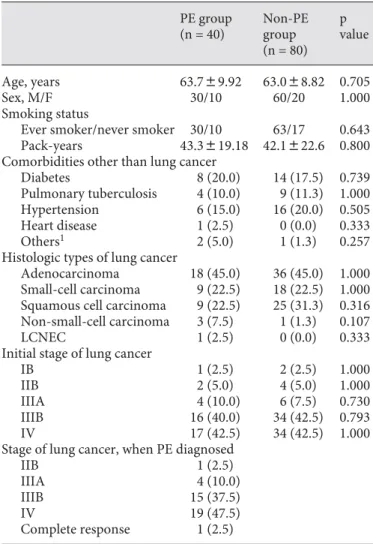
The demographic data are summarized in table 1 . The smoking status, including the frequency of ever-smokers and pack-years, and comorbid conditions did not differ between the PE and non-PE groups. Adenocarcinoma (45.0%, n = 18) was the most common histological type of lung cancer, followed by small cell carcinoma (22.5%, n = 8) and squamous cell carcinoma (22.5%, n = 8). Ac- cording to TNM staging [15] , when PE was diagnosed, most of the lung cancer patients were in stages IIIB (37.5%, n = 15) and IV (47.5%, n = 19).
Clinical Characteristics
The clinical characteristics of the study subjects are presented in table 2 . The most common presenting man- ifestation in the PE group was dyspnea (50.0%), followed by incidental CT findings of PE (37.5%). The median du- ration of the presenting symptom was 7 days (range, 2–30 days). Immobilization and trauma were noted as the fac- tors predisposing to PE, excluding factors related to lung cancer and anticancer treatment. The prevalence of deep- venous thrombosis was 75% (15/20) based on CT venog- raphy.
Laboratory Findings
The median values of serum troponin I (n = 32) and D-dimer (n = 37) were 0.04 ng/ml (0.01–0.40 ng/ml) and 449.0 mg/dl (0.2–1,896.0 mg/dl), respectively ( table 3 ).
The variables related to thrombophilia in the PE group are summarized in table 3 . Antithrombin III deficiency,
Table 1. Demographic data of the subjects PE group (n = 40)
Non-PE group (n = 80)
p value
Age, years 63.789.92 63.088.82 0.705
Sex, M/F 30/10 60/20 1.000
Smoking status
Ever smoker/never smoker 30/10 63/17 0.643
Pack-years 43.3819.18 42.1822.6 0.800
Comorbidities other than lung cancer
Diabetes 8 (20.0) 14 (17.5) 0.739
Pulmonary tuberculosis 4 (10.0) 9 (11.3) 1.000
Hypertension 6 (15.0) 16 (20.0) 0.505
Heart disease 1 (2.5) 0 (0.0) 0.333
Others1 2 (5.0) 1 (1.3) 0.257
Histologic types of lung cancer
Adenocarcinoma 18 (45.0) 36 (45.0) 1.000 Small-cell carcinoma 9 (22.5) 18 (22.5) 1.000 Squamous cell carcinoma 9 (22.5) 25 (31.3) 0.316 Non-small-cell carcinoma 3 (7.5) 1 (1.3) 0.107
LCNEC 1 (2.5) 0 (0.0) 0.333
Initial stage of lung cancer
IB 1 (2.5) 2 (2.5) 1.000
IIB 2 (5.0) 4 (5.0) 1.000
IIIA 4 (10.0) 6 (7.5) 0.730
IIIB 16 (40.0) 34 (42.5) 0.793
IV 17 (42.5) 34 (42.5) 1.000
Stage of lung cancer, when PE diagnosed
IIB 1 (2.5)
IIIA 4 (10.0)
IIIB 15 (37.5)
IV 19 (47.5)
Complete response 1 (2.5)
Values are means 8 SDs, or numbers and percentages (in pa- rentheses). LCNEC = Large cell neuroendocrine carcinoma.
1 Others include renal vein thrombosis, end-stage renal dis- ease, interstitial lung disease, and chronic obstructive pulmonary disease.
Table 2. Clinical characteristics of the pulmonary embolism group (n = 40)
Presenting manifestation
Dyspnea 20 (50.0)
Incidental findings on chest CT 15 (37.5)
Leg swelling or pain 2 (5.0)
Chest pain 1 (2.5)
Duration of symptom, days 7 (2–30) Risk factors other than lung cancer
Immobilization1 6 (7.5)
Trauma 1 (1.3)
Deep-vein thrombosis on CT scan 15/20 (75.0)
Values are medians and ranges (in parentheses), means 8 SDs, or numbers and percentages (in parentheses).
1 Immobilization for >3 consecutive days (bed rest except for going to bathroom) or surgery in previous 4 weeks [15].
lupus anticoagulant, protein C deficiency and hyperho- mocysteinemia were observed in 28.6% (8/28), 25.0%
(5/20), 22.2% (6/27) and 14.3% (3/21) of the patients, re- spectively.
Electrocardiographic and Echocardiographic Findings The most common electrocardiographic finding was sinus rhythm (47.5%, n = 19) followed by sinus tachycar-
dia (37.5%, n = 15). T-inversions on the precordial leads of electrocardiograms and right bundle branch block or S1Q3T3 were observed in 3 patients (7.5%); atrial fibrilla- tion, premature ventricular contraction and sinus brady- cardia were noted in 1 patient (2.5%). Of 9 patients who had echocardiography, 1 (11.1%) had right-ventricular dysfunction and the median right-ventricular systolic pressure was 33.1 mm Hg (28–74 mm Hg).
Imaging Studies
The most commonly used first imaging modality in the diagnosis of PE was a CT scan (97.5%, n = 39/40). The frequencies of the PE-involved largest pulmonary arter- ies were as follows: 6 main pulmonary artery, 35.9%
(14/39); lobar artery, 30.8% (12/39), and ^ segmental ar- tery, 33.3% (13/39).
Clinical Course and Survival
The median time from the diagnosis of lung cancer to the identification of PE was 3.4 months (–5.8 to 120.3 months). Twenty-eight (70.0%) and 34 (85.0%) patients were diagnosed with PE within 6 and 12 months of the lung cancer diagnosis, respectively ( fig. 2 a). Thirty-two patients (80.0%) in the PE group received unfractionated heparin intravenously or low-molecular-weight heparin subcutaneously, followed by an oral anticoagulant. Nine- teen (79.2%) of the 24 patients in the PE group who un-
Table 3. Laboratory findings of pulmonary embolism group ABGA
PaO2, mm Hg (n = 33) 72.7817.46 P(A-a)O2, mm Hg (n = 33) 39.0821.19 Biomarkers
Troponin I, ng/ml (n = 32) 0.04 (0.01–0.40) D-dimer, mg/dl (n = 37) 449.0 (0.2–1,896.0) Thrombophilia
Antithrombin III deficiency 8/28 (28.6) Positive lupus anticoagulant 5/20 (25.0)
Protein C deficiency 6/27 (22.2)
Hyperhomocysteinemia 3/21 (14.3)
Positive antiphospholipid IgM 1/19 (5.3) Positive anticardiolipin IgG 1/22 (4.5)
Protein S deficiency 1/28 (3.6)
Factor V Leiden mutation 0/22 (0.0) Positive antiphospholipid IgG 0/19 (0.0) Positive anticardiolipin IgM 0/22 (0.0)
Values are mean 8 SD, medians and ranges in parentheses or numbers and percentages in parentheses.
NT-proBNP = N-terminal-pro-B-type natriuretic peptide.
0 10 20 30 40 50 60 70
% n = 28
<6
n = 6
n = 3 n = 3
6–12 12–24 >24
Months a
0 10 20 30 40 50 60 70
%
n = 14
Normalization
n = 5
n = 2
n = 1
Impr ovement
No change
Undet ermined
Aggravation n = 2
b
Fig. 2. a The time interval from the diagnosis of lung cancer to the detection of PE. Most patients (85%) were diagnosed with PE within 12 months of lung cancer diagnosis. b The changes in pul- monary emboli upon follow-up CT scan. The majority of patients took a favorable (normalization + improvement) clinical course in the PE group.
derwent a follow-up CT scan demonstrated improvement or normalization of the PE ( fig. 2 b).
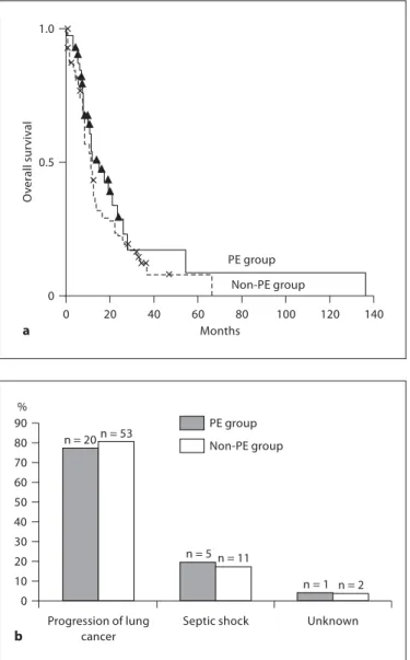
Comparison of survival rates revealed no significant difference between the PE and non-PE groups (p = 0.249);
median survival was 15.3 and 11.4 months, respectively ( fig. 3 a). However, the median survival from the diagno- sis of PE was merely 3.5 months in the PE group. In the PE and non-PE groups, the most common cause of death was lung cancer progression (76.9 and 80.3%, respective- ly), followed by chemotherapy-related septic shock (19.2 and 16.7%, respectively) ( fig. 3 b). After excluding patients whose cause of death was not identifiable (3.8 and 3%, respectively), all deaths in both groups were caused di- rectly by lung cancer or indirectly by complications re- lated to anticancer treatment.
Discussion
In lung cancer patients with PE, adenocarcinoma was the most common histologic type and most patients (82.5%) had advanced-stage disease (IIIB/IV). The me- dian survival time of lung cancer patients with PE was 3.5 months, suggesting that PE may be a poor prognostic factor in lung cancer patients. No difference in survival was noted between lung cancer patients with and without PE. In addition, as with lung cancer patients without PE, most patients in the PE group died of lung cancer or treat- ment-related complications, and not directly from the PE. Thus, PE may be one of an array of complications which lung cancer patients experience, rather than a pri- mary cause of mortality.
The incidence of VTE in patients hospitalized with cancer varies according to the type of malignancy [16] . It was the highest in those with carcinoma of the pancreas (4.3%) [16] . To date, adenocarcinoma, especially the mu- cin-producing type, is considered to be the most common histologic type of cancer in VTE patients, regardless of the primary sites [9, 17] . Recently, Blom et al. [3] demon- strated that the risk of VTE in lung cancer patients in- creased 20-fold compared to the general population and that patients with adenocarcinoma have a higher risk of VTE than patients with squamous cell carcinoma. Like- wise, adenocarcinoma was the most frequent histological type of lung cancer in the present study.
Most patients in the PE group had advanced stages of lung cancer, and the median survival from the diagnosis of PE was merely 3.5 months. These results imply that PE in lung cancer patients is a poor prognostic factor. They are in agreement with previous studies [8, 18] which dem-
onstrated that patients with cancer and VTE have a low- er survival rate than those with cancer without VTE.
Similarly, in a registry study, ‘fatal PE’ was more common in cancer patients with VTE than in those without can- cer, and metastatic disease was one of the risk factors for fatal PE [19] . Very recently, the same study group con- firmed that cancer is one of the independent clinical pre- dictors of fatal PE [20] .
However, there was no significant difference in the survival curve between the lung cancer patients with and
0 1.0
Months
Overallsurvival
0.5
0 20 40 60 80 100 120 140
PE group Non-PE group
a
0 90
%
n = 20
Progression of lung cancer 10
20 30 50 60 70 80
40
n = 53
n = 5
n = 1
Septic shock Unknown n = 11
n = 2 PE group
Non-PE group
b
Fig. 3. a A survival curve illustrating no significant difference in the survival rate in the PE and non-PE groups (p = 0.249). b A frequency distribution illustrating the causes of death. The main causes of death were lung cancer progression and septic shock related to anticancer therapy in the PE and non-PE groups.
References
1 Heit JA, Silverstein MD, Mohr DN, Petterson TM, O’Fallon WM, Melton LJ 3rd: Risk fac- tors for deep vein thrombosis and pulmo- nary embolism: a population-based case- control study. Arch Intern Med 2000; 160:
809–815.
2 Rosendaal FR: Risk factors for venous throm- bosis: Prevalence, risk, and interaction.
Semin Hematol 1997; 34: 171–187.
3 Blom JW, Osanto S, Rosendaal FR: The risk of a venous thrombotic event in lung cancer patients: Higher risk for adenocarcinoma than squamous cell carcinoma. J Thromb Haemost 2004; 2: 1760–1765.
4 Falanga A, Rickles FR: Pathophysiology of the thrombophilic state in the cancer pa- tient. Semin Thromb Hemost 1999; 25: 173–
182.
5 Gale AJ, Gordon SG: Update on tumor cell procoagulant factors. Acta Haematol 2001;
106: 25–32.
6 Donati MB, Falanga A: Pathogenetic mecha- nisms of thrombosis in malignancy. Acta Haematol 2001; 106: 18–24.
7 Tesselaar ME, Osanto S: Risk of venous thromboembolism in lung cancer. Curr Opin Pulm Med 2007; 13: 362–367.
8 Sorensen HT, Mellemkjaer L, Olsen JH, Baron JA: Prognosis of cancers associated with venous thromboembolism. N Engl J Med 2000; 343: 1846–1850.
9 Levitan N, Dowlati A, Remick SC, Tahsildar HI, Sivinski LD, Beyth R, Rimm AA: Rates of initial and recurrent thromboembolic dis- ease among patients with malignancy versus those without malignancy. Risk analysis us- ing medicare claims data. Medicine 1999; 78:
285–291.
10 Blom JW, Doggen CJ, Osanto S, Rosendaal FR: Malignancies, prothrombotic muta- tions, and the risk of venous thrombosis.
JAMA 2005; 293: 715–722.
11 O’Connell CL, Boswell WD, Duddalwar V, Caton A, Mark LS, Vigen C, Liebman HA:
Unsuspected pulmonary emboli in cancer patients: clinical correlates and relevance. J Clin Oncol 2006; 24: 4928–4932.
12 Gladish GW, Choe DH, Marom EM, Sabloff BS, Broemeling LD, Munden RF: Incidental pulmonary emboli in oncology patients:
prevalence, CT evaluation, and natural his- tory. Radiology 2006; 240: 246–255.
13 Sebastian AJ, Paddon AJ: Clinically unsus- pected pulmonary embolism – an important secondary finding in oncology CT. Clin Ra- diol 2006; 61: 81–85.
14 The PIOPED Investigators: Value of the ven- tilation/perfusion scan in acute pulmonary embolism. Results of the prospective investi- gation of pulmonary embolism diagnosis (PIOPED). JAMA 1990; 263: 2753–2759.
15 Mountain CF: Revisions in the international system for staging lung cancer. Chest 1997;
111: 1710–1717.
16 Stein PD, Beemath A, Meyers FA, Skaf E, Sanchez J, Olson RE: Incidence of venous thromboembolism in patients hospitalized with cancer. Am J Med 2006; 119: 60–68.
17 Lee AY, Levine MN: Venous thromboembo- lism and cancer: Risks and outcomes. Circu- lation 2003; 107:I17–I21.
18 Carson JL, Kelley MA, Duff A, Weg JG, Fulkerson WJ, Palevsky HI, Schwartz JS, Thompson BT, Popovich J Jr, Hobbins TE, Spera MA, Alavi A, Terrin ML: The clinical course of pulmonary embolism. N Engl J Med 1992; 326: 1240–1245.
without PE. In addition, the primary cause of death in the PE and non-PE groups was lung cancer progression or chemotherapy-induced events in our study. These are consistent with the findings of a previous paper [18] , which found that when properly diagnosed and treated, clinically evident PE was not a common cause of death and that most deaths were attributable to underlying dis- eases. Despite the one-year mortality rate of nearly 24%
among the 399 patients with clinically apparent PE, death from PE itself occurred in only 2.5% of the patients. Fur- thermore, in a recent study of patients with cancer who died over the 19-year study period [21] , PE was the cause of death in 1% or less of the patients depending on the type of cancer. Consequently, the findings of this study indicate that PE is one of the common complications in lung cancer patients, rather than the primary cause of death [22] .
This study has a number of limitations. Specifically, since our study was performed retrospectively, a selection bias cannot be avoided. In addition, the patients in the lung cancer group were not enrolled from consecutive lists, but were selected from cases diagnosed in clinical practice. Secondly, pharmacologic thromboprophylaxis is not routinely given in Korea, except during admission to an intensive care unit, although this is against the cur-
rent clinical guidelines for VTE prophylaxis in patients with cancer [23] . This policy may result from the belief that Asians traditionally have a lower risk of overall or cancer-related VTE compared to Western populations as evidenced by several studies [24–26] . Not giving pharma- cologic thromboprophylaxis is not thought to have af- fected our conclusions because no difference in survival between PE and non-PE groups was observed and no pa- tient died of PE. However, the incidence of VTE in Asians is not rare as noted in the present study. Therefore, phar- macologic thromboprophylaxis is thought to be neces- sary in Asian lung cancer patients as well. Next, as noted earlier, many patients not suspected to have PE were in- cluded in the lung cancer group, which exhibited less se- vere clinical manifestations. Lastly, the causes of death were determined clinically but not based on the autopsy.
To what extent PE actually contributed to the patients’
death is difficult to answer confidently.
In conclusion, PE occurred more frequently in ad- vanced stages of lung cancer, and the most common his- tological type was adenocarcinoma. No difference in sur- vival was observed between lung cancer patients with and without PE. In addition, despite suggesting a poor prog- nosis, PE is not believed to be the primary cause of death but one of the various complications of lung cancer.
19 Monreal M, Falga C, Valdes M, Suarez C, Ga- briel F, Tolosa C, Montes J: Fatal pulmonary embolism and fatal bleeding in cancer pa- tients with venous thromboembolism: find- ings from the RIETE registry. J Thromb Hae- most 2006; 4: 1950–1956.
20 Laporte S, Mismetti P, Decousus H, Uresan- di F, Otero R, Lobo JL, Monreal M: Clinical predictors for fatal pulmonary embolism in 15,520 patients with venous thromboembo- lism: findings from the RIETE registry. Cir- culation 2008; 117: 1711–1716.
21 Stein PD, Beemath A, Meyers FA, Kayali F, Skaf E, Olson RE: Pulmonary embolism as a cause of death in patients who died with can- cer. Am J Med 2006; 119: 163–165.
22 Sallah S, Wan JY, Nguyen NP: Venous throm- bosis in patients with solid tumors: determi- nation of frequency and characteristics.
Thromb Haemost 2002; 87: 575–579.
23 Lyman GH, Khorana AA, Falanga A, Clarke- Pearson D, Flowers C, Jahanzeb M, Kakkar A, Kuderer NM, Levine MN, Liebman H, Mendelson D, Raskob G, Somerfield MR, Thodiyil P, Trent D, Francis CW: American Society of Clinical Oncology guideline: rec- ommendations for venous thromboembo- lism prophylaxis and treatment in patients with cancer. J Clin Oncol 2007; 25: 5490–
5505.
24 White RH, Dager WE, Zhou H, Murin S: Ra- cial and gender differences in the incidence of recurrent venous thromboembolism.
Thromb Haemost 2006; 96: 267–273.
25 Chew HK, Wun T, Harvey D, Zhou H, White RH: Incidence of venous thromboembolism and its effect on survival among patients with common cancers. Arch Intern Med 2006; 166: 458–464.
26 Oh SY, Kim JH, Lee KW, Bang SM, Hwang JH, Oh D, Lee JS: Venous thromboembolism in patients with pancreatic adenocarcinoma:
Lower incidence in Asian ethnicity. Thromb Res 2008; 122: 485–490.