• 검색 결과가 없습니다.

Risk Factor and Mortality in Patients with Pulmonary Embolism Combined with Infectious Disease

N/A
N/A
Protected

Academic year: 2021

Share "Risk Factor and Mortality in Patients with Pulmonary Embolism Combined with Infectious Disease"

Copied!
10
0
0

로드 중.... (전체 텍스트 보기)

전체 글

(1)

Risk Factor and Mortality in Patients with Pulmonary Embolism Combined with Infectious Disease

Gi Dong Lee, M.D.

1

, Sunmi Ju, M.D.

2

, Ju-Young Kim, M.D.

3

, Tae Hoon Kim, M.D., Ph.D.

3

, Jung-Wan Yoo, M.D.

2

, Seung Jun Lee, M.D., Ph.D.

2

, Yu Ji Cho, M.D., Ph.D.

2

, Yi Yeong Jeong, M.D., Ph.D.

2

, Kyung Nyeo Jeon, M.D. Ph.D.

4

, Jong Deog Lee, M.D., Ph.D.

2

and Ho Cheol Kim, M.D., Ph.D.

3

1

Department of Internal Medicine, Saint Carollo Hospital, Suncheon,

2

Department of Internal Medicine, Gyeongsang National University Hospital, Gyeongsang National University School of Medicine, Jinju, Departments of

3

Internal Medicine and

4

Diagnostic Radiology, Gyeongsang National University Changwon Hospital, Gyeongsang National University School of Medicine, Changwon, Korea

Background: Infectious conditions may increase the risk of venous thromboembolism. The purpose of this study was to evaluate the risk factor for combined infectious disease and its influence on mortality in patients with pulmonary embolism (PE).

Methods: Patients with PE diagnosed based on spiral computed tomography findings of the chest were retrospectively analyzed. They were classified into two groups: patients who developed PE in the setting of infectious disease or those with PE without infection based on review of their medical charts.

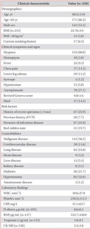
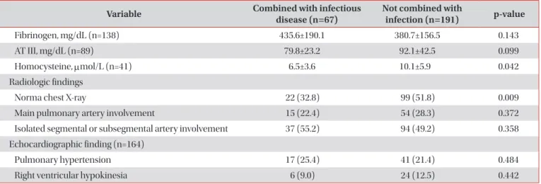
Results: Of 258 patients with PE, 67 (25.9%) were considered as having PE combined with infectious disease. The sites of infections were the respiratory tract in 52 patients (77.6%), genitourinary tract in three patients (4.5%), and hepatobiliary tract in three patients (4.5%). Underlying lung disease (odds ratio [OR], 3.69; 95% confidence interval [CI], 1.926–7.081;

p<0.001), bed-ridden state (OR, 2.84; 95% CI, 1.390–5.811; p=0.004), and malignant disease (OR, 1.867; 95% CI, 1.017–

3.425; p=0.044) were associated with combined infectious disease in patients with PE. In-hospital mortality was higher in patients with PE combined with infectious disease than in those with PE without infection (24.6% vs. 11.0%, p=0.006). In the multivariate analysis, combined infectious disease (OR, 4.189; 95% CI, 1.692–10.372; p=0.002) were associated with non-survivors in patients with PE.

Conclusion: A substantial portion of patients with PE has concomitant infectious disease and it may contribute a mortality in patients with PE.

Keywords: Pulmonary Embolism; Infectious Disease; Mortality

Address for correspondence: Ho Cheol Kim, M.D., Ph.D.

Department of Internal Medicine, Gyeongsang National University Changwon Hospital, Gyeongsang National University School of Medicine, 11 Samjeongja-ro, Seongsan-gu, Changwon 51472, Korea

Phone: 82-55-214-3730, Fax: 82-55-214-8618, E-mail: [email protected]

Received: May. 2, 2019, Revised: Aug. 24, 2019, Accepted: Jan. 21, 2020, Published online: Mar. 10, 2020

cc It is identical to the Creative Commons Attribution Non-Commercial License (http://creativecommons.org/licenses/by-nc/4.0/).

Copyright © 2020

The Korean Academy of Tuberculosis and Respiratory Diseases.

(2)

Introduction

Venous thromboembolism (VTE), including deep vein thrombosis (DVT) and pulmonary embolism (PE), is a fre- quently overlooked, serious disorder

1

. Its incidence is increas- ing according to recent reports in western countries and in Korea

2-4

.

There are various well-defined risk factors of VTE, including recent major surgery, immobilization, cancer, old age, obesity, use of oral contraceptives, hormonal factors, a previous his- tory of thrombosis, and inherited risk factors

5,6

. In addition to known risk factors, infectious disease and the associated systemic inflammatory response may increase thrombotic events of the venous system

7,8

caused by one or more of the three precipitants of venous thrombosis, as proposed by Vir- chow: venous stasis, hypercoagulability, and endothelial dam- age

9,10

. Several reports have documented that the risk of VTE was increased in patients with certain infectious conditions, such as cytomegalovirus, Chlamydia pneumoniae, human im- munodeficiency virus (HIV), hepatitis C virus infection, and a recent respiratory tract infection

11-18

. Despite several reports of recent infection and VTE, its independent relationship is un- clear, and clinical data regarding PE combined with infectious conditions are lacking. Therefore, the aim of the study was to evaluate the clinical characteristics and source of infection in patients with PE combined with infectious disease. And, the risk factor associated with combined infectious disease and its influence on mortality in patients with PE were assessed.

Materials and Methods

1. Study design and population

Patients with PE found on computed tomography (CT) within 48 hours after admission were enrolled in this study.

The study was conducted at regional university affiliated hospital (890 beds) between September 2010 and May 2014.

Patients were excluded if they were admitted for pregnancy, preexisting PE or DVT, or were <18 years of age. The diagnosis of PE was confirmed based on chest findings obtained using a 64-detector CT, including enhanced CT and CT pulmonary angiogram, scanner (Brilliance-64; Philips Medical Systems, Eindhoven, Netherlands). A pulmonary radiologist assessed the existence of thrombosis in the pulmonary artery. This study was approved by the hospital’s institutional review board (No. GNUCH-2018-07-001). The need for informed consent was waived because of the retrospective nature of the study.

2. Data collection and clinical outcomes

We retrospectively investigated patients’ demographics, physiologic data, and laboratory data and the causes of infec-

Table 1. Clinical characteristics of enrolled patients with PE Clinical characteristic Value (n=258) Demographics

Age, yr 68.2±13.0

Age >65 yr 171 (66.3)

Male sex 143 (55.4)

BMI (n=233) 22.76±3.9

BMI >30 kg/m

2

13 (5.0)

Current smoking history 17 (6.5)

Clinical symptoms and signs

Dyspnea 155 (60.0)

Hemoptysis 10 (3.8)

Fever 24 (9.3)

Chest pain 37 (14.3)

Lower leg edema 29 (11.2)

Syncope 3 (1.2)

Hypotension 15 (5.8)

Asymptomatic 70 (27.1)

Revised Geneva score 6.0±1.6

Died 37 (14.3)

Risk factors

History of recent operation (<3 mo) 67 (25.9)

Previous history of VTE 20 (7.7)

Presence of infectious disease 67 (25.9)

Bed-ridden state 51 (19.7)

Comorbidities

Malignant disease 145 (56.2)

Cerebrovascular disease 30 (11.6)

Lung disease 64 (24.8)

Heart disease 9 (3.5)

Liver disease 14 (5.4)

Kidney disease 8 (3.1)

Diabetes 56 (21.7)

Hypertension 85 (32.9)

Autoimmune disease 3 (1.2)

Laboratory findings

WBC, mm

3

/L 10.6±27.8

Platelet, mm

3

/L 239.2±111.5

CRP, mg/L 51.1±62.7

D-dimer, µg/mL (n=195) 6.6±6.3

BNP, pg/mL (n=147) 332.7±440.0

Troponin-I, ng/mL (n=155) 1.0±9.1

CK-MB (n=158) 3.5±3.8

(3)

tion, underlying diseases, and clinical outcomes. We divided patients into one of two groups based on information ob- tained from their medical charts: patients with PE combined with infectious disease or those with PE without infection.

Patients’ characteristics were compared between those two groups, and between survivors and non-survivors. Finally, we analyzed the risk factors associated with combined infec- tious disease in PE patients and compared mortality between those two groups. And we assessed the factor associated non- survivors in patients with PE.

3. Definition

The definition of infection was based on microbiologic data and the patient’s clinical course upon medical chart review.

Infection was classified according to the Centers for Disease Control and Prevention’s definitions

19

.

4. Statistical analysis

Continuous variables are reported as the mean±standard deviation. Categorical variables are expressed as a number (%).

Differences between the groups were statistically assessed

using Student’s t test or the chi-square test, as appropriate.

Univariate and multivariate logistic regression analyses were performed to examine the risk factors for the development of infectious disease and mortality. A p-value of <0.05 was considered statistically significant in all tests. All data were analyzed using SPSS version 22.0 for Window (IBM Corp., Ar- monk, NY, USA).

Results

1. Patients’ characteristics

Two hundred fifty-eight patients (male:female, 143:115;

mean age, 68.2±13.0 years) were included. The most com- mon symptom was dyspnea in up to 60% of patients, and 70 patients (26.9%) were asymptomatic. Twenty patients (7.7%) had a previous history of VTE, and 145 (55.8%) had an under- lying malignant disease. One hundred seventy-one patients (65.8%) were older than 65 years, and 13 (5%) had a body mass index more than 30 kg/m

2

. Fifty-eight patients (22.3%) showed echocardiographic evidence of pulmonary hyperten- sion, such as right ventricular hypokinesia or an increased pulmonary arterial pressure more than 20 mm Hg. The main Table 1. Continued

Clinical characteristic Value (n=258) Fibrinogen, mg/dL (n=138) 393.4±165.7

AT III, mg/dL (n=89) 89.4±39.4

Homocysteine, µmol/L (n=41) 9.3±5.6

Radiologic findings

Norma chest X-ray 121 (46.8)

Involvement of pulmonary artery on chest CT

Main pulmonary artery, right 52 (20.2) Main pulmonary artery, left 39 (15.1) Interlobar artery, right 103 (39.9)

Interlobar artery, left 69 (26.7)

Segmental artery, right 158 (61.2)

Segmental artery, left 95 (36.8)

Subsegmental artery, right 88 (34.1)

Subsegmental artery, left 51 (19.7)

Echocardiographic finding (n=164)

Pulmonary hypertension 58 (22.4)

Right ventricular hypokinesia 30 (11.6) Values are presented as mean±SD or number (%).

BMI: body mass index; VTE: venous thromboembolism; WBC:

white blood cell; CRP: C-reactive protein; BNP: B-type natriuretic peptide; CK-MB: creatine kinase-muscle/brain; AT: antithrombin;

CT: computerized tomography.

Table 2. The site and cause of infection in patients with pulmonary embolism combined with infectious disease

Cause of infection No. (%) (n=67) Respiratory tract infection 52 (77.6)

Pneumonia 44 (65.6)

Acute bronchiolitis 4 (5.9)

Pulmonary tuberculosis 4 (5.9) Genitourinary tract infection 3 (4.4) Hepatobiliary tract infection 3 (4.4)

Acute cholecystitis 1 (1.5)

Acute pancreatitis 1 (1.5)

Perihepatic abscess 1 (1.5)

Gastrointestinal tract infection 2 (2.9)

Ischemic colitis 2 (2.9)

Bone and joint infection 3 (4.4)

Osteomyelitis of spine 3 (4.4)

Skin and soft tissue infection 2 (2.9)

Shoulder abscess 1 (1.5)

Necrotizing fasciitis 1 (1.5)

Miscellaneous infection 2 (2.9)

Infective endocarditis 1 (1.5)

Tuberculous pericarditis 1 (1.5)

(4)

Table 3. Comparison of clinical characteristics in patients with PE combined with infectious disease and in patients with PE without infection

Variable Combined with infectious

disease (n=67) Not combined with

infection (n=191) p-value Demographics

Age, yr 71.6±12.2 67.2±13.2 0.028

Age >65 yr 52 (77.6) 119 (62.3) 0.018

Male sex 21 (31.3) 122 (63.9) 0.972

BMI (n=233) 21.8±4.2 23.1±3.7 0.053

BMI >30 kg/m

2

4 (6.0) 9 (4.7) 0.458

Current smoking history 1 (1.5) 16 (8.3) 0.208

Clinical symptoms and signs

Dyspnea 51 (76.1) 104 (54.4) 0.002

Hemoptysis 4 (6.0) 6 (3.1) 0.294

Fever 12 (17.9) 12 (6.2) 0.004

Chest pain 7 (10.4) 30 (15.7) 0.304

Lower leg edema 7 (10.4) 22 (11.5) 0.831

Syncope 0 (0) 3 (1.6) 0.305

Hypotension 3 (4.5) 12 (6.2) 0.599

Asymptomatic 15 (22.4) 55 (28.8) 0.331

Revised Geneva score 5.9±1.6 6.1±1.5 0.302

Died 15 (22.4) 22 (11.5) 0.027

Risk factors

History of recent operation (<3 mo) 7 (10.4) 20 (10.4) 0.458

Previous history of VTE 5 (7.5) 15 (7.8) 0.497

Bed-ridden state 21 (31.3) 30 (15.7) 0.001

Comorbidities

Malignant disease 27 (40.3) 98 (51.3) 0.343

Cerebrovascular disease 12 (17.9) 18 (9.4) 0.012

Lung disease 39 (58.2) 25 (13.1) 0.001

Heart disease 9 (13.4) 29 (15.1) 0.103

Liver disease 5 (7.5) 9 (4.7) 0.451

Kidney disease 3 (4.5) 6 (3.1) 0.454

Diabetes 12 (17.9) 44 (23.0) 0.402

Hypertension 16 (23.9) 69 (36.1) 0.074

Autoimmune disease 2 (3.0) 1 (0.5) 0.164

Laboratory findings

WBC, mm

3

/L 16.4±54.0 8.6±5.4 0.048

Platelet, mm

3

/L 269.3±115.4 228.7±108.4 0.013

CRP, mg/L 69.1±63.0 44.9±61.7 0.008

D-dimer, µg/mL (n=195) 5.9±5.9 6.9±6.5 0.299

BNP, pg/mL (n=147) 445.8±543.7 275.0±366.3 0.025

Troponin-I, ng/mL (n=155) 0.3±1.2 1.3 ±11.0 0.521

CK-MB (n=158) 3.9±4.4 3.4±3.6 0.496

(5)

pulmonary artery was involved in 88 patients (33.8%) based on CT findings. Thirty-seven patients (14.2%) died and the cause of death were underlying malignant disease in 27 pa- tients (10.4%), uncontrolled infectious disease with multi- organ failure in six patients (2.3%), progression of underlying lung disease in three patients (1.1%), and heart failure in one patient (Table 1). Overall, 67 patients (25.9%) had a concomi- tant infectious disease. The infections involved the respiratory tract in 52 patients (77.6%), genitourinary tract in three (4.5%), hepatobiliary tract in three (4.5%), gastrointestinal tract in two (2.98%), bones and joints in three (4.5%), and other organ sys- tems in four (5.4%) (Table 2).

2. Comparison of clinical characteristics in PE patients with infectious disease and without infection

The proportion of patients with dyspnea was greater among patients with PE combined with infectious disease than among those with PE without infection (76.1% vs. 53.9%, p<0.05). The frequency of fever was more common in patients with PE combined with infectious disease than in those with PE without infection (17.9% vs. 6.2%, p<0.05). The proportion of patients older than 65 years was higher among patients with PE combined with infectious disease than among those with PE without infection (77.6% vs. 61.7%, p<0.05). The revised Geneva score

20

was not different between the two groups. The white blood cell count, serum C-reactive protein level, B-type natriuretic peptide level, and platelet count were significantly higher in patients with PE combined with infectious disease Table 3. Continued

Variable Combined with infectious

disease (n=67) Not combined with

infection (n=191) p-value

Fibrinogen, mg/dL (n=138) 435.6±190.1 380.7±156.5 0.143

AT III, mg/dL (n=89) 79.8±23.2 92.1±42.5 0.099

Homocysteine, µmol/L (n=41) 6.5±3.6 10.1±5.9 0.042

Radiologic findings

Norma chest X-ray 22 (32.8) 99 (51.8) 0.009

Main pulmonary artery involvement 15 (22.4) 54 (28.3) 0.372

Isolated segmental or subsegmental artery involvement 37 (55.2) 94 (49.2) 0.358

Echocardiographic finding (n=164)

Pulmonary hypertension 17 (25.4) 41 (21.4) 0.484

Right ventricular hypokinesia 6 (9.0) 24 (12.5) 0.442

Values are presented as mean±SD or number (%).

PE: pulmonary embolism; BMI: body mass index; VTE: venous thromboembolism; WBC: white blood cell; CRP: C-reactive protein; BNP: B- type natriuretic peptide; CK-MB: creatine kinase-muscle/brain; AT: antithrombin.

Table 4. Factor associated with infectious disease in patients with PE

Variable Univariate analysis Multivariate analysis

OR 95% CI p-value OR 95% CI p-value

Age >65 yr 2.15 1.133–4.103 0.019 1.922 0.977–3.779 0.058

BMI >30 kg/m

2

1.27 0.378–4.268 0.699 - - -

Bed-ridden state 2.48 1.299–4.735 0.006 2.842 1.390–5.811 0.004

Lung disease 3.44 1.879–6.317 <0.001 3.693 1.926–7.081 0.000

Cerebrovascular disease 2.12 0.964–4.672 0.062 - - -

Malignant disease 2.331 1.321–4.111 0.003 1.867 1.017–3.425 0.044

Kidney disease 1.043 0.205–5.296 0.960 - - -

Liver disease 1.649 0.532–5.106 0.386 - - -

Diabetes 1.353 0.666–2.751 0.403 - - -

PE: pulmonary embolism; OR: odds ratio; CI: confidence interval; BMI: body mass index.

(6)

Table 5. Comparison of clinical characteristics between survivors and non-survivors in patients with PE

Variable Survivors (n=221) Non-survivors (n=37) p-value

Demographics

Age, yr 62.0±15.4 69.3±12.3 0.002

Male sex 122 (55.2) 21 (56.8) 0.817

Age >65 yr 155 (70.1) 16 (43.2) 0.002

BMI (n=233) 22.8±3.9 22.1±3.9 0.333

BMI >30 kg/m

2

12 (5.4) 1 (2.7) 0.552

Current smoking history 17 (7.6) 0 (0) 0.316

Clinical symptoms and signs

Dyspnea 129 (58.3) 26 (70.3) 0.154

Hemoptysis 9 (4.0) 1 (2.7) 0.696

Fever 19 (8.5) 5 (13.5) 0.331

Chest pain 31 (14.0) 6 (16.2) 0.709

Lower leg edema 19 (8.5) 10 (27.0) 0.001

Syncope 3 (1.3) 0 (0) 0.478

Hypotension 11 (5.0) 4 (10.8) 0.156

Asymptomatic 63 (28.5) 7 (18.9) 0.236

Revised Geneva score 6.0±1.5 6.2±1.4 0.655

Risk factors

History of recent operation (<3 mo) 22 (9.9) 2 (5.4) 0.458

Previous history of VTE 15 (6.8) 5 (13.2) 0.186

Bed-ridden state 38 (17.1) 13 (35.1) 0.010

Comorbidities

Malignant disease 114 (51.5) 31 (83.8) <0.001

Cerebrovascular disease 27 (12.2) 3 (8.1) 0.481

Lung disease 53 (23.9) 11 (29.7) 0.436

Heart disease 37 (16.7) 3 (7.9) 0.225

Liver disease 12 (5.4) 2 (5.4) 0.995

Kidney disease 5 (2.2) 3 (8.1) 0.454

Diabetes 48 (21.7) 8 (21.6) 0.989

Hypertension 70 (31.6) 15 (40.5) 0.272

Autoimmune disease 3 (1.3) 0 0.478

Infectious disease 52 (23.5) 15 (40.5) 0.027

Laboratory findings

WBC, mm

3

/L 10,521±3,001 11,197±6,334 0.765

Platelet, mm

3

/L 244,257±11,322 209,354±96,246 0.052

CRP, mg/L 48.3±62.8 68.3±60.7 0.073

D-dimer, µg/mL (n=195) 6.69±6.29 6.75±6.65 0.964

BNP, pg/mL (n=147) 317.5±431.4 442.9±497.5 0.320

Troponin-I, ng/mL (n=155) 0.30±0.99 5.3±24.28 0.018

CK-MB (n=158) 3.72±3.99 2.54±2.57 0.70

Fibrinogen, mg/dL (n=138) 392.4±167.4 398.59±160.7 0.871

(7)

than in those with PE without underlying infection (p<0.05).

The D-dimer level was not significantly different between two groups. The frequency of main pulmonary artery involvement based on CT findings was not different between PE patients with infectious disease and without infection (28% and 22.4%, respectively). The sole segmental or subsegmental pulmonary artery involvement was also not different between PE patients with infectious disease and without infection (48.7% and 55.2%, respectively). In-hospital mortality was higher in pa- tients with PE combined with infectious disease than in those with PE without infection (22.4% vs. 11.4%, p=0.006) (Table 3).

3. Factors associated with PE combined with infectious disease

In multivariate analysis, the results showed that underlying lung disease (odds ratio [OR], 3.69; 95% confidence interval [CI], 1.926–7.081; p<0.001), bed-ridden state (OR, 2.84; 95%

CI, 1.390–5.811; p=0.004), and malignant disease (OR, 1.867;

95% CI, 1.017–3.425; p=0.044) were associated with combined infectious disease in patients with PE (Table 4).

4. Comparison of clinical characteristics between survivors and non-survivors in patients with PE The clinical characteristics and laboratory findings were compared between survivors and non-survivors in patients with PE (Table 5). The mean age and proportion of patients older than 65 years were higher in non-survivors than in survivors (p<0.05). Clinical symptoms and signs were not dif- ferent between the two groups. Bed-ridden state, underlying malignant and cerebrovascular diseases were more com- mon in non-survivors than in survivors (p<0.05). Combined infectious diseases were also common in non-survivors than in survivors (44.7% vs. 23.4%, p<0.05). The troponin-I were

higher in non-survivors than in survivors (p<0.05). Radiologi- cally, main pulmonary artery involvement was more common in non-survivors than in survivors (43.2% vs. 23.8%, p<0.05).

Right ventricular hypokinesia detected by echocardiography was also more common in non-survivors than in survivors (21.6% vs. 9.9%, p<0.05).

5. Factors associated with non-survivors in patients with PE

In multivariate analysis, the result showed that age >65 years (OR, 2.575; 95% CI, 1.125–5.892; p=0.025), bed-ridden state (OR, 3.487; 95% CI, 1.397–8.703; p=0.007), malignant disease (OR, 9.275; 95% CI, 3.181–27.040; p<0.001), main pul- monary artery involvement (OR, 3.032; 95% CI, 1.283–7.162;

p=0.011), and concomitant infectious disease (OR, 4.189; 95%

CI, 1.692–10.372; p=0.002) were associated with non-survivors in patients of PE (Table 6).

Discussion

This study showed that a substantial portion of patients with PE had concomitant infectious disease. The underlying lung disease, bed-ridden state and malignant disease were in- dependent risk factor for PE with combined infectious disease and presence of infectious disease may contribute mortality in patients with PE.

Several reports have shown that various infectious condi- tions were associated with the development of VTE, and over- all, 11%–26% of patients with VTE may have an underlying acute infectious conditions

21-25

. For example, cytomegalovirus antigen positivity was significantly higher in patients with VTE than in age-matched and sex-matched controls

13

. Addition- ally, in 312,147 patients with inflammatory bowel disease, Table 5. Continued

Variable Survivors (n=221) Non-survivors (n=37) p-value

AT III, mg/dL (n=89) 92.2±41.1 73.61±22.4 0.023

Homocysteine, µmol/L (n=41) 9.4±5.7 7.2±3.0 0.476

Radiologic findings

Norma chest X-ray 111 (49.8) 10 (27.0) 0.010

Main pulmonary artery involvement 53 (23.8) 16 (43.2) 0.013

Isolated segmental or subsegmental artery involvement 113 (50.7) 18 (48.6) 0.820 Echocardiographic finding (n=164)

Pulmonary hypertension 49 (22.0) 9 (24.3) 0.750

Right ventricular hypokinesia 22 (9.9) 8 (21.6) 0.038

Values are presented as mean±SD or number (%).

PE: pulmonary embolism; BMI: body mass index; VTE: venous thromboembolism; WBC: white blood cell; CRP: C-reactive protein; BNP: B-

type natriuretic peptide; CK-MB: creatine kinase-muscle/brain; AT: antithrombin.

(8)

the presence of VTE was 2-fold higher in patients with Clos- tridium difficile infection (CDI) than in those without CDI

26

.

It has been also demonstrated that HIV infection and hepa- titis virus infection are associated with an increased risk of VTE

11,12

. In a review of a primary care database of 11,557 cases of first-time PE or VTE, Clayton et al.

27

reported that a preced- ing respiratory infection may increase the risk of PE or VTE by more than 2.5-folds for up to a year. A prospective study of 5,451 patients with ultrasound-confirmed DVT showed that beside known risk factors, osteoarthritis, pneumonia, sepsis, and other infectious diseases were present in more than 30%

of patients1. In addition, in acutely ill, hospitalized, general medical patients, the presence of acute infectious diseases was significantly associated with an increased risk of VTE (relative risk, 1.47)

24

. Moreover, the epidemiologic study, a case control study involving patients with a first documented VTE and controls subjects, influenza vaccination during the previous 12 months reduced the risk of VTE by about 50% in those younger than 52 years and had a similar effect on DVT and PE

28

. Further, when heparin was routinely administered in hospital-admitted patients with underlying infectious disease, the occurrence of non-thromboembolic events was signifi- cantly lower in these patients with various infectious diseases than in control patients with infectious diseases

22

.

However, it is unclear whether the infection itself increases the risk of developing VTE or whether secondary problems, such as immobility caused by infection, increase the inci- dence of VTE. Although the mechanism was not elucidated here, there are possible explanations for the mechanism un- derlying infection and VTE. Acute infectious processes may

cause endothelial injury, promoting the genesis of thrombosis in the venous system, which is usually much more vulnerable to stasis than is arterial blood. From experimental study, en- dothelial dysfunction in vein of healthy volunteer exposed to endotoxin may increase the risk of developing cardiovascular thromboticevent

10

.

Respiratory tract infection was the most common cause of infection in the present study. Cohoon et al.

29

reported that genitourinary and lower respiratory tract infection was the most common infections in their study. They also reported a relatively higher risk of VTE in intra-abdominal infection and blood stream infection. This is a reason why patients with lower respiratory tract infection often undergo chest CT for evaluation of the disease. Thus, PE may be detected inciden- tally by chest CT. The increasing incidence of chest CT is often correlated with an increasing incidence of PE, indicating that PE is incidentally detected by chest CT

30,31

. In this study, over two-thirds patients were diagnosed in chest CT, not embolism CT. It may indicate that PE is incidentally detected in clinical practice.

As expected, patients with PE combined with infectious conditions had a poor outcome, resulting in a higher mortality in these patients. This has not been reported previously. Other factors associated with an increased risk of mortality, such as older age, malignant disease, right ventricular dysfunction, and immobilization

32,33

, were similar to those identified in our study. Additionally, main pulmonary artery involvement is as- sociated with a poor short-term outcome in patients with PE

34

. This study has a several limitations. First, because of the study’s retrospective design, infectious disease could not be Table 6. Factors associated with non-survivors in patients with PE

Variable Univariate analysis Multivariate analysis

OR 95% CI p-value OR 95% CI p-value

Age >65 yr 2.992 1.471–6.086 0.002 2.575 1.125–5.892 0.025

BMI >30 kg/m

2

2.087 0.263–16.547 0.486 - - -

Bed-ridden state 2.637 1.233–5.638 0.012 3.487 1.397–8.703 0.007

Lung disease 1.357 0.629–2.929 0.437 - - -

Cerebrovascular disease 1.561 0.449–5.434 0.484 - - -

Malignant disease 4.940 1.983–12.306 0.001 9.275 3.181–27.040 0.000

Kidney disease 3.847 0.879–16.837 0.074 - - -

Liver disease 1.005 0.216–4.683 0.995 - - -

Diabetes 1.006 0.432–2.342 0.989 - - -

Infectious disease 2.242 1.085–4.634 0.029 4.189 1.692–10.372 0.002

Hypotension 2.336 0.702–7.769 0.166 - - -

Troponin-I 1.045 0.958–1.141 0.319 - - -

Main pulmonary artery involvement 2.444 1.190–5.020 0.015 3.032 1.283–7.162 0.011

Right ventricular hypokinesia 2.520 1.027–6.187 0.044 1.997 0.696–5.735 0.199

PE: pulmonary embolism; OR: odds ratio; CI: confidence interval; BMI: body mass index.

(9)

clearly classified based on the medial record review and the exact incidence of infectious disease couldn’t estimate from this study in all PE patients. Second, some proportion of pa- tients with PE was incidentally detected by chest CT for evalu- ating respiratory tract infection. So, other infectious disease like intraabdominal and genitourinary tract infection may be underestimated. Third, most of patients had a severe underly- ing disease which may significantly contribute mortality in these patients. The adjustment of severe underlying disease needed to exact contribution of infectious disease to mortality in patients with PE.

Authors’ Contributions

Conceptualization: Kim HC. Methodology: Lee GD, Kim HC. Formal analysis: Lee GD, Kim HC. Data curation: Lee GD, Kim HC. Software: Lee GD, Kim HC. Validation: Lee GD, Kim HC. Investigation: Ju S, Kim JY, Kim TH, Yoo JW, Lee SJ, Cho YJ, Jeong YY, Jeon KN, Lee JD. Writing - original draft preparation:

Lee GD. Writing - review and editing: Lee GD, Kim HC. Ap- proval of final manuscript: all authors.

Conflicts of Interest

No potential conflict of interest relevant to this article was reported.

Funding

No funding to declare.

References

1. Goldhaber SZ. Pulmonary embolism. Lancet 2004;363:1295- 305.

2. Heit JA. Epidemiology of venous thromboembolism. Nat Rev Cardiol 2015;12:464-74.

3. Hong J, Lee JH, Yhim HY, Choi WI, Bang SM, Lee H, et al. In- cidence of venous thromboembolism in Korea from 2009 to 2013. PLoS One 2018;13:e0191897.

4. Jang MJ, Bang SM, Oh D. Incidence of venous thromboembo- lism in Korea: from the Health Insurance Review and Assess- ment Service database. J Thromb Haemost 2011;9:85-91.

5. Anderson FA Jr, Spencer FA. Risk factors for venous thrombo- embolism. Circulation 2003;107(23 Suppl 1):I9-16.

6. Heit JA. Risk factors for venous thromboembolism. Clin Chest Med 2003;24:1-12.

7. Smeeth L, Cook C, Thomas S, Hall AJ, Hubbard R, Vallance P. Risk of deep vein thrombosis and pulmonary embolism

after acute infection in a community setting. Lancet 2006;367:

1075-9.

8. Grainge MJ, West J, Card TR. Venous thromboembolism dur- ing active disease and remission in inflammatory bowel dis- ease: a cohort study. Lancet 2010;375:657-63.

9. Cervantes J, Rojas G. Virchow’s Legacy: deep vein thrombosis and pulmonary embolism. World J Surg 2005;29 Suppl 1:S30- 4.

10. Bhagat K, Moss R, Collier J, Vallance P. Endothelial “stunning”

following a brief exposure to endotoxin: a mechanism to link infection and infarction? Cardiovasc Res 1996;32:822-9.

11. Copur AS, Smith PR, Gomez V, Bergman M, Homel P. HIV infection is a risk factor for venous thromboembolism. AIDS Patient Care STDS 2002;16:205-9.

12. Wijarnpreecha K, Thongprayoon C, Panjawatanan P, Un- gprasert P. Hepatitis C virus infection and risk of venous thromboembolism: a systematic review and meta-analysis.

Ann Hepatol 2017;16:514-20.

13. Schimanski S, Linnemann B, Luxembourg B, Seifried E, Jilg W, Lindhoff-Last E, et al. Cytomegalovirus infection is associated with venous thromboembolism of immunocompetent adults:

a case-control study. Ann Hematol 2012;91:597-604.

14. Paran Y, Halutz O, Swartzon M, Schein Y, Yeshurun D, Justo D.

Venous thromboembolism and cytomegalovirus infection in immunocompetent adults. Isr Med Assoc J 2007;9:757-8.

15. Yildiz H, Zech F, Hainaut P. Venous thromboembolism associ- ated with acute cytomegalovirus infection: epidemiology and predisposing conditions. Acta Clin Belg 2016;71:231-4.

16. Kiser KL, Badowski ME. Risk factors for venous thrombo- embolism in patients with human immunodeficiency virus infection. Pharmacotherapy 2010;30:1292-302.

17. Lozinguez O, Arnaud E, Belec L, Nicaud V, Alhenc-Gelas M, Fiessinger JN, et al. Demonstration of an association between Chlamydia pneumoniae infection and venous thromboem-

bolic disease. Thromb Haemost 2000;83:887-91.

18. Koster T, Rosendaal FR, Lieuw-A-Len DD, Kroes AC, Emm- erich JD, van Dissel JT. Chlamydia pneumoniae IgG seroposi- tivity and risk of deep-vein thrombosis. Lancet 2000;355:1694- 5.

19. Horan TC, Andrus M, Dudeck MA. CDC/NHSN surveillance definition of health care-associated infection and criteria for specific types of infections in the acute care setting. Am J In- fect Control 2008;36:309-32.

20. Le Gal G, Righini M, Roy PM, Sanchez O, Aujesky D, Bou- nameaux H, et al. Prediction of pulmonary embolism in the emergency department: the revised Geneva score. Ann In- tern Med 2006;144:165-71.

21. Gardlund B. Fatal pulmonary embolism in hospitalized non- surgical patients. Acta Med Scand 1985;218:417-21.

22. Gardlund B. Randomised, controlled trial of low-dose heparin for prevention of fatal pulmonary embolism in patients with infectious diseases. The Heparin Prophylaxis Study Group.

Lancet 1996;347:1357-61.

(10)

23. Belch JJ, Lowe GD, Ward AG, Forbes CD, Prentice CR. Preven- tion of deep vein thrombosis in medical patients by low-dose heparin. Scott Med J 1981;26:115-7.

24. Alikhan R, Cohen AT, Combe S, Samama MM, Desjardins L, Eldor A, et al. Risk factors for venous thromboembolism in hospitalized patients with acute medical illness: analysis of the MEDENOX Study. Arch Intern Med 2004;164:963-8.

25. Alikhan R, Cohen AT, Combe S, Samama MM, Desjardins L, Eldor A, et al. Prevention of venous thromboembolism in medical patients with enoxaparin: a subgroup analysis of the MEDENOX study. Blood Coagul Fibrinolysis 2003;14:341-6.

26. Bhandari S, Mohammed Abdul MK, Dhakal B, Kreuziger LB, Saeian K, Stein D. Increased rate of venous thromboembo- lism in hospitalized inflammatory bowel disease patients with Clostridium difficile infection. Inflamm Bowel Dis 2017;

23:1847-52.

27. Clayton TC, Gaskin M, Meade TW. Recent respiratory in- fection and risk of venous thromboembolism: case-control study through a general practice database. Int J Epidemiol 2011;40:819-27.

28. Zhu T, Carcaillon L, Martinez I, Cambou JP, Kyndt X, Guillot K, et al. Association of influenza vaccination with reduced risk of venous thromboembolism. Thromb Haemost 2009;102:1259- 64.

29. Cohoon KP, Ashrani AA, Crusan DJ, Petterson TM, Bailey KR, Heit JA. Is infection an independent risk factor for venous thromboembolism? A population-based, case-control study.

Am J Med 2018;131:307-16.

30. Winston CB, Wechsler RJ, Salazar AM, Kurtz AB, Spirn PW.

Incidental pulmonary emboli detected at helical CT: effect on patient care. Radiology 1996;201:23-7.

31. Dentali F, Ageno W, Becattini C, Galli L, Gianni M, Riva N, et al. Prevalence and clinical history of incidental, asymp- tomatic pulmonary embolism: a meta-analysis. Thromb Res 2010;125:518-22.

32. Conget F, Otero R, Jimenez D, Marti D, Escobar C, Rodriguez C, et al. Short-term clinical outcome after acute symptomatic pulmonary embolism. Thromb Haemost 2008;100:937-42.

33. Goldhaber SZ, Visani L, De Rosa M. Acute pulmonary em- bolism: clinical outcomes in the International Cooperative Pulmonary Embolism Registry (ICOPER). Lancet 1999;353:

1386-9.

34. Alonso Martinez JL, Anniccherico Sanchez FJ, Urbieta Eche-

zarreta MA, Garcia IV, Alvaro JR. Central versus peripheral

pulmonary embolism: analysis of the impact on the physi-

ological parameters and long-term survival. N Am J Med Sci

2016;8:134-42.

수치

Table 1. Clinical characteristics of enrolled patients with PE  Clinical characteristic Value (n=258) Demographics     Age, yr  68.2±13.0    Age &gt;65 yr 171 (66.3)    Male sex 143 (55.4)    BMI (n=233) 22.76±3.9    BMI &gt;30 kg/m 2 13 (5.0)
Table 2. The site and cause of infection in patients with  pulmonary embolism combined with infectious disease
Table 3. Comparison of clinical characteristics in patients with PE combined with infectious disease and in patients with PE  without infection
Table 4. Factor associated with infectious disease in patients with PE
+2

참조

관련 문서

surveillance study of patients with Emergency Department injuries under the control of the Korea Centers for Disease Control and Prevention (KCDC).Statistical

Serum uric acid levels and risk for vascular disease in patients with metabolic syndrome... Prevalence if the metabolic syndrome in a Turkish

Incident major cardiovascular events (coronary artery disease, ischemic stroke, hemorrhagic stroke and cardiovascular mortality) were set as primary end points.

Anti‑cyclic citrullinated peptide antibody testing is used to diagnose rheumatoid arthritis and associated with interstitial lung disease in RA.. Herein, we

Chronic kidney disease (partial update): Early identification and management of chronic kidney disease in adults in primary and secondary care (Clinical

Elevated body mass index as a causal risk factor for symptomatic gall- stone disease: a Mendelian randomization study.. Banim PJ, Luben RN, Bulluck

Severe disease activity and cy- tomegalovirus colitis are predictive of a nonresponse to infliximab in patients with ulcerative colitis. Guidelines for the management of

Local and global asymptotic stability of the disease steady state Firstly, we present sufficient conditions leading to locally asymptotically stable disease steady