계명대학교 의과대학 1내과학교실, 2영상의학교실
박재석1, 최원일1, 민보람1, 박지혜1, 채진녕1, 전영준1, 유호정2, 김지영2, 김경주2, 고성민2
Assessment of Two Clinical Prediction Models for a Pulmonary Embolism in Patients with a Suspected Pulmonary Embolism
Jae Seok Park, M.D.
1, Won-Il Choi, M.D.
1, Bo Ram Min, M.D.
1, Jie Hae Park, M.D.
1, Jin Nyeong Chae, M.D.
1, Young June Jeon, M.D.
1, Ho Jung Yu, M.D.
2, Ji-Young Kim, M.D.
2, Gyoung-Ju Kim, M.D.
2, Sung-Min Ko, M.D.
2Departments of
1Internal Medicine and
2Diagnostic Radiology, Keimyung University School of Medicine, Daegu, Korea
Background: Estimation of the probability of a patient having an acute pulmonary embolism (PE) for patients with a suspected PE are well established in North America and Europe. However, an assessment of the prediction rules for a PE has not been clearly defined in Korea. The aim of this study is to assess the prediction rules for patients with a suspected PE in Korea.Methods: We performed a retrospective study of 210 inpatients or patients that visited the emergency ward with a suspected PE where computed tomography pulmonary angiography was performed at a single institution between January 2005 and March 2007. Simplified Wells rules and revised Geneva rules were used to estimate the clinical probability of a PE based on information from medical records.
Results: Of the 210 patients with a suspected PE, 49 (19.5%) patients had an actual diagnosis of a PE. The proportion of patients classified by Wells rules and the Geneva rules had a low probability of 1% and 21%, an intermediate probability of 62.5% and 76.2%, and a high probability of 33.8% and 2.8%, respectively. The prevalence of PE patients with a low, intermediate and high probability categorized by the Wells rules and Geneva rules was 100%
and 4.5% in the low range, 18.2% and 22.5% in the intermediate range, and 19.7% and 50% in the high range, respectively. Receiver operating characteristic curve analysis showed that the revised Geneva rules had a higher accuracy than the Wells rules in terms of detecting PE. Concordance between the two prediction rules was poor (κ coefficient=0.06).
Conclusion: In the present study, the two prediction rules had a different predictive accuracy for pulmonary embolisms. Applying the revised Geneva rules to inpatients and emergency ward patients suspected of having PE may allow a more effective diagnostic process than the use of the Wells rules. (Tuberc Respir Dis 2008;64:
266-271)
Key Words: Pulmonary embolism, Diagnosis, Computed tomography
Address for correspondence: Won-Il Choi, M.D.
Department of Internal Medicine, Keimyung University School of Medicine, 194, Dongsan-dong, Jung-gu, Daegu 700-712, Korea
Phone: 82-53-250-7572, Fax: 82-53-250-7434 E-mail: [email protected]
Received: Feb. 5, 2008 Accepted: Mar. 28, 2008
서 론
폐색전증은 미국에서 1,000,000인 년 당 23예가 발생하 는 비교적 드물지 않은 질환이며1, 치료하지 않을 경우 병
원 내 치사율이 30%에 이르는 치명적 질환이나 별다른 증상이 없거나 대부분의 환자들이 비 특이적인 임상양상 을 나타내어 진단이 지연될 수 있다. 이에 폐색전증의 진 단에 도움을 주는 객관적인 척도의 필요성이 대두되었고, 이러한 목적을 만족하는 대표적인 평가척도로 Wells 그리 고 Geneva 예측 모형이 개발 되었다2,3. 이와 같은 객관적 으로 폐색전증의 진단을 예측하는 임상 모형에 관한 연구 에서, 폐색전증의 진단은 주로 폐 환기 및 관류 스캔을 이용하였으나, 근래에는 민감도와 특이도가 높으며 비교 적 시행에 용이한 multidetector computed tomography (MDCT)로 폐색전증을 진단하고 있다4. 따라서 진단방법
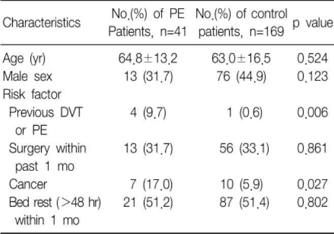
Table 1. General characteristics of 210 patients Characteristics No.(%) of PE
Patients, n=41
No.(%) of control patients, n=169 p value
Age (yr) 64.8±13.2 63.0±16.5 0.524
Male sex 13 (31.7) 76 (44.9) 0.123
Risk factor Previous DVT or PE
4 (9.7) 1 (0.6) 0.006
Surgery within past 1 mo
13 (31.7) 56 (33.1) 0.861
Cancer 7 (17.0) 10 (5.9) 0.027
Bed rest (>48 hr) within 1 mo
21 (51.2) 87 (51.4) 0.802
PE: pulmonary embolism; DVT: deep vein thrombosis.
에 따라 폐색전증의 가능성을 예측하는 임상 모형의 정확 도 또한 변할 것으로 예상할 수 있다.
폐색전증 위험인자로 알려진 혈전성향증(thrombophi- lia) 질환의 빈도가 서구에 비해 국내에서는 낮으며5, 국내 에서 폐색전증 발생빈도도 서구에 비해 낮은 것으로 보인 다6. 따라서 국내 폐색전증 환자에 대해 진단 예측 모형의 유용성에 대한 연구가 필요할 것으로 생각한다. 이에 본 연구는 폐색전증이 의심되어 MDCT를 촬영한 국내 환자 를 대상으로 Wells 및 개정된 Geneva예측 모형에 대한 유용성에 대하여 평가하고자 한다.
대상 및 방법
1. 연구 대상
2005년 1월부터 2007년 3월까지 계명의대 동산병원의 입원 환자 및 응급실 방문 환자 중에서 갑작스런 호흡곤란 이나 호흡곤란의 악화가 있었으며, 임상적으로 폐색전증 을 의심하여 CTPA (Computed Tomography Pulmonary Angiography)를 시행한 환자 210명을 대상으로 하였다.
2. 연구 방법
전공의들의 의무기록을 기초로 폐색전증의 위험인자, 임상양상을 조사하였고, 이를 바탕으로 Wells2 그리고 개 정된 Geneva7 예측 모형에 적용하여 폐색전증의 가능성 에 대해 저위험군, 중등도 위험군, 고위험군으로 분류하였 다.Wells 예측 모형에서 0∼1점을 저위험군, 2∼6점을 중 등도 위험군, 7점 이상을 고위험군으로 분류하였고 개정 된 Geneva 예측 모형에서 0∼3점을 저위험군, 4∼10점 을 중등도 위험군, 11점 이상을 고위험군으로 분류하였 다. Wells 예측 모형을 적용할 때에, 3점에 해당하는 '폐색 전증 외 다른 진단이 없음'의 항목에 대해서는 담당 전공 의의 의무기록에서 추정 주 진단명으로 폐색전증이 기재 된 경우 3점을 부여하였다.
CTPA는 Siemens 사의 16 또는 64개의 검출기(detector) 를 가진 스캐너를 이용하여 시행하였고, 판독은 폐동맥 중심부 혹은 주변부의 충만결손(filling defect)이 있을 때 폐색전증으로 진단하였다.
혈장 D-dimer는 latex agglutination turbidimetric im- munoassay method를 사용하여 정량 측정(STAR-Lia- testRD-Di, Diagnostica Stago, Asnieres, France)하였다.
3. 통계 처리
위험인자에 대한 비 연속 변수의 비교는 chi-square test 로, 연속 변수의 비교는 t-test를 이용하여 검증하였으며, 유의 수준 0.05 미만일 때 통계적으로 유의한 것으로 판정 하였다. 두 가지 폐색전증 예측 임상모형에 대한 진단 유 용성 비교는 receiver operating characteristic (ROC) 곡선 을 이용하여 분석하였고, 두 가지 임상모형에 대한 임상적 평가의 일치율은 k 통계로 평가하였다. 통계프로그램은 SPSS Inc.의 SPSS 13.0을 사용하였다.
결 과
1. 환자의 임상적 특성 및 위험인자
210명의 환자의 평균 연령은 63.3±15.9세, 남자가 90명 (42.9%), 여자가 120명(57.1%)이었다. 210명 중 폐색전증 으로 진단된 환자는 41명(19.5%) 이었다. 응급실에서 폐 색전증이 의심되어 CTPA를 시행한 89명 중 19명(21.3%) 이 폐색전증으로 진단되었고, 병실에서는 121명 중 22명 (18.1%)에서 폐색전증으로 진단되었다.
폐색전증으로 진단된 환자와 폐색전증이 없었던 환자 에 대하여 폐색전증의 위험인자로 알려진 요소들에 대하 여 비교한 결과, 폐색전증이 진단된 환자에서 대조군에 비해 심부정맥혈전증 병력의 빈도 및 악성종양의 빈도가 유의하게 높았다(Table 1). 임상적 특성 비교에서 수축기 혈압이 폐색전증 환자 군에서 대조군에 비해 유의하게 낮 았고, D-dimer는 폐색전증 환자군에서 대조군에 비해 유 의하게 높았으나, C반응성단백(CRP)과 섬유소원(fibrino-
Table 2. Clinical and laboratory findings of 210 patients Clinical findings PE patients
(n=41)
Control patients (n=169)
p value Respiratory
frequency (breath/min)
23.2±5.3 23.6±5.8 0.718
Heart
rate (breath/min)
94.4±16.9 91.7±19.2 0.418
Systolic blood pressure (mmHg)
117.1±23.7 128.3±27.4 0.017
Temperature (oC) 36.5±0.3 36.7±0.7 0.091 Leg pain
or edema
9 (21.9) 24 (14.2) 0.221
PaO2 (mmHg) 66.4±19.0 67.7±20.6 0.728
WBC (/μl) 10,881±5,081 11,814±6,376 0.321 Fibrinogen (sec) 301.5±101.7 370.8±128.3 0.008
CRP (mg/dl) 4.2±5.0 8.9±9.6 0.000
D-dimer (μg/ml) 10.3±11.8 5.6±9.3 0.029
Pro-BNP (pg/ml) 5,303.0±5,776.3 2,750.1±4,390.6 0.058 BMI (kg/m2) 24.2±3.9 22.8±3.8 0.058 Values are mean±SD.
PE: pulmonary embolism.
Table 3. Proportions of patients and frequency of pulmo- nary embolism in the two clinical probabilities
Clinical probability category
Wells score (n=210)
Revised Geneva score (n=210) No. (%)
Proprotion of patients in category
Low 2 (1.0) 44 (21)
Intermediate 137 (65.2) 160 (76.2)
High 71 (33.8) 6 (2.8)
Frequency of pulmonary embolism*
Low 2 (100) 2 (4.5)
Intermediate 25 (18.2) 36 (22.5)
High 14 (19.7) 3 (50)
*The denominators for the percentages can be found in the first tree rows of the table.
gen)은 대조군에 비해 유의하게 낮았다(Table 2).
2. 폐색전증의 예측도 평가 및 폐색전증의 발생빈도
임상적으로 폐색전증이 의심된 210명의 환자를 Wells 예측 모형과 개정된 Geneva 예측 모형을 이용하여 평가 하였다. Wells 예측 모형으로 평가하면, 2명(1%)이 저위 험군, 137명(65.2%)이 중등도 위험군, 71명(33.8%)이 고 위험군으로 분류되었다. 저위험군으로 분류된 2명(100%) 은 모두 폐색전증으로 진단되었고, 중등도 위험군으로 분 류된 환자들 중 25명(18.2%), 고위험군으로 분류된 환자 들 중 14명(19.7%)에서 폐색전증이 발생하였다. 개정된 Geneva 예측 모형으로 평가하면, 44명(21%)이 저위험군, 160명(76.2%)이 중등도 위험군, 6명(2.8%)이 고위험군으 로 분류되었다. 저 위험군으로 분류된 환자들 중 2명(4.5%), 중등도 위험군으로 분류된 환자들 중 36명(22.5%), 고 위험 군으로 분류된 환자들 중 3명(50%)에서 폐색전증이 발생하 였다(Table 3).3. 폐색전증 예측 모형의 민감도, 특이도, 및 유용성
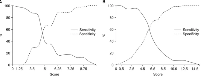
Wells 예측모형에서는 4.7점 정도에서 민감도와 특이 도가 약 50% 정도였고, 개정된 Geneva 예측모형에서는 5.2점 정도에서 민감도와 특이도가 약 60%로 관찰되었다(Figure 1).
두 가지 진단 예측 임상모형에 대한 유용성 비교를 위 해 ROC (Receiver Operating Characteristic) 곡선을 사용 하였고, ROC 곡선 하부 영역(area under the ROC curve) 은 Wells 모형의 경우 0.56에 비해, 개정된 Geneva 모형 에서 0.64로 높게 관찰되었다(Figure 2).
4. 폐색전증 진단 모형의 일치도
Wells와 개정된 Geneva 모형을 이용한 위험도 분류에 서 110명(52.4%)의 위험도가 서로 다르게 분류되었고, 일 치도는 불량하였다(Table 4,
k
=0.06).고 찰
본 연구는 국내에서 폐색전증이 의심되는 입원 및 응급 실 환자에 대해 Wells 그리고 개정된 Geneva 예측 모형을 후향적으로 적용하여 이들 임상모형의 유용성에 대하여 분석하였다. 폐색전증이 의심되어 MDCT를 시행한 210명 중에서 폐색전증으로 진단된 환자는 41(19.5%)명이었다.
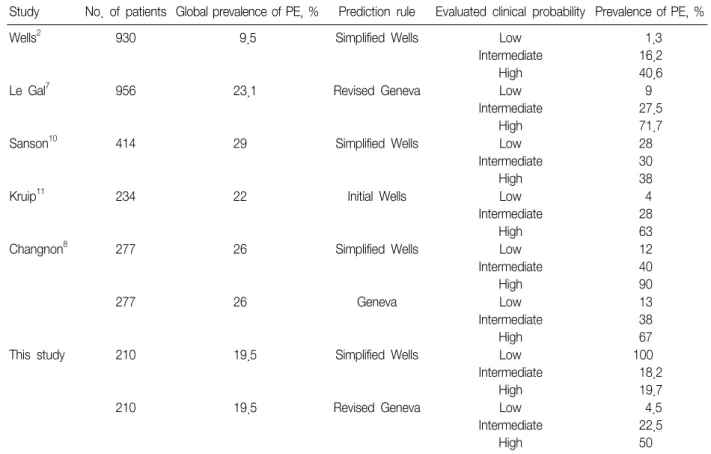
폐색전증이 의심된 환자에서 폐색전증 진단율이 외래환 자를 포함하지 않은 다른 연구에 비해 본 연구에서 낮았다 (Table 5). 이는 환자를 직접 대면하는 의사들의 폐색전증 에 대한 경험과 인지도가 부족해서 나온 결과로 추정된다.
본 연구에서는 Wells 예측 모형을 적용할 경우 210명 중에서 2명이 저위험군으로 분류되었고 고위험군이 71명
Figure 1. Comparison of the predictive accuracy of the two methods for pulmonary embolism. (A) Specificity and sensi- tivity of Wells score, (B) Sensitivity and specificity of revised Geneva score.
Figure 2. ROC curve (Receiver Operating Characteristic curve) of Wells scoreand revised Geneva score. Area un- der the ROC curve=0.56 (95% CI: 0.46 to 0.66) for Wells score, area under the ROC curve=0.64 (95% CI: 0.55 to 0.73) for revised Geneva score.
Table 4. Concordance of clinical probability category as- signment by the Geneva and the Wells scores
Wells score clinical probability
Revised Geneva score clinical probability
Low Intermediate High
No. of patients
Low 0 2 0
Intermediate 43 94 0
High 1 64 6
κcoefficient=0.06.
(33.8%)으로 대부분의 환자가 중등도 위험에 속하고 저위 험군이 거의 없고 고위험군이 다른 연구에 비해 지나치게 많았다8. 본 연구에서 개정된 Geneva 예측 모형으로 대상 환자를 분류한 결과 저위험군이 44명(21%), 고위험군이 6명(2.8%)으로 분류가 되었다. 이전 연구에서 Wells 예측 모형과 Geneva 예측 모형이 높은 일치율을 보인 점을8 고려한다면, 본 연구에서는 임상적으로 폐색전증을 과다 하게 의심하여 Wells 예측모형에서 중등도 및 고위험군이 증가한 것으로 추측되며, 이로 인해 두 예측 모형 사이의 일치율은 불량했던 것으로 보인다(Table 4, κ coefficient=
0.06). 또한 Receiver operating characteristic (ROC) 곡선 분석에서 개정된 Geneva 예측 모형에서 ROC 곡선 아래 의 면적이 0.64로 Wells 예측 모형의 0.56에 비해 더 커서 이는 개정된 Geneva 예측 모형의 정확도가 높음을 의미 한다(Figure 2).
Wells 예측 모형에서는 의사의 주관적인 판단인 '폐색전 증외 다른 진단이 없음'의 항목이 3점을 차지하여 이 3점 만으로도 중등도 환자군 이상으로 분류가 되어 실제 임상 경험이 적은 수련의가 사용하기에는 문제가 제기된바 있 다9. 본 연구에서도 의무기록에서 추정 주 진단으로 폐색 전증으로 기재된 경우에 3점을 부여하였지만 진료 의사의 숙련도에 따라 오류가 발생한 것으로 보인다.
Geneva 예측 모형은 Wells 예측 모형과는 다르게 주관 적인 항목을 배제하고 객관적인 항목만으로 구성되어 있 다3. 객관적인 항목 중에서 동맥혈 검사가 항상 이용 가능 하지 않다는 문제점을 보완하여 개정된 Geneva 예측 모
Table 5. Accuracy of clinical prediction rules for pulmonary embolism in previous study and this study
Study No. of patients Global prevalence of PE, % Prediction rule Evaluated clinical probability Prevalence of PE, %
Wells2 930 9.5 Simplified Wells Low
Intermediate High
1.3 16.2 40.6
Le Gal7 956 23.1 Revised Geneva Low
Intermediate High
9 27.5 71.7
Sanson10 414 29 Simplified Wells Low
Intermediate High
28 30 38
Kruip11 234 22 Initial Wells Low
Intermediate High
4 28 63
Changnon8 277 26 Simplified Wells Low
Intermediate High
12 40 90
277 26 Geneva Low
Intermediate High
13 38 67
This study 210 19.5 Simplified Wells Low
Intermediate High
100 18.2 19.7
210 19.5 Revised Geneva Low
Intermediate High
4.5 22.5 50 PE: pulmonary embolism.
형7이 발표되었다. 본 연구에서도 모든 환자에서 동맥혈 검사가 이루어 지지 않았으며, 이미 산소 치료를 시작한 후 동맥혈 검사가 시행된 경우도 포함되어 개정된 Geneva 예측 모형을 사용하게 되었다.
본 연구가 후향적으로 연구가 이루어진 점, 단일 의료 기관에 내원한 환자를 대상으로 한 점, 그리고 적은 대상 환자 수 등으로 인하여 오류가 발생했을 수 있다.
본 연구의 결과로 미루어 보면, 임상적으로 폐색전증의 발병이 의심될 경우, 우선 객관적인 항목으로 평가하는 개정된 Geneva 예측 모형의 적용을 하는 것이 진단의 정 확도를 높일 것으로 기대되며, 특히 의사의 임상 경험이 적을 경우에 Geneva 예측 모형이 폐색전증 진단에 많은 도움을 줄 것으로 생각된다. 나아가 많은 환자를 대상으 로 전향적인 다 기관 연구를 통해 국내 폐색전증의 위험인 자에 대한 분석 및 국내에서 유용하게 사용할 수 있는 새 로운 폐색전증 진단 예측 임상 모형의 개발이 필요할 것이 다.
요 약
연구배경: 급성 폐색전증의 발생을 예측하는 Wells 및 Geneva 예측 모형은 서구에서 잘 확립되어 있다. 폐색전 증의 역학이 서구와 다를 것으로 보이는 국내에서의 예측 모형의 유용성에 대해서 평가 하고자 한다.
방 법: 단일 의료기관에서 폐색전증 의심 하에 multi- detector computed tomography (MDCT)를 시행한 환자 210명을 대상으로 후향적으로 조사하였다. 성별 구성은 남자 90명(42.9%), 여자 120명(57.1%)이었고, 평균 연령 은 63.3±15.9세였다. 의무기록을 바탕으로 Wells 및 개정 된 Geneva 예측 모형으로 폐색전증의 가능성에 대해 저 위험군, 중등도 위험군, 고위험군으로 분류하였다.
결 과: 폐색전증으로 진단된 환자는 210명 중 41명 (19.5%)이었다. Wells 예측 모형을 적용한 폐색전증 발병 가능성 평가에서는, 2명(1%)이 저위험군, 137명(62.5%) 이 중등도 위험군, 71명(33.8%)이 고위험군으로 분류되었 고, 각 군에서 폐색전증의 발생률은 100%, 18.2%, 19.7%
였다. 개정된 Geneva 예측 모형을 적용할 경우 44명(21%) 이 저위험군, 160명(76.2%)이 중등도 위험군, 6명(2.8%)이 고위험군으로 분류되었고, 각 군에서 폐색전증의 발생률은 4.5%, 22.5%, 50%로 나타났다. Receiver operating charac- teristic (ROC) 곡선 분석에서 개정된 Geneva 예측 모형이 Wells 예측 모형에 비해 정확도가 높았다. 두 예측 모형 사 이의 일치율은 불량했다(κ coefficient=0.06).
결 론: 본 연구에서는 폐색전증이 의심되는 환자에서 개정된 Geneva 예측모형과 Wells 예측 모형으로 평가하 여 두 모형 사이에 일치율이 불량하였으며, 개정된 Geneva 모형이 Wells 모형에 비해 폐색전증 진단 예측이 더 정확 하였다.
참 고 문 헌
1. Anderson FA Jr, Wheeler HB, Goldberg RJ, Hosmer DW, Patwardhan NA, Jovanovic B, et al. A pop- ulation-based perspective of the hospital incidence and case-fatality rates of deep vein thrombosis and pulmo- nary embolism. The Worcester DVT Study. Arch Intern Med 1991;151:933-8.
2. Wells PS, Anderson DR, Rodger M, Ginsberg JS, Kearon C, Gent M, et al. Derivation of a simple clinical model to categorize patients probability of pulmonary embo- lism: increasing the models utility with the SimpliRED D-dimer. Thromb Haemost 2000;83:416-20.
3. Wicki J, Perneger TV, Junod AF, Bounameaux H, Perrier A. Assessing clinical probability of pulmonary embolism in the emergency ward: a simple score. Arch Intern Med 2001;161:92-7.
4. Stein PD, Fowler SE, Goodman LR, Gottschalk A, Hales CA, Hull RD, et al. Multidetector computed tomog-
raphy for acute pulmonary embolism. N Engl J Med 2006;354:2317-27.
5. Kim TW, Kim WK, Lee JH, Kim SB, Kim SW, Suh C, et al. Low prevalence of activated protein C resistance and coagulation factor V Arg506 to Gln mutation among Korean patients with deep vein thrombosis. J Korean Med Sci 1998;13:587-90.
6. Choi WI, Park JS, Min BR, Park JH, Chae JN, Jeon YJ, et al. Estimated incidence of acute pulmonary embo- lism in a university teaching hospital. Tuberc Respir Dis 2007;63(suppl 2):68.
7. Le Gal G, Righini M, Roy PM, Sanchez O, Aujesky D, Bounameaux H, et al. Prediction of pulmonary embo- lism in the emergency department: the revised Geneva score. Ann Intern Med 2006;144:165-71.
8. Chagnon I, Bounameaux H, Aujesky D, Roy PM, Gourdier AL, Cornuz J, et al. Comparison of two clin- ical prediction rules and implicit assessment among pa- tients with suspected pulmonary embolism. Am J Med 2002;113:269-75.
9. Moores LK, Collen JF, Woods KM, Shorr AF. Practical utility of clinical prediction rules for suspected acute pulmonary embolism in a large academic institution.
Thromb Res 2004;113:1-6.
10. Sanson BJ, Lijmer JG, Mac Gillavry MR, Turkstra F, Prins MH, Buller HR. Comparison of a clinical proba- bility estimate and two clinical models in patients with suspected pulmonary embolism. ANTELOPE-Study Group. Thromb Haemost 2000;83:199-203.
11. Kruip MJ, Slob MJ, Schijen JH, van der Heul C, Buller HR. Use of a clinical decision rule in combination with D-dimer concentration in diagnostic workup of patients with suspected pulmonary embolism: a prospective management study. Arch Intern Med 2002;162:1631-5.