Introduction
Intramuscular injection, which is widely performed for acute treatment in an emergency department, may induce some complications such as injection pain, hematoma and infection around an injection area. Nicolau syndrome was reported as one of the rare complications following injection, which had some characteristic clinical manifestations including
severe pain immediately after injection, cutaneous lesions, and variable degrees of tissue necrosis. With its rarity, correct diagnosis may be delayed, and fur- thermore tissue necrosis may be worsened by inap- propriate treatment. We report a 34 year-old female who developed Nicolau syndrome requiring a surgi- cal reconstruction following intramuscular diclofenac injection.
Case report
A 34-year-old woman revisited our emergency department due to pain and rash on her right but- tock. Her symptoms developed about 2 hours after intramuscular injection of 90 mg diclofenac (beta dimethyl amino-ethanol) at her right upper outer but- tock for headache. She experienced severe dull pain immediately after the injection. She had neither spe- cific past illness nor previous medication issues. Her vital signs were within normal limits. Physical exami-
응급실에서 디클로페낙 근주 후 발생한 니콜라우 증후군 1례
관동대학교 의과대학 응급의학교실
정상원∙강지훈∙여준모∙고재욱
Nicolau Syndrome following Diclofenac Injection in an Emergency Department
Sangwon Chung, M.D., Jihoon Kang, M.D., Junmo Yeo, M.D., Jaiwoog Ko, M.D.
Department of Emergency Medicine, Kwandong University College of Medicine
Nicolau syndrome is a rare adverse reaction at the site of an intramuscular injection, and is characterized by severe pain immediately after the injection and rapid development of distinct skin lesions. As this syndrome is rare, it may be overlooked at the early clinical phase and subsequently, clinical outcomes may be worse due to delay in treat- ment. We report on a female who developed Nicolau syndrome following intramuscular diclofenac injection, which required surgical reconstruction. Understanding the characteristics of Nicolau syndrome and careful surveillance for relevant clinical features may help physicians to more quickly diagnose and treat this condition.
Key Words: Diclofenac, Injections, Adverse effect, Wounds, Soft tissue injury 증
증 례례
투고일: 2011년 6월 20일 게재승인일: 2011년 8월 18일 책임저자: 고 재 욱
경기도 고양시 덕양구 화정동 697-24 관동대학교 의과대학 명지병원 응급의학과 Tel: 031) 810-5534, Fax: 031) 810-7013 E-mail: jupitor@kd.ac.kr
* 본 증례는 어떠한 이해관계 없이 작성되었으며, 지상 발표에 따른 환자 동의를 구하였습니다.
* 본 증례는 2011년 제 6회 지중해 응급의학회 (MEMC VI)에서 지상 발표 되었음.
J KOREANSOCCLINTOXICOL/ 101 Journal of The Korean Society of Clinical Toxicology 대한임상독성학회지 2011:9(2):101~104
Volume 9, Number 2, December, 2011
nation revealed an abnormal finding in the right but- tock area with erythematous reticular rash, swelling (measuring 20×15 cm), and tenderness (Fig. 1A).
Blood tests revealed leukocytosis (12600/uL, 70.5%
segmented neutrophil), increased creatine kinase (969, reference range: 51-188 IU/L), serum glutamic oxalacetic transaminase (31, reference range: 8-28 IU/L) and D-dimer (698.56, reference range: <500 ng/mL). Erythrocyte sedimentation rate, C-reactive protein, prothrombin time and activated partial thromboplastin time were within normal limits. There
was no specific finding in a chest x-ray and electro- cardiography. Fibrous tissues and coagulated ery- throcytes were reported after percutaneous needle aspiration biopsy at the lesion area. Sonographic examination revealed diffuse thickening of skin and a subcutaneous fat layer with heterogeneous echogenicity (Fig. 2). Extensive edema involving gluteal and piriformis muscle was detected in mag- netic resonance image (Fig. 3). Necrotizing fasciitis or myositis following injection was assumed as an initial diagnosis. She was admitted and received conserva-
102 / J KOREANSOCCLINTOXICOL
대한임상독성학회지 제 9 권 제 2 호 2011
Fig. 2. Ultrasonographic study at both buttocks. (A) Left buttock. (B) Right buttock (injection area). Comparing to left buttock (A), the image (B) of right buttock shows diffuse thickening of subcutaneous fat layer with heterogeneous echogenicity.
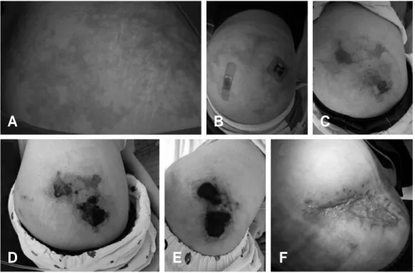
Fig. 1. Change of skin lesions in Nicolau syndrome following diclofenac injection. Livedoid reticular rash appeared 2 hours after injec- tion (A). The amount of rash was diminished the next day (B). Vesicles (C) were found at the fourth day after injection. Skin necrosis appeared at the eighth day (D) and progressed until the twenty-second day after injection (E). Wound healed with atroph- ic scar after surgical debridement and reconstruction (F).
A
A B
D E F
B C
tive managements including ice bag application, dressing with a topical antibiotic agent, pain control, and intravenous antibiotics. Her vital signs were sta- ble, but the buttock pain was not controlled well despite the use of non-steroidal anti-inflammatory drugs and even fentanyl. No pathogens were isolated in blood, urine or the lesion area. The rash seemed to be lessened the next day (Fig. 1B), but vesicles appeared on the fourth day after injection (Fig. 1C).
Necrotic skin appeared on the eighth (Fig. 1D) and progressed until the twenty-second day after the injection (Fig. 1E). On the twenty-fourth day, surgical debridement of necrotic tissue and reconstruction was performed by plastic surgeons. Two and a half months later, the lesion completely healed, leaving an atrophic scar (Fig. 1F).
Discussion
Nicolau syndrome is defined as a local aseptic tis- sue necrosis, and characterized by intractable pain, distinct skin lesions, and various degrees of tissue destruction at the injection site
1). Various drugs were reported to induce Nicolau syndrome: benzathine penicillin and other penicillins, non-steroidal anti- inflammatory drugs, local anesthetics, antihistamines, corticosteroids, and the DTP vaccine
1-4). Although the pathogenesis is not clear, the vascular hypothesis is thought to be most reasonable. Pain and skin lesions
may result from either embolism induced by oleous drug preparation, sympathetic nerve stimulation, aseptic inflammation, and direct damage of vessel after injection (embolia cutis medicamentosa)
1,2). Dull pain occurs immediately after an injection and may persist for weeks. Its severity can be much greater than that of other typical injections. Livedoid and reticular rashes occur usually several hours to days after an injection. The rashes have a distinct border with a high possibility of skin blanching (livedoid dermatitis). Vesicles and necrotic skin change may follow the rashes (Fig. 1). There are some recom- mendations for prevention and treatment. The upper outer quadrant of the buttock has fewer blood ves- sels and thus is recommended for site of injection, and Lesser’s sign (to inject the drug only after having aspirated with the syringe) should be checked before an injection
5). Pain control and wound care are the mainstay of treatment. Conservative treatment with analgesics and dressings is generally recommended.
Antibiotics should be used in the event of secondary infection. Tissue damage seems to be irreversible, and the effect of vasoactive treatments such as vasodilation, anti-inflammation and anti-coagulation has not been validated
1,5). Surgical debridement and reconstruction may be required in case of necrotic progression and wound infection. The outcome is variable, as the wound may be healed with no scar to atrophic change.
정상원 외: 응급실에서 디클로페낙 근주 후 발생한 니콜라우 증후군 1례
J KOREANSOCCLINTOXICOL/ 103 Fig. 3. Magnetic resonance image.
T1 (A)/T2-weighted image (B) 28 hours after injection shows severe edema of the muscular and fat layer with some fluid. MRI study help to determine the extent of injury in Nicolau syndrome.
A B
In the early clinical phase, it may be difficult for physicians to differentiate between Nicolau syn- drome and necrotizing fasciitis. Although fever, hypotension, and altered mentality are known as classic signs in necrotizing fasciitis in spite of its low incidence, local erythema and swelling with pain are most common signs in necrotizing fasciitis
6). Injection history is characteristic in Nicolau syndrome, but necrotizing fasciitis has been reported following injection of non-steroidal anti-inflammatory drugs, which impair granulocyte chemotaxis and phagocy- tosis
7). There are some different strategies for treat- ment. Early surgical debridement and an adequate use of antibiotics are known as the corner stone of therapy for necrotizing fasciitis. Cold application is generally used to suppress pain and inflammation, but it may aggravate tissue necrosis with vasospasm in Nicolau syndrome
8). Differentiation is essential for proper management and better outcome, but may require time.
Unfortunately, radiologic and laboratory studies are not powered nor validated for differentiation between Nicolau syndrome and necrotizing fasciitis
6). Culture study may not be helpful at the early clinical phase. Unusual severe dull pain and characteristic rash following injection can be a signs of Nicolau syndrome, but careful vigilance of clinical signs should be required in the event of necrotizing fasci- itis. The use of antibiotics may be reasonable until ruling out necrotizing fasciitis or other infectious
causes at the early clinical phase.
Nicolau syndrome is rare and not well understood.
Recognizing the characteristic signs and careful sur- veillance may allow emergency physicians to make the correct diagnosis and provide proper treatment.
REFERENCES
01. Luton K, Garcia C, Poletti E, Koester G. Nicolau Syndrome: three cases and review. Int J Dermatol 2006;45 :1326-8.
02. Faucher L, Marcoux D. What syndrome is this? Nicolau syndrome. Pediatr Dermatol 1995;12:187-90.
03. Cherasse A, Kahn MF, Mistrih R, Maillard H, Strauss J, Tavernier C. Nicolau’s syndrome after local glucocor- ticoid injection. Joint Bone Spine 2003;70:390-2.
04. Ezzedine K, Vadoud-Seyedi J, Heenen M. Nicolau syn- drome following diclofenac administration. Br J Dermatol 2004;150:385-7.
05. Corazza M, Capozzi O, Virgilit A. Five cases of livedo- like dermatitis (Nicolau’s syndrome) due to bismuth salts and various other non-steroidal anti-inflammatory drugs. J Eur Acad Dermatol Venereol 2001;15:585-8.
06. Sarani B, Strong M, Pascual J, Schwab CW. Necrotizing fasciitis: current concepts and review of the literature. J Am Coll Surg 2009;208:279-88.
07. Souyri C, Olivier P, Grolleau S, Lapeyre-Mestre M. Severe necrotizing soft-tissue infections and nonsteroidal anti- inflammatory drugs. Clin Exp Dermatol 2008;33:249-55.
08. Senel E, Ada S, Gulec AT, Caglar B. Nicolau syndrome aggravated by cold application after i.m. diclofenac. J Dermatol 2008;35:18-20.
104 / J KOREANSOCCLINTOXICOL
대한임상독성학회지 제 9 권 제 2 호 2011