궤양의 방사선 근접치료 시, 인접한 정상조직 보호 및 재현성 향상과 궤양 체적내의 선량 균등성을 개선하기 위하여 mold을 자체 제작 한 후 유용성을 평가하였다.
대상 및 방법: Mold는 상악용, 하악용으로 분리하여 제작하였고, 치료 시 인접한 잇몸과 치아를 보호하기 위하여 2 mm의 납 을 mold에 삽입하였다. Simulation을 통하여 얻어진 orthogonal image를 바탕으로 치료계획을 하였고, 치료는 하루 200 cGy씩 총 3,000 cGy의 선량을 조사하였다. 차폐의 유용성을 평가하기 위하여 조직등가물질(bolus)을 이용하여 혀와 잇몸 phantom을 만들고, 납으로 차폐한 경우와 납을 제거 후 차폐하지 않은 경우 TLD를 이용하여 잇몸의 5개 지점 선량을 각각 3회씩 반복 측정하였다.
결 과: Mold의 유용성 평가를 위한 선량측정 결과 평균값은 차폐하지 않은 경우 A: 33.9 cGy, B: 30.1 cGy, C: 31.8 cGy, D:
23.3 cGy, E: 24.1 cGy로 나타났고, 납으로 차폐한 경우의 선량 평균값은 A: 20.6 cGy, B: 18.8 cGy, C: 19.6 cGy, D: 14.7 cGy, E: 15.1 cGy로 측정되었다.
결 론: 구강 내 국소적으로 발생한 과다혈관성 궤양에 mold brachytherapy를 시행하여 치료 시 높은 정확성과 재현성을 유지 할 수 있었고, 궤양 체적내의 선량 분포도의 균등성을 효율적으로 개선할 수 있었다. 또한 인접한 잇몸과 치아의 선량을 약 7.35∼10.3%까지 감소시킴으로서 방사선 치료로 인하여 수반되는 부작용 발생 확률을 현저하게 줄일 수 있었다.
핵심용어: paraneoplastic autoimmune multiorgan syndrome, mold brachytherapy, 잇몸과 치아의 선량
이 논문은 2008년 12월 13일 접수하여 2009년 2월 2일 채택되었음.
책임저자:차석용, 전북대학교병원 방사선종양학과 Tel: 063)250-1187, Fax: 063)250-1192 E-mail: [email protected]
서 론
최근 두경부암에서의 방사선치료는 선형가속기를 이용한 외부조사방사선치료(external beam radiation therapy, EBRT) 인 3차원 입체조형치료(3-D conformal radiation therapy) 또 는 세기조절방사선치료(intensity modulated radiation therapy, IMRT),1) 영상유도방사선치료(image guided radiation ther- apy, IGRT), 입체세기조절회전방사선치료(volumetric modu- lated arc therapy, VMAT) 등의 최신 기법의 방사선치료가 시행되어지고 있다.2,3) 그러나 외부조사방사선치료의 경우 주위 정상조직이 한계 선량 이상으로 방사선이 조사되어 구 강 건조증 및 치아의 부식, 주변 뼈의 방사선 괴사 등의 부작 용이 보고 되고 있다.4,5) 이런 부작용을 피하기 위하여 국소
적인 병변인 경우 근접치료(brachytherapy)를 시행하여 근치 적 목적의 방사선 치료를 시행 할 수 있으며, 또한 근접 조사 는 외부조사방사선치료 이후 남아있는 종괴에 추가적으로 치료 효과를 증가시키기 위하여 외부 조사 치료와 병행하여 시행할 수 있다.4-7) 현재 근접치료는 두경부에서 입술, 편도, 인두, 목, 비인두, 부비동, 혀등의 경우 종양에 HDR (high dose tare) remote afterloading system을 이용하여 치료를 시 행하고 있으며, 종양에는 고 선량의 방사선을 국소적으로 집 중 조사할 수 있는 반면, 종양 주변에 있는 정상조직을 최대 한 보호함으로써 방사선치료로 인하여 수반되는 합병증을 최소한으로 줄일 수 있는 치료법으로, 전체 치료기간을 단축 할 수 있고, 원하는 부위에 균일한 선량을 줄 수 있는 치료법 중의 하나이다. 근접 치료의 여러 가지 방법 중에서 종양의 형태에 맞는 mold를 만들고 그 안에 치료용 카테터를 고정시 켜 치료하는 mold brachytherapy는 국소적으로 존재하는 표 재성 종양을 치료하는데 높은 재현성을 제공할 뿐만 아니라,
Fig. 1. Hypervascular ulcer lesion in oral cavity with paraneoplastic autoimmune multiorgan syndrome.
주변 장기 보호를 위하여 방사선치료 전에 종양 및 주변 장 기의 선량 분포에 대한 적절한 치료 선량을 계획 하는데 유 용한 치료방법이다.8-10) 이에 본원에서는 paraneoplastic auto- immune multiorgan syndrome으로 인하여 구강 내의 혀와 경 구개에 국소적으로 발생한 과다혈관성 궤양환자(Fig. 1)를 대상으로 mold brachytherapy를 시행하는 과정에서, 치아를 교정할 때 사용하는 acrylic resin을 이용하여 구강을 고정할 수 있는 mold를 자체 제작하여 주변 장기의 보호 및 종양의 선량 분포 유용성을 평가 하고자 한다.
대상 및 방법 1. Mold의 제작
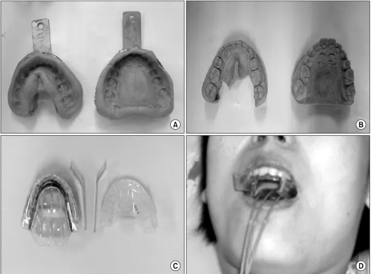
Mold의 제작은 치과에서 치아를 교정할 때 주로 사용하는 재료인 acrylic resin (Dentaurum, Germany)을 이용하여 본원 보철과에서 이루어졌다. Acrylic resin은 polymethylmetha- crylate: PMMA와 methylmethacrylate: MMA로 구성되어 있 고 PMMA는 powder, MMA는 liquid 형태로 되어있다. 먼저 인상획득(acquisition of impression)을 하게 되는데 비가역성 하이드로콜로이드(irreversible hydrocolloide) 인상재료인 알 지네이트(Alginate: Aroma Fine DF III Normal Set, GC, Japan)를 이용하여 구강 내 모양을 획득하였다. 국소적으로 존재하는 혀와 경구개의 과다혈관성 궤양을 포함할 수 있도 록 구강 전체를 고정하기 위하여 상악과 하악 각각의 모양을 획득하였다(Fig. 2A). 그 후 알지네이트 인상체로부터 경석 고(Silky Rock, Whip Mix, USA) 모형을 제작하였고(Fig.
2B), 경석고 모형에 acrylic resin의 powder와 liquid를 교차로 도포하여 상악용과 하악용 mold를 구분하여 제작하였다. 각
각 분리된 mold를 일정한 거리와 각도를 유지하기 위하여 좌, 우 대구치(molar) 부분에 홈을 내어 얇은 두 개의 고정 클립을 끼울 수 있게 하였고, 혀에 해당되는 부분에는 acrylic resin을 최대한 얇게 덮어서 mold 착용 시 혀의 움직임을 최 소화 하였다. 또한 치료용 카테터를 궤양에 최대한 근접시키 기 위하여 하악용에 3개의 6F catheter hole을 만들었다. 그리 고 치료 시 인접한 잇몸과 치아 및 주변 장기에 치료 중 부 작용 및 불편함을 주지 않는 범위 내에서 2 mm의 납을 삽입 하였다.
2. 치료과정
자체 제작한 mold를 이용하여 환자의 상악과 하악을 일정 한 간격과 각도로 고정시키고, 치료용 카테터인 Lumeancath (6F 150 cm Neucletron, Netherlands)에 dummy source를 삽 입한 후 conventional simulation을 통한 치료계획을 위하여 orthogonal image를 획득하였다(Fig. 3).
치료계획은 Plato planing system (BPS v14.2.6 Neucletron, Netherlands)으로 orthogonal film을 이용하였고, 선원으로부 터 0.7 cm 떨어진 지점에 선량표준화(normalization)를 하였 다. 그리고 동일한 지점에 200 cGy의 선량이 조사되도록 하 였다(Fig. 4). 환자의 치료는 Ir-192 HDR micro selectron af- terloading system을 이용하여 10회(20 Gy) 실시한 후, 종괴 의 감소에 따른 치료반응을 확인하고, 부분 반응을 보여, 5회 (10 Gy)를 추가로 조사하여 총 30 Gy를 조사하였다. 치료 시 인접한 잇몸과 치아의 보호를 위해 삽입한 납의 유용성을 평 가하기 위하여 조직등가물질(bolus)을 이용하여 혀와 잇몸 phantom을 만들고, 납으로 차폐한 경우와 납을 제거한 후 차 폐하지 않은 경우 열형광선량계(TLD)를 이용하여 중절치의
Fig. 3. Orthogonal image by conventional simulation with dummy source in Lumencath catheter. (A) Anterior - Posterior view. (B) Right - Left lateral view.
Fig. 2. Mold customizing process. (A) Acquisition of impression with alginate. (B) Yellow model stone. (C) Acrylic resin mold. (D) Mold adapted in patient's mouth.
Fig. 4. The geometrically optimized dose generated using Plato planning system. 200 cGy was given to the normalization point. (A) 3D view. (B) Sagittal view. (C) Axial view. (D) Coronal view.
TLD
No. Gum of teeth TLD dose (cGy)
TLD dose (%) A Gum of central
incisor
33.9 16.95
B Gum of left first premolar
30.1 15.05
C Gum of right first premolar
31.8 15.90
D Gum of left first molar
23.3 11.65
E Gum of right first molar
24.1 12.05
Table 1. Measurement without 2 mmPb shielding the front of gums measured with TLD
잇몸(gum of central incisor): A, 좌측 제1소구치의 잇몸(left gum of first premolar): B, 우측 제1소구치의 잇몸(right gum of first premolar): C, 좌측 제1대구치의 잇몸(left gum of first molar): D, 우측 제1대구치의 잇몸(right gum of first molar): E의 선량을 측정하였다. TLD의 측정오차를 감안하 여 통계상의 오차를 줄이기 위해 각각 3회씩 반복하여 측정 한 후 평균값을 얻어 표시하였고, 각 부위의 측정값을 200 cGy를 100%로 하여 상대적인 비율 값으로 나타내었다. 측정 에 사용한 TLD소자는 TLD-100 (7LiF;Mg,Ti)형 식의 칩이며, 크기는 0.31⨉0.31⨉0.09 cm3으로 장기간 사용 시 감도 (sensitivity)가 안정하고 재현성(변동계수 3% 미만)이 양호하 여 선량계로서 보편적으로 사용되고 있는 선량계를 이용하 였다.11) TLD의 판독은 Harshaw 3500 (Saint-Gobain Crystals
& Detectors, USA)을 이용하여 실시하였다.
TLD
No. Gum of teeth TLD dose (cGy)
TLD dose (%) A Gum of central
incisor
20.6 10.30
B Gum of left first premolar
18.8 9.40
C Gum of right first premolar
19.6 9.80
D Gum of left first molar
14.7 7.35
E Gum of right first molar
15.1 7.55
Table 2. Measurement with 2 mmPb shielding the front of gums measured with TLD
TLD
No. Gum of Teeth TLD dose (cGy)
TLD dose (%) A Gum of central
incisor
13.3 6.65
B Gum of left first premolar
11.3 5.65
C Gum of right first premolar
12.2 6.10
D Gum of left first molar
8.6 4.30
E Gum of right first molar
9 4.50
Table 3. Difference between those with 2 mmPb and those without 2 mmPb
15.90%, D: 11.65%, E: 12.05%이었고, 차폐를 했을 때에는 A:
10.30%, B: 9.40%, C: 9.80%, D: 7.35%, E: 7.55%이었다. 그리 고 납으로 차폐를 하지 않았을 때와 차폐를 했을 때의 선량
만 아니라 비 침습적인 치료법인 방사선치료는 암 치료의 중 요 방법 중 하나로서 다양한 선형가속기의 개발과 함께 치료 기술의 향상으로 역할은 더욱 더 확대될 것이다. 이와 함께 방사선방어 목표인 ALARA 개념이 확대되면서 주변 정상 장기의 방사선 조사선량을 줄이면서 종양을 효과적으로 제 어 할 수 있는 여러 외부조사 방사선 기법이 개발되고 있기 는 하나, 국소적인 병변의 경우 병변의 위치가 근접 조사를 시행하기에 적합하다면, 근접 방사선 치료를 고려할 수 있다.
이에 본원의 paraneoplastic autoimmune multiorgan syndrome 환자의 방사선 치료에서 구강 내 혀와 경구개의 국소적인 과 다혈관성 궤양에 mold를 자체 제작하여 근접 치료를 시행함 으로서 치료 시 높은 정확성과 재현성을 유지할 수 있었고, 궤양 체적내의 선량 분포도의 균등성 효율적으로 개선할 수 있었다. 또한 인접한 잇몸과 치아 부분 및 정상 장기의 방사 선량은 납으로 차폐를 하지 않은 경우에는 치료 선량의 약 11.65∼16.95%이었지만, 차폐를 통하여 치료 선량의 약 7.35
∼10.3%까지 감소시킴으로서 방사선 치료로 인한 부작용 발 생 확률을 효과적으로 줄일 수 있었다. 본원의 환자 또한 치 료 후 경도의 방사선 구내 점막염을 제외하고 다른 부작용은 관찰되지 않았으며 치료 후 종양 반응률 또한 90%의 감소율 이 관찰되었다. 따라서 추후 근접 방사선 치료를 시행 하는 환자의 정확성 및 재현성 향상과 정상조직의 효과적인 차폐 를 위한 다양한 노력을 통하여 효과적으로 방사선 근접 치료 를 여러 질환에 근치적 목적의 치료로 적용할 수 있을 것으 로 사료된다.
참고문헌
1. Hansen E, Xia P, Quivery J, et al.: The roles of repeat CT imaging and re-planning during the course of IMRT for patients with head and neck cancer. Paper presented at:
ASTRO's 46th Annual Meeting; October 3-7, 2004; Atlanta.
GA
2. Garsa AA, Andrade RS, Heron DE, et al.: Four-dimensional computed tomograhy-based respiratory-gated whole-abdominal intensity-modulated radiation therapy for ovarian cancer: a feasibility study. Int J Gynecol Cancer 2007;17:55-60 3. van der Geld YG, van Triest B, Verbakel WF, et al.:
Evaluation of four-dimensional computed tomography-based intensity- modulated and respiratory-gated radiotherapy techniques for pancreatic carcinoma. Int J Radiat Oncol Biol Phys 2008;72:1215-1220
4. Obinata K, Ohmori K, Shirato H, Nakamura M: Experience of high-dose-rate brachytherapy for head and neck cancer treated by a customized intraoral mold technique. Radiat Ned 2007;25:181-186
5. Cengiz M, Ozyar E, Ersu B, Akyol FH, Atahan IL: High dose rate mold brachytherapy of early gingival carcinoma: a clinical report. J Prosthet Dent 1999;82:512-514
6. Garran C, Montesdeoca N, Martinez MR: Treatment of upper
gum carcinoma with high-dose-rate customized-mold brachy- therapy. Brachytherapy 2008;7:267-269
7. Ariji E, Hayashi N, Kimura Y, Uchida T, Hayashi K, Nakamura T: Customized mold Brachytherapy for oral carci- nomas through use of high-dose-rate remote afterloading apparatus. Oral Surg Oral Med Pathol Oral Radiol Endod 1999;87:508-512
8. Minsley GE, Rothenberg S: Use of the afterloading technique for intraoral radiation carriers. J Prosthet Dent 1985;53:767- 768
9. Rustgi SN, Cumberlin RL: An afterloading 192-Ir surface mold. Med Dosim 1993;19:37-42
10. Takeda M, Shibuya H, Inoue T: The efficacy of gold-198 grain mold therapy for mucosal carcimomas of the oral cavity. Acta Oncol 1996;35:463-467
11. Kron T, Metcalfe P, Wong T: Thermoluminescence dosimetry of therapeutic x-rays with LiF ribbons and rods. Phys Med Biol 1993;38:833-845
Purpose: Evaluate the mold we have made to improve the reproducibility of the patient position and make homogeneous dose distribution to the treatment volume effectively when treating the patient who has hypervascular ulcer on her tongue caused by paraneoplastic autoimmune multiorgan syndrome by mold brachytherapy.
Materials and Methods: The mold is consisted of upper and lower parts. We inserted 2 mm of lead sheet on the gums toward the oral cavity to protect them from unnecessary irradiation during the treatment. We had planned on orthogonal images obtained the patient. 200 cGy was delivered in every fraction with a total dose of 3000 cGy. To evaluate the effect of the lead sheet, we made a measurement with a phantom that has gums and tongue made of tissue with an equivalent material (bolus). Five of TLDs were placed on the interesting points of gums to measure the dose during irradiation with lead sheet and without lead sheet for three times respectively.
Results: The result of the measurement without lead sheet are A: 33.9 cGy, B: 30.1 cGy, C: 31.8 cGy, D: 23.3 cGy, E: 24.1 cGy. The results of measurement with lead sheet are A: 20.6 cGy, B: 18.8 cGy, C: 19.6 cGy, D:
14.7 cGy, E: 15.1 cGy.
Conclusion: Since we are using the mold made in our department during the treatment of the patient with hypervascular ulcer on tongue, we could deliver a proper dose to the treatment volume. In addition, the mold provided highly accurate and reproducible treatment and reduced the dose to the gums and teeth. Therefore, the possibility of side effects could be decreased significantly.
Key words: paraneoplastic autoimmune multiorgan syndrome, mold brachytherapy, dose of gums and teeth