Actinomycosis is a chronic, suppurative, and granulomatous disease caused by an anaerobic Gram-positive bacterium, Actinomyces israelii, manifesting itself as fistula, sinus, inflammatory pseudotumor, or abscess formation.1Humans are natural reservoirs and there is no documented person-to-person transmission of the disease, and it is commonly cultured from carious teeth, tonsilar crypts.2It is characterized by a tendency to feign malignancy due to its capacity to invade surrounding tissues and to form masses.3Therefore, there are multiple clinical presentations, often leading to misdiagnosis. The three main clinical forms of this disease are cervicofacial, thoracic, and abdominopelvic. The cervicofacial region accounts for 50% to 65%, followed by abdomen (20%).4-6
The disease usually shows an indolent course with clinical symtoms and signs that are not specific, resulting in delayed diagnosis. Actinomyces are sensitive to penicillin, but the duration of treatment varies from several weeks to months to achieve permanent recovery.7-10The aim of this study was to evaluate the charac- teristic clinical features with short literature review on the topic.
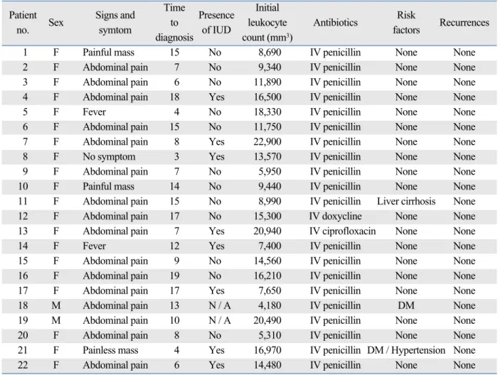
Between January 2000 and January 2006, 22 patients with abdominopelvic actinomycosis were identified. Patient’s demographic data and outcome are
Clinical Features of Abdominopelvic Actinomycosis:
Report of Twenty Cases and Literature Review
Myung-Min Choi,
1Jeong Heum Beak,
1Jung Nam Lee,
1Sanghui Park,
2and Won-Suk Lee
1Departments of 1 Surgery, 2 Pathology, Gil Medical Center, Gachon University of Medicine and Science, Seoul, Korea.
Purpose:Intrabdominal actinomycosis is difficult to diagnose preoperatively. This chronic infection has a propensity to mimic many other diseases and may present with a wide variety of symptoms. The aim of this study was to evaluate the characteristic clinical features with review of the literature. Materials and Methods: We retrospectively analyzed 22 patients with intrabdominal actinomycosis between January 2000 and January 2006.
Results:There were two men and 20 women with a mean age of 42.8 years (range, 24 - 69). Twelve patients presented with masses or abdominal pain, whereas 3 patients presented with acute appendicitis. The rate of performing an emergency surgery was 50% due to symptoms of peritonitis. The mean size of tumor was 5.5 cm (range, 2.5 - 11.0). Sixty percent (n = 12) of female patients had intrauterine device (IUD). The average time to definite diagnosis was 10.6 days. Conclusion:Intrabdominal abdominal actinomycosis must first be suspected in any women with a history of current or recent IUD use who presents abdominal pain. If recognized preoperatively, a limited surgical procedure, may spare the patient from an extensive operation.
Key Words : Actinomycosis, surgery, intrauterine device
Received: September 24, 2008 Revised: November 1, 2008 Accepted: November 10, 2008 Corresponding author: Dr. Won-Suk Lee, Department of Surgery, Gil Medical Center, Gachon University of Medicine and Science, 1198 Guwol-dong, Namdong-gu, Incheon 405-760, Korea.
Tel: 82-32-460-1234, Fax: 82-32-460-3009 E-mail: [email protected]
∙The authors have no financial conflicts of interest.
© Copyright:
Yonsei University College of Medicine 2009
INTRODUCTION
MATERIALS AND METHODS
summarized in Table 1. The clinical data including age, gender, mass size, preoperative diagnosis, presence and duration of intrauterine device (IUD) were retrospectively analyzed. Intrabdominal mass assessments consisted of physical examination, colonoscopy, ultrasonography, and abdominopelvic CT scan.
The clinical details of these patients are presented in Table 2. There were two men and twenty women with a mean age of 42.8 (range, 24 - 69) years. Twelve patients presented with masses or abdominal pain, whereas three patients presented with acute appendicitis (Table 3).
Among the twenty two patients, only two patients presented with a colonic mass mimicking colon cancer. Fifteen patients (68.2%) had leukocytosis with a mean WBC count of 12,765 mm3 (range; 4,180 - 22,900 mm3). None of the patients presented with small bowel or colon obstruc- tion. However, emergency surgery rate was 50% due to peritonitis symptoms. A preoperative abdominal CT scan
RESULTS
Table 1. Summary of 22 Patients with Intrabominal Actinomycosis
Patient Signs and Time
Presence Initial
Risk
no. Sex
symtom to
of IUD leukocyte Antibiotics
factors Recurrences diagnosis count (mm3)
1 F Painful mass 15 No 8,690 IV penicillin None None
2 F Abdominal pain 7 No 9,340 IV penicillin None None
3 F Abdominal pain 6 No 11,890 IV penicillin None None
4 F Abdominal pain 18 Yes 16,500 IV penicillin None None
5 F Fever 4 No 18,330 IV penicillin None None
6 F Abdominal pain 15 No 11,750 IV penicillin None None
7 F Abdominal pain 8 Yes 22,900 IV penicillin None None
8 F No symptom 3 Yes 13,570 IV penicillin None None
9 F Abdominal pain 7 No 5,950 IV penicillin None None
10 F Painful mass 14 No 9,440 IV penicillin None None
11 F Abdominal pain 15 No 8,990 IV penicillin Liver cirrhosis None
12 F Abdominal pain 17 No 15,300 IV doxycline None None
13 F Abdominal pain 7 Yes 20,940 IV ciprofloxacin None None
14 F Fever 12 Yes 7,400 IV penicillin None None
15 F Abdominal pain 9 No 14,560 IV penicillin None None
16 F Abdominal pain 19 No 16,210 IV penicillin None None
17 F Abdominal pain 17 Yes 7,650 IV penicillin None None
18 M Abdominal pain 13 N / A 4,180 IV penicillin DM None
19 M Abdominal pain 10 N / A 20,490 IV penicillin None None
20 F Abdominal pain 8 No 5,310 IV penicillin None None
21 F Painless mass 4 Yes 16,970 IV penicillin DM / Hypertension None
22 F Abdominal pain 6 Yes 14,480 IV penicillin None None
IUD, intrauterine device.
Table 2. Patients’ Characteristics
Intrabdominal actinomycosis (n = 22)
Age (yrs)
Mean (range) 42.8 (24 - 69)
Gender: M / F 2 : 20
IUD (n = 20)
Yes 12 (60%)
No 8 (40%)
Emergency vs. elective operation
Yes 11 (50%)
No 11 (50%)
WBC, mm3
Mean (range) 12,765 (4,180 - 22,900) GI obstruction
Yes 0 (0.0%)
No 100 (0.0%)
Mass size, cm
Mean (range) 5.5 (2.5 - 11.0) IUD, intrauterine device; WBC, white blood cell; GI, gastrointestinal.
or ultrasonography was done in all patients and detected intrabdominal mass or abscess but failed to give a definite diagnosis. The median operative time was 140 (range, 90 - 420) minutes and the median blood loss was 250 (range, 150 - 800) mL. The mean size of tumor was 5.5 (range, 2.5 - 11.0) cm. Sixty percent (n = 12) of female patients had IUD. The patients had been wearing IUD for an average of 7 years, and 15% had been wearing an IUD for 3 years or less. Confirmation of the diagnosis of actinomycosis was done by histology in all cases. Microscopically, each of the specimens showed chronic inflammatory reactions with sulfur granules (Figs. 1 and 2). None of the patients under- went percutaneous biopsy. There were no cancer cells found in all patients. The average time to definitive diagnosis was 10.6 days (range, 4 - 19 days).
After a median follow up of 37.5 months (range, 6.6 - 23.1 months), recurrence was not seen in any patients. The antibiotic of choice was IV penicillin, however, one patient was given ciprofloxacin due to penicillin allergy. The duration of treatment was 3 months in twelve patients, 6 months in five patients, 4 months in two patients and 1 month in two patients who refused to continue.
Actinomycosis was first diagnosed in a live patient by Ponfick in 1879.11It is a chronic suppurative disease caused by an anaerobic, filamentous Gram positive bacteria.12 Actinomycosis Israelii is a constant part of the microflora in the human oral cavity, gastrointestinal and genital tracks.12,13 The organism is unable to cross normal mucosal barrier, therefore, opportunistic infections can occur only in context of underlying local disease such as trauma, surgery, or a foreign body which is significant enough to penetrate this barrier. Once the organisms have penetrated the mucosa, spread by continuity seems to be the primary method of intrabdominal propagation. Lymphatic and hematogenous spread is uncommon,14,15although there is a hematogenous and lymphatic spread with nodal involvement reported.16
Clinically, the disease follows an indolent course and the initial presentation usually includes lower abdominal pain and fever with or without a palpable mass as was seen in our series. Since symptoms and signs are not specific, the diagnosis is often delayed and only 10% of cases are diag-
DISCUSSION
Fig. 1. 10.3 ×9.3 cm ovoid mass on the serosal surface of the cecum and ascending colon with ulceration. The cut surface demonstrates typical light gray color with necrosis.
Fig. 2. (A) A actinomycotic abscesses containing sulfur granules with radiating filaments (H & E, ×100). (B) A magnified view of the characteristic sulfur granule (H & E, ×200).
A
B
Table 3. Pre-Existing Diagnosis before Intrabdominal Acti- nomycosis
Pre-existing diagnosis No. of patients (n = 22), %
Diverticulitis 2 (9.0)
PID 2 (9.0)
Pelvic mass 5 (22.8)
Tubovarian abscess* 5 (22.8)
Appendicitis 3 (13.6)
Lymphoma 3 (13.6)
Endometriosis 1 (4.5)
Pelvic abscess 1 (4.5)
*One patient with combined sigmoid colon fistula. PID, pelvic inflammatory disease.
nosed preoperatively.16In our series, only one patient (4.5%) was diagnosed before surgery.
Intrabdominal actinomycosis can appear as an abdo- minal mass of ambiguous benignity and can mimic a malignant tumor. For example, actinomycosis of the colon or the greater omentum is a rare differential diagnosis of colonic carcinoma or peritoneal tumor.17-19The pathogenesis of abdominal actinomycosis is not yet well understood.
There are two possibilities suggested that can affect intrabdominal organs: through blood-borne infection or by swallowing.20 Actinomyces can normally inhabit colon, predominating in areas of stagnation i.e. the cecum and appendix. Actinomyces requires injury to the normal mucosa to penetrate and cause disease. Predisposing factors may include appendicitis and diverticulitis, gast- rointestinal perforations, previous surgery, foreign bodies, or neoplasia.21Thus, intrabdominal actinomycosis should be included in differential along with other inflammatory diseases such as ulcerative colitis, Crohn’s disease, tuber- culosis, diverticulitis and pelvic inflammatory disease.
Pelvic actinomycosis has recently become more preva- lent and is associated almost exclusively with women who use IUDs.16Orogentital tract is thought to be an important mode of acquiring this type of infection in lower genital tract.22In the uterine cavity, the microorganisms are appa- rently confined to the superficial layers of the mucosa, a fact perhaps related to its cyclic shedding. The most likely route of spread with subsequent development of pelvic abscess appears to be through patent fallopian tubes. The use of IUD may increase the risk of infection through injury to the normal uterine mucosa.13,23Pelvic actinomy- cosis associated with the use of IUDs can mimick pelvic malignancy.24For such reason, it is often surgically excised.
However, if a diagnosis can properly be made preopera- tively, antibiotic treatment and removal of IUD may lead to complete remission, avoiding unnecessary surgery. In our series, none of the patients with IUDs was diagnosed of actinomycosis preoperatively. Fiorino25reported 92 patients with actinomycotic abscess in 63 case reports. In this study, the average duration of IUD implantation was 8 years, and only 16% had been using an IUD for less than 3 years. Likewise, the average duration of IUD was 7 years and only 15% of those patients had IUD for less than 3 years in our study. Although IUD is strongly correlated with intrabdominal actinomycosis, a definite duration of IUD implantation and the risk of developing actinomy- cosis infection has not yet been established.
The modern principle of therapy for actinomycosis began with Peabody and Seabury in 1960,26who recom- mended abscess drainage in combination with high dose antibiotics. Smith et al.27reported that ciprofloxacin and tetracyclines showed poor performance in antimicrobial
susceptibility testing of actinomyces species. Actinomyces species appear to be susceptible to a wide range of beta- lactam agents and, when combined with beta-lactamase inhibitors, they should be regarded as agents of first choice.27
Uncomplicated actinomycosis can be medically treated by antibiotics, although there are differing opinions in the literature about dosage and duration of antibiotic treat- ment.28,29A prolonged treatment course is required because of the poor penetration of antibiotics into the fibrotic tissues.
Thus, when there are more avascular spaces present due to severe tissue reactions, medical therapy may be less effec- tive, resulting in longer duration of antibiotic treatment, regardless of the site of actinomycosis. Interestingly, two patients in our series (9.0%) who were treated with iv anti- biotics less than 4 weeks did not recur during the last 34 months of follow-up. Hence, the clinical impact of surgical resection followed by short-term antibiotics merits for further study.
Even though intrabdominal actinomycosis is very rare in its frequency, it should be included in a list of differential diagnosis, especially in any women with a history of IUD use who presents with abdominal pain or a pelvic mass. If actinomycosis is suspected preoperatively, appropriate handling of cultures will increase the diagnostic yield which may spare the patient from an extensive surgery.
1. Koren R, Dekel Y, Ramadan E, Veltman V, Dreznik Z.
Periappendiceal actinomycosis mimicking malignancy report of a case. Pathol Res Pract 2002;198:441-3.
2. Uchiyama N, Ishikawa T, Miyakawa K, Iinuma G, Nakajima H, Ushio K, et al. Abdominal actinomycosis: barium enema and computed tomography findings. J Gastroenterol 1997;32:89-94.
3. Milach J, Zió kowski P, Orze W. [A case of actinomycosis of the sigmoid in a 41-year-old woman with a clinical appearance of cancer.] Wiad Lek 1989;42:895-8.
4. Belmont MJ, Behar PM, Wax MK. Atypical presentations of actinomycosis. Head Neck 1999;21:264-8.
5. Berchtenbreiter C, Brüning R, Auernhammer A, Reiser M.
Misleading diagnosis of retroperitoneal actinomycosis. Eur Radiol 1999;9:1869-72.
6. Ferrari TC, Couto CA, Murta-Oliveira C, Conceiçäo SA, Silva RG. Actinomycosis of the colon: a rare form of presentation.
Scand J Gastroenterol 2000;35:108-9.
7. Anteby E, Milvidsky A, Goshen R, Ben-Chetrit A, Ron M. [IUD- associated abdominopelvic actinomycosis.] Harefuah 1991;121:
150-3.
8. Atad J, Hallak M, Sharon A, Kitzes R, Kelner Y, Abramovici H.
Pelvic actinomycosis. Is long-term antibiotic therapy necessary? J Reprod Med 1999;44:939-44.
9. Hinnie J, Jaques BC, Bell E, Hansell DT, Milroy R. Actinomy- cosis presenting as carcinoma. Postgrad Med J 1995;71:749-50.
l l
REFERENCES
10. Turnbull AE, Cohen ME. Case report: pelvic actinomycosis with the development and resolution of a recto-sigmoid stricture. Clin Radiol 1991;43:420-2.
11. Stringer MD, Cameron AE. Abdominal actinomycosis: a forgot- ten disease? Br J Hosp Med 1987;38:125-7.
12. Scribner DR Jr, Baldwin J, Johnson GA. Actinomycosis mimic- king a pelvic malignancy. A case report. J Reprod Med 2000;45:
515-8.
13. Cintron JR, Del Pino A, Duarte B, Wood D. Abdominal actino- mycosis. Dis Colon Rectum 1996;39:105-8.
14. Klaaborg KE, Kronborg O, Olsen H. Enterocutaneous fistulization due to Actinomyces odontolyticus. Report of a case. Dis Colon Rectum 1985;28:526-7.
15. Piper MH, Schaberg DR, Ross JM, Shartsis JM, Orzechowski RW. Endoscopic detection and therapy of colonic actinomycosis.
Am J Gastroenterol 1992;87:1040-2.
16. Harris LF, Kakani PR, Selah CE. Actinomycosis. Surgical aspects.
Am Surg 1985;51:262-4.
17. Baierlein SA, Wistop A, Looser C, Peters T, Riehle HM, von Flüe M, et al. Abdominal actinomycosis: a rare complication after laparoscopic gastric bypass. Obes Surg 2007;17:1123-6.
18. Huang CJ, Huang TJ, Hsieh JS. Pseudo-colonic carcinoma caused by abdominal actinomycosis: report of two cases. Int J Colorectal Dis 2004;19:283-6.
19. Rose G, Franke FE, Weimar B, Buhr J, Padberg W. [Actinomy- cosis of the colon as a rare differential diagnosis of colonic carci- noma.] Chirurg 2000;71:93-7.
20. Alvarado-Cerna R, Bracho-Riquelme R. Perianal actinomycosis-- a complication of a fistula-in-ano. Report of a case. Dis Colon Rectum 1994;37:378-80.
21. Fowler RC, Simpkins KC. Abdominal actinomycosis: a report of three cases. Clin Radiol 1983;34:301-7.
22. Gupta PK, Woodruff JD. Actinomyces in vaginal smears. JAMA 1982;247:1175-6.
23. Luff RD, Gupta PK, Spence MR, Frost JK. Pelvic actinomycosis and the intrauterine contraceptive device. A cyto-histomorpho- logic study. Am J Clin Pathol 1978;69:581-6.
24. Spagnuolo PJ, Fransioli M. Intrauterine device-associated actino- mycosis simulating pelvic malignancy. Am J Gastroenterol 1981;
75:144-7.
25. Fiorino AS. Intrauterine contraceptive device-associated actinomy- cotic abscess and Actinomyces detection on cervical smear.
Obstet Gynecol 1996;87:142-9.
26. Peabody JW Jr, Seabury JH. Actinomycosis and nocardiosis. A review of basic differences in therapy. Am J Med 1960;28:99-115.
27. Smith AJ, Hall V, Thakker B, Gemmell CG. Antimicrobial susceptibility testing of Actinomyces species with 12 antimic- robial agents. J Antimicrob Chemother 2005;56:407-9.
28. Kaya E, Yilmazlar T, Emiroglu Z, Zorluoglu A, Bayer A. Colonic actinomycosis: report of a case and review of the literature. Surg Today 1995;25:923-6.
29. Udagawa SM, Portin BA, Bernhoft WH. Actinomycosis of the colon and rectum: report of two cases. Dis Colon Rectum 1974;
17:687-95.