The frog appliance for upper molar distalization: a case report
11
0
0
전체 글
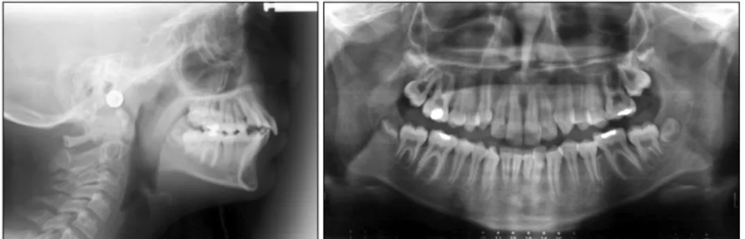
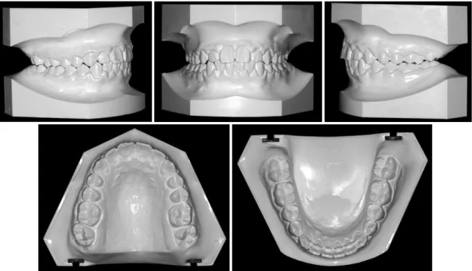
(2) Vol. 40, No. 1, 2010. Korean J Orthod. teeth. She had no significant medical and dental history in terms of orthodontic treatment. She was in the permanent dentition stage. Her gingival health was moderate and the radiographs did not reveal any periodontal problem or other pathology. Clinical examination revealed normal jaw function with no signs of temporomandibular joint dysfunction. Pretreatment facial photographs indicated that her face was symmetrical from the front and the profile was mildly convex (Fig 1). The maxillary and mandibular dental midline was coincident with the soft tissue facial midline. The dental casts and intraoral examination revealed that she had a bilateral Class II molar and canine relationship, mild upper and lower crowding, 4 mm overjet, 50 per cent overbite, and no Bolton dis-. The frog appliance for upper molar distalization: a case report. crepancy (Fig 2). There was no transverse discrepancy. The initial panoramic radiograph showed no missing teeth, and alveolar bone and root formation were within normal limits (Fig 3). Cephalometrically, the patient had an SNA angle of 76o, an SNB angle of 71o, and an ANB angle of 5o (Table 1). The upper incisors had o a 115 angle relative to the palatal plane and the lower incisors had a 110o angle relative to the mandibular plane. The patient did not want to wear an extraoral appliance, and she and her parents requested full retraction of the upper anterior teeth, without extractions.. Fig 1. Facial and intraoral photographs of the case before treatment (age 11 years).. 51.
(3) Mehmet Bayram, Metin Nur, Dogan Kilkis. 대치교정지 40권 1호, 2010년. Fig 2. Pretreatment dental casts of the case.. Fig 3. Pretreatment lateral cephalometric and panoramic radiographs of the case.. TREATMENT OBJECTIVES. TREATMENT ALTERNATIVES. The treatment objectives, based on the clinical examination and the cephalometric analysis, were to 1. Distalize the maxillary molars to establish a well-intercuspated bilateral Class I molar and canine relationship. 2. Retract the upper incisors for overjet reduction. 3. Ideally align the fully erupted lower and upper permanent teeth.. The patient’s chief concern was the protruding upper incisors, and her parents wanted complete retraction of the upper anterior teeth. There were six treatment alternatives for this case: (1) distalization of upper molars using an extraoral traction, (2) distalization of upper molars using an intraoral appliance, (3) extraction of two upper first premolars, (4) extraction of four first premolars, (5) extraction of maxillary first and mandibular second premolars, and (6) fixed orthodontic treatment with extraction followed by growth modification with a functional appliance.. 52.
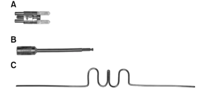
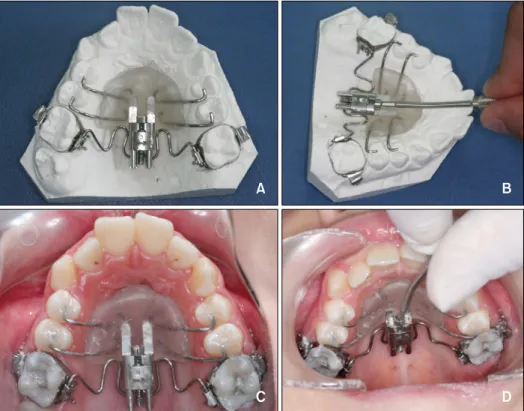
(4) Vol. 40, No. 1, 2010. Korean J Orthod. The frog appliance for upper molar distalization: a case report. Table 1. Cephalometric measurements of the patient Before. After. After. treatment distalization treatment o. 76. 76. 76. o. SNB ( ). 71. 71. 71. ANB (o). 5. 5. 5. 35. 36. 36. FMA ( ). 24. 27. 26. ANS-PNS/GoGn (o). 20. 22. 22. S-Go (mm). 76.5. 78. 81. 120. 123. SNA ( ). o. SN-GoGn ( ) o. N-Me (mm). 118. ANS-Me (mm). 63.5. 66. 70.5. S-Go/N-Me (%). 64. 65. 66. Wits appraisal (mm). 3. 2. 3.5. 1-NA (mm). 6. 8. 3. 25. 29. 22. 115. 119. 112. 9. 10. 9. o. 36. 35. 34. o. 110. 110. 109. 54. 56. 52. 24. 20. 23. 6-ANS/PNS ( ). 103. 106. 99. Interincisal angle (o). 113. 108. 117. o. 1-NA ( ) o. 1-ANS/PNS ( ) 1-NB (mm) 1-NB ( ) IMPA ( ) 1-PTV (mm) 6-PTV (mm) o. Overjet (mm). 3.5. 5. 2. Overbite (mm). 4.5. 3. 2. Lower lip-E line (mm). 2.5. 3. 0. Upper lip-E line (mm). 0.5. 1.5. 1. o. Nasolabial angle ( ). 118. 112. 115. Usage of an extraoral appliances and extraction therapy were rejected by the patient and her parents because they were against extraction of healthy teeth for orthodontic purposes and the patient was concerned about her facial appearance with an extraoral appliance. Thus, another alternative involving distalization of maxillary molars using an intraoral appliance and enmasse retraction of the anterior teeth was evaluated. This treatment plan was selected by the patient and her parents.. Fig 4. Parts of the Frog Appliance. A, Screw; B, screw driver; C, preformed spring.. Construction of the Frog Appliance A frog appliance kit consists of a screw, a preformed spring and a screw driver (Fig 4). Firstly, molar bands with lingual sheaths were fitted to the upper first molars for construction of the appliance. During the same visit, alginate impression was taken and molar bands were transferred to the impression. An accurate model cast of the maxillary arch was obtained. Anchor wires were bent from 0.028" stainless steel wire for the maxillary premolars. The anchor wires should lie in the embrasures distal to the anchor teeth. The frog screw is placed on the model with the distal of the appliance being aligned anteroposteriorly with the mesial of the lingual sheaths. According to the manufacturer, occluso-apically the frog screw should be placed approximately 10 mm to 12 mm from the occlusal surface. This will place the appliance at approximately the center of resistance of the molars for bodily tooth movement. Therefore, we placed the frog screw 10 mm from the occlusal surface of the upper first molars. An acrylic Nance button with anterior extensions was fabricated and the frog screw was embedded in it (Figs 5A and B). The body of the frog screw was removed from the button to allow easier polishing. The preformed 0.032 inch stainless steel spring was adjusted to customize the distalizing spring (frog-legs). The polished appliance was secured with an elastic roundel for delivery to the patient.. TREATMENT PROGRESS Treatment began with the cementation of the frog. 53.
(5) Mehmet Bayram, Metin Nur, Dogan Kilkis. 대치교정지 40권 1호, 2010년. Fig 5. Occlusal views of the Frog Appliance. A, During activation; B, on the dental cast and immediately after the cementation (C, D).. appliance into the oral cavity (Figs 5C and D). According to the manufacturer’s instructions, the following procedures were followed: (1) cemented the upper first molar bands using a multi-cure glass ionomer orthodontic band cement (3M Unitek, Monrovia, CA, USA); (2) thoroughly cleaned, etched, and rinsed the anchor teeth; (3) inserted the ends of the distalizing spring into the lingual sheaths of the molar bands and pressed the Nance Button against the palate; (4) painted unfilled composite resin on the etched surfaces of the anchor teeth; (5) placed filled composite resin over the anchor wires; (6) with finger pressure held the appliance firmly in place against the palate and light cured the composite resin; (7) removed the securing elastic and activated the appliance. According to the manufacturer, one complete rotation around the axis of the activation screw opens the appliance 0.4 mm. Three rotations are recommended for four to five-week intervals and five rotations are recommended for eight-week intervals. If second molars are erupted, three revolutions with five to six-week. 54. intervals are recommended. More than three rotations are not recommended when second molars are erupted. In our patient, the upper second molars were not erupted fully into the oral cavity so three rotations were performed to the appliance at four-week intervals. At each appointment, the stability of the appliance, progress of distalization, and oral hygiene were evaluated. The maxillary first molars were distalized until a super Class I molar relationship was achieved (Figs 6 and 7). This was completed after four months. Soon after the maxillary first molar distalization, the device was left in place as a retention appliance after cutting of the premolar anchor wires to increase maxillary molar anchorage for three months. In this way the premolars and the canines drifted distally by means of the pull of the transeptal fibers. Preadjusted fixed appliances (0.022 × 0.028-inch, MBT system) were placed in both arches for leveling and alignment. Maxillary premolars and canines were completely distalized by using sectional arches and power chains. After the Class I canine relationship was.
(6) Vol. 40, No. 1, 2010. Korean J Orthod. The frog appliance for upper molar distalization: a case report. Fig 6. Upper occlusal view of the patient immediately after the distalization (A), and intraoral photographs after cutting of the anchor wires of premolars (B-D) (after 4 months of distalization).. Fig 7. Lateral cephalometric and panoramic radiographs of the case taken immediately after the distalization.. obtained, 0.017 × 0.025 inch stainless steel retraction archwire formed individually with reverse closing loops were used in the maxillary arch to retract the anterior teeth. At the end of active treatment, finishing procedures were applied for final alignment of the teeth and detailing of the occlusion. The orthodontic appliances were removed after active treatment was completed. A maxillary removable Hawley retainer and a canine to canine mandibular fixed lingual retainer were constructed for the patient and delivered after debonding (Figs 8, 9 and 10).. RESULTS After 16 months of treatment with the Frog and pre-adjusted fixed appliances, a bilateral Class I molar and canine relationship with optimal alignment of both arches was obtained. Additionally, a favorable occlusal outcome with acceptable intercuspation was gained. Acceptable overjet and overbite were also achieved. After distalization, cephalometric analysis revealed that the maxillary first molars were moved 4 mm (aco cording to PTV) and tipped 3 (according to ANS/ PNS) distally. As for anchorage loss, the upper central. 55.
(7) Mehmet Bayram, Metin Nur, Dogan Kilkis. 대치교정지 40권 1호, 2010년. Fig 8. Facial and intraoral photographs of the case at the end of the fixed orthodontic treatment (age 12 years 4 months).. incisors exhibited a mesial movement of 2 mm, associated with a proclination of 4°. At the end of treatment, final cephalometric analysis and superimposition of pre- and post-treatment tracings showed that the skeletal Class II relationship had been maintained, and that she had an antero-inferior growth pattern (Fig 11). Cephalometrically, the dramatic changes were observed at overjet, overbite, and upper incisor’s position at the end of treatment. After the completion of active treatment, centric relation coincided with centric occlusion, and the patient reported no temporomandibular joint problems. The final panoramic radiograph showed good root parallel-. 56. ism, and the developing four third molars will be monitored.. DISCUSSION Several methods exist for the correction of Class II malocclusion, none of which work for all patients in all situations. The availability of several methods to correct different Class II malocclusions is valuable for orthodontists. Compliance-dependent appliances such as headgear2 or removable plate appliance16 were traditionally used for upper molar distalization in treatment of Class II malocclusions. For over a decade, various.
(8) Vol. 40, No. 1, 2010. Korean J Orthod. The frog appliance for upper molar distalization: a case report. Fig 9. Postreatment dental casts of the case.. Fig 10. Postreatment lateral cephalometric and panoramic radiographs of the case.. innovative noncompliance intraoral molar distalization appliances have been described. These appliances derive their anchorage in an intramaxillary manner and act only in the maxillary arch to move molars distally: 3-7 eg, the pendulum appliance, the sectional jig assem8-10 11-13 bly, the distal jet, the Keles slider,14 or the first class appliance.15 One of the important goals of molar distalizing therapy is to obtain bodily tooth movement of the molars with minimal rotation and distal inclination. For this purpose, the vector of effective distalizing force ideally should pass through the center of resistance of upper. molar or the heavy rods should be used for better con11-13 trol of the direction of the force. In the distal jet and the Keles slider,14 the force producers (closed coil springs) are placed at the level of center of resistance of upper first molar to obtain bodily distal movement. Similarly, the Frog appliance was positioned approximately 10 to 12 mm apically to the occlusal surface of the maxillary molar with parallel orientation to the occlusal plane in our case. In this manner, a vector of effective force passing through the centre of resistance of the first molar was obtained. The distalization force was produced by the activation of the screw. The pre-. 57.
(9) Mehmet Bayram, Metin Nur, Dogan Kilkis. 대치교정지 40권 1호, 2010년. Fig 11. Local and total superimpositions of the lateral cephalometric tracings before treatment (solid line), after distalization (dotted line) and after treatment (dashed line).. o. Fig 12. Schematic drawing of distalization effect on the maxillary dentition clearly shows an explicit distal molar movement with a slight anchorage loss on the premolars and the incisors.. formed spring was not activated before inserting the appliance as pendulum springs. In the current case, the correction of the Class II molar relationship was achieved by a 4 mm distal movement of maxillary first molar into a Class I relationship with a slight distal tipping of 3o after four months of distalization. There was also some anchorage loss as defined by maxillary incisor proclination (2 mm and. 58. 4 ), increase in overjet (1.5 mm), and a mesial movement of the anchoring premolars at the end of distalization (Fig 12). The distal jet and the pendulum are two of the more commonly used “noncompliance intraoral appliances” for upper molar distalization. Previous studies3-7,11-13 have indicated that the pendulum appliance produces on average greater molar distalization (3.14 - 6.1 mm) than the distal jet appliance (2.1 - 3.2 mm). The distal o o jet produces better bodily movement (1.8 - 5 of molar distal tipping) than the pendulum (8.4o - 15.7o) because the distalizing force is directed close to the level of the maxillary first molar’s center of resistance. The amounts of anchorage loss that can be expected as a result of the mesial reciprocal force on the premolars are similar for both appliances (1.8 - 2.5 mm for the pendulum; 1.3 - 2.6 mm for the distal jet). In the Jones jig studies,8-10 in addition to the distal tipping and movement of molars; there were also significant mesial movement and tipping of the anchoring premolar and increase in the overjet after distalization. 10 Brickman et al. found that an average distal movement of 2.51 mm and distal tipping of 7.53o in maxillary first molar and an average mesial movement of 2 o mm and mesial tipping of 4.76 in maxillary premolar at the end of distalization with the Jones jig appliance. Anchorage control is a vexing problem during molar.
(10) Vol. 40, No. 1, 2010. Korean J Orthod. distalizing therapy; not only is it required for efficient molar distalization, but there is also the necessity of holding the distalized molars while the anterior dentition is subsequently retracted. Recently, intraoral molar distalizing appliances have been combined with various implants to achieve osseous anchorage and overcome the limitations of tooth and/or palate-supported appliances. Satisfactory distalization results without anchorage loss have been achieved in these implant or minis1,17-21 crew supported molar distalization studies. We could implement the Frog appliance with an implant or a miniscrew to achieve osseous anchorage but we decided to apply it alone following the manufacturer’s recommendation. Thus we aimed to determine the effects of the appliance applied alone. In further studies, the appliance can be combined with an osseous anchorage unit to eliminate the side effects on anchoring teeth. Minor irritation of the palatal mucosa was determined after the removal of the appliance. This kind of soft tissue irritation was also reported with the use of a pendulum appliance and a Nance button. This situation can be prevented with maintenance of optimum oral hygiene.. The frog appliance for upper molar distalization: a case report. - 국문초록 -. Frog appliance를 이용한 상악 대구치의 원심 이동: 증례 보고 Mehmet Bayram,a Metin Nur,a Dogan Kilkisb. 본고의 목적은 2급 1류 치열안면 구조를 가지는 한 환자 에 적용된 새로이 고안된 상악 대구치 원심 이동 장치인 Frog appliance의 효과를 평가하기 위함이다. 11세의 여자 환자가 교정 치료를 위해 본 진료실로 의뢰되었다. 환자는 미약한 2급 골격관계와 2급의 대구치 및 견치 관계를 양측 모두에서 보이고 있었다. 고정성 장치 치료를 통해 양측 상 악 제1대구치를 원심 이동시키는 방법을 포함하는 치료 계 획을 수립하였으며 상악 대구치를 원심 이동하기 위해 새로 이 고안된 Frog appliance를 제작 및 적용하였다. 측모 두부 방사선 사진으로 치료 결과를 평가하였으며 상악 제1대구치 의 원심 이동이 4개월의 치료 기간 동안 이루어 졌고 1급의 구치 관계가 얻어졌다. 총 치료 기간은 16개월이 소요되었 다. 두부방사선 사진을 평가한 결과 약간의 고정원 상실과 함께 대구치의 원심 이동이 치축 이동에 가깝게 일어난 것을 확인하였다. 결론적으로 Frog appliance는 환자의 협조를 요 하지 않는 장치로서 간단하고 효과적으로 양측 대구치의 원 심이동을 이룰 수 있는 구내 장치이다.. CONCLUSION 주요 단어: 원심이동, 2급 부정교합, 고정원, 장치. The Frog is a fixed appliance, which does not rely on patient compliance and is doctor-controlled. Successful distalization of maxillary molars into a Class I position was achieved in 4 months. The results from this study indicate that the Frog appliance is an effective and reliable method for the distalization of maxillary molars. Unfortunately, reciprocal anchorage loss in the premolars and incisors occurred during distalization. The easy assembling and activation, lack of need for patient compliance, invisibility (palatal placement), patient acceptance and bodily molar distalization are the main advantages of the appliance. Additionally, this appliance eliminates the need to construct a new Nance appliance to stabilize the molars in their new positions after distalization. However, further studies with large samples are needed to determine the effects of it on dentofacial structures.. REFERENCES 1. Gelgör IE, Büyükyilmaz T, Karaman AI, Dolanmaz D, Kalayci A. Intraosseous screw-supported upper molar distalization. Angle Orthod 2004;74:838-50. 2. Cangialosi TJ, Meistrell ME Jr, Leung MA, Ko JY. A cephalometric appraisal of edgewise Class II nonextraction treatment with extraoral force. Am J Orthod Dentofacial Orthop 1988; 93:315-24. 3. Hilgers JJ. The pendulum appliance for Class II non-compliance therapy. J Clin Orthod 1992;26:706-14. 4. Ghosh J, Nanda RS. Evaluation of an intraoral maxillary molar distalization technique. Am J Orthod Dentofacial Orthop 1996; 110:639-46. 5. Byloff FK, Darendeliler MA. Distal molar movement using the pendulum appliance. Part 1: clinical and radiological evaluation. Angle Orthod 1997;67:249-60. 6. Bussick TJ, McNamara JA Jr. Dentoalveolar and skeletal changes associated with the pendulum appliance. Am J Orthod Dentofacial Orthop 2000;117:333-43. 7. Kinzinger GS, Fritz UB, Sander FG, Diedrich PR. Efficiency. 59.
(11) Mehmet Bayram, Metin Nur, Dogan Kilkis. 8.. 9.. 10.. 11. 12.. 13.. 14.. 60. of a pendulum appliance for molar distalization related to second and third molar eruption stage. Am J Orthod Dentofacial Orthop 2004;125:8-23. Gulati S, Kharbanda OP, Parkash H. Dental and skeletal changes after intraoral molar distalization with sectional jig assembly. Am J Orthod Dentofacial Orthop 1998;114:319-27. Haydar S, Uner O. Comparison of Jones jig molar distalization appliance with extraoral traction. Am J Orthod Dentofacial Orthop 2000;117:49-53. Brickman CD, Sinha PK, Nanda RS. Evaluation of the Jones jig appliance for distal molar movement. Am J Orthod Dentofacial Orthop 2000;118:526-34. Carano A, Testa M. The distal jet for upper molar distalization. J Clin Orthod 1996;30:374-80. Ngantung V, Nanda RS, Bowman SJ. Posttreatment evaluation of the distal jet appliance. Am J Orthod Dentofacial Orthop 2001;120:178-85. Bolla E, Muratore F, Carano A, Bowman SJ. Evaluation of maxillary molar distalization with the distal jet: a comparison with other contemporary methods. Angle Orthod 2002;72: 481-94. Keles A, Pamukcu B, Tokmak EC. Bilateral maxillary molar. 대치교정지 40권 1호, 2010년. 15.. 16. 17.. 18.. 19. 20.. 21.. distalization with sliding mechanics: keles Slider. World J Orthod 2002;3:57-66. Fortini A, Lupoli M, Giuntoli F, Franchi L. Dentoskeletal effects induced by rapid molar distalization with the first class appliance. Am J Orthod Dentofacial Orthop 2004;125:697-704. Cetlin NM, Ten Hoeve A. Nonextraction treatment. J Clin Orthod 1983;17:396-413. Kim SJ, Chun YS, Jung SH, Park SH. Three dimensional analysis of tooth movement using different types of maxillary molar distalization appliances. Korean J Orthod 2008;38:376-87. Karaman AI, Basciftci FA, Polat O. Unilateral distal molar movement with an implant-supported distal jet appliance. Angle Orthod 2002;72:167-74. Keles A, Erverdi N, Sezen S. Bodily distalization of molars with absolute anchorage. Angle Orthod 2003;73:471-82. Kinzinger GS, Diedrich PR, Bowman SJ. Upper molar distalization with a miniscrew-supported Distal Jet. J Clin Orthod 2006;40:672-8. Escobar SA, Tellez PA, Moncada CA, Villegas CA, Latorre CM, Oberti G. Distalization of maxillary molars with the bone-supported pendulum: a clinical study. Am J Orthod Dentofacial Orthop 2007;131:545-9..
(12)
수치

+5


관련 문서
The Dutch physicist Pieter Zeeman showed the spectral lines emitted by atoms in a magnetic field split into multiple energy levels... With no magnetic field to align them,
Modern Physics for Scientists and Engineers International Edition,
Five days later, on 15 January 1975, the Portuguese government signed an agreement with the MPLA, FNLA and UNITA providing for Angola to receive its independence on 11
We derive a mathematical formulation and a process for an appro- priate control along the portion of the boundary to minimize the vorticity motion due to the flow in the
5. To enter the descaling mode, while the machine is turned on, press both the espresso and Lungo buttons for 3 seconds. Refill the water tank with the used
Ross: As my lawfully wedded wife, in sickness and in health, until
glen plaids 글렌 플레이드와 캐시미어 카디건, 캐리지 코트, 그리고 케이프 -> 격자무늬의 캐시미어로 된 승마용 바지, 마부용 코트, 말 그림이 수
다양한 번역 작품과 번역에 관한 책을 읽는 것은 단순히 다른 시대와 언어, 문화의 교류를 넘어 지구촌이 서로 이해하고 하나가
