Changes in Coronary Flow Reserve Assessed by Transthoracic Echocardiography after Lipid-Lowering Therapy in
Patients with Hypercholesterolemia
Myung-A Kim, MD, Dae-Won Sohn, MD, Young-Seok Cho, MD, Yong-Jin Kim, MD, Young-Bae Park, MD and Yun-Shik Choi, MD
Division of Cardiology, Department of Internal Medicine, Seoul National University College of Medicine, Seoul, Korea
ABSTRACT
Background:The coronary flow reserve is known to be reduced in patients with hypercholesterolemia, and has also been reported to improve after lipid-lowering therapy. Using transthoracic Doppler echocardiography, the changes in the coronary flow reserve were evaluated after lipid-lowering therapy in hypercholesterolemic patients. Methods:The coronary flow reserve was determined by pulsed-wave Doppler examination at the distal left anterior descending coronary artery before and after five months of lipid-lowering therapy in 14 hyper- cholesterolemic patients (total cholesterol ≥230 mg/dL) with no other modifiable risk factors of coronary heart disease. Results:In all patients, the total cholesterol and low-density lipoprotein (LDL) cholesterol were signi- ficantly decreased after therapy (from 273±27 mg/dL to 199±22 mg/dL, p=0.001, from 182±25 mg/dL to 110±27 mg/dL, p=0.001, respectively). However, there was no significant change in the coronary flow reserve after lipid-lowering therapy (from 2.4±0.5 to 2.5±0.5, p=0.875). The Baseline LDL-cholesterol showed an inverse correlation with the baseline coronary flow reserve (r =-0.649, p=0.012). Conclusions:In the present study, no significant change in the coronary flow reserve was noted after lipid-lowering therapy in hypercholesterolemic patients with no other risk factors of coronary heart disease, although the baseline LDL-cholesterol levels were found to correlate well with the baseline coronary flow reserve. Transthoracic Doppler echocardiography can be used to easily and non-invasively evaluate the changes in the coronary flow velocity, coronary flow reserve and other related parameters. Therefore, a controlled trial using transthoracic Doppler echocardiography relating to the effect of lipid-lowering therapy on patients showing a wider range of baseline risk factors and LDL- cholesterol level is required. (Korean Circulation J 2004; 34 (7):670-676)
KEY WORDS:Coronary circulation;Hypercholesterolemia;Echocardiography.
Introduction
It is well known that hypercholesterolemia is asso- ciated with a reduced coronary vasodilatory reserve in response to hyperemic stress.1)2) and has been reported
to improve after lipid-lowering therapy.3-5) However, most of the subjects studied in previous reports had other modifiable risk factors of coronary heart disease, such as diabetes, hypertension and smoking, in addition to hypercholesterolemia, which can affect the baseline coronary flow reserve levels and its changes.3-5) In addi- tion, in previous studies with mildly hypercholestero- lemic patients, six months of lipid-lowering therapy had no significant effect on the coronary vasomotor function measured by cardiac catheterization6) or positron emis- sion tomography.7)
Transthoracic echocardiography is now able to mea- Received:March 2, 2004
Accepted:July 5, 2004
Correspondence:Dae-Won Sohn, MD, Division of Cardiology, Department of Internal Medicine, Seoul National University College of Medicine, 28 Yongon-dong, Chongno-gu, Seoul 110-744, Korea
Tel:82-2-760-2855, Fax:82-2-764-4281 E-mail:[email protected]
sure the coronary blood flow and coronary flow reserve,8) and allows the coronary flow reserve to be determined with the same accuracy as coronary Doppler wire9) and positron emission tomography.10)
Using transthoracic Doppler echocardiography, the changes in the coronary flow reserve were evaluated after lipid-lowering therapy in hypercholesterolemic patients with no other risk factors of coronary heart disease.
Methods
Study subjects and design
Hypercholesterolemic patients (total cholesterol ≥230 mg/dL) without any past history, symptoms or signs of coronary artery disease were prospectively studied. Pa- tients with other risk factors of coronary artery disease, such as current- or ex-smokers, diabetes mellitus (fasting blood glucose ≥126 mg/dL or hypoglycemic treatment), hypertension (blood pressure ≥140/90 or antihyperten- sive treatment) or a family history of early coronary heart disease, were excluded.
Fifteen patients were enrolled in this study. After obtaining baseline serum lipid measurements, trans- thoracic echocardiography was performed to measure the coronary flow reserve. The patients were then trea- ted with hydroxymethyl-glutaryl coenzyme A (HMG- CoA) reductase inhibitors for six months. The dosage and selection of HMG-CoA reductase inhibitors were decided by the preference of the physician. The addi- tion of another lipid-lowering agent was permitted on an individual basis. Of the 15 patients enrolled, one was excluded because of a gastrointestinal side effect attributed to the lipid-lowering agent. Fourteen patients successfully completed the study protocol, including follow-up lipids and coronary flow reserve measure- ments after the five months of lipid-lowering therapy (155±89 days).
Eleven of the 14 patients received simvastatin (one at 10 mg/day, nine at 20 mg/day and one at 40 mg/day), two received atorvastatin (10 and 20 mg/day, respec-
tively) and one received lovastatin (20 mg/day). Two patients took bezafibrate at 400 mg/day, in addition to simvastatin at 10 and 40 mg/day, respectively. The study protocol was approved by the Institutional Review Board of Seoul National University Hospital and written in- formed consent was obtained from all patients.
Determination of coronary flow reserve
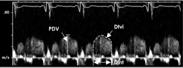
The coronary flow reserve was assessed using an Acuson Sequoia C 256 (Acuson Inc., Mountain View, CA, USA) echocardiographic machine with a 7.0-MHz transducer. High-frequency pulsed-wave Doppler was performed with the sample volume at the distal left anterior descending artery (LAD) during end-expiratory apnea. The peak diastolic velocity (PDV) of the coronary artery flow, the diastolic time-velocity integral (Dtvi) and the diastolic flow interval (Dint) were measured during three consecutive beats, and the results averaged (Figure 1). The mean diastolic velocity (MDV) was calculated from Dtvi/Dint. The coronary flow reserve was defined as the ratio of the hyperemic MDV, which was obtained after maximal inducible vasodilatation by the intrave- nous administration of a standard dose of dipyridamole (0.56 mg/kg of body weight) over a 4-minute period, to that of the MDV at rest.
Other echocardiographic parameters were also mea- sured, including the left ventricular end-systolic diameter, left ventricular end-diastolic diameter, left ventricular ejection fraction, end-diastolic thickness of the interven- tricular septum and of the left ventricular posterior wall,
Figure 1. Parameters measured in the coronary flow velocity curve. The mean diastolic velocity (MDV) was calculated from Dtvi/Dint. Dint: diastolic flow interval, Dtvi: diastolic time-velocity integral, LAD: the left ante- rior descending coronary artery and PDV: peak diastolic velocity of coronary artery flow.
Dtvi
Dint PDV
diameter of the aortic root and left atrium, mitral E and A wave velocities and deceleration time (DT) of the mitral inflow E wave.
Laboratory measurements
The serum lipid levels were measured in the venous blood, after 14-hours fasting, at the baseline and at the end of the study period. The total cholesterol, trigly- ceride and high-density lipoprotein (HDL) cholesterol levels were determined, and the low-density lipoprotein (LDL) cholesterol levels were calculated using Friede- wald’s formula.
Statistical analysis
All statistical tests were performed using the SPSS for Windows (version 10.0) package. Values are expressed as the mean±standard deviation. Differences between the baseline and after-therapy values for the lipid profiles, coronary blood flow parameters and coronary flow reserves were analyzed using Wilcoxon signed rank tests.
Spearman’s linear correlation analysis was used to test for correlations between the baseline lipid profiles and the changes in the coronary flow reserve. A value of p<
0.05 was considered statistically significant.
Results
Patients’ characteristics
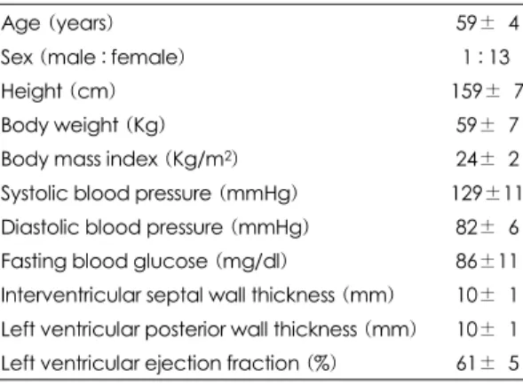
The 14 patients enrolled in this study consisted of 13 women and one man, with a mean age of 59±4 years (Table 1). All patients had normal systolic and diastolic blood pressures, fasting blood glucose levels, left vent- ricular wall thicknesses and left ventricular systolic functions.
Lipid profiles
The serum total cholesterol and LDL-cholesterol levels were significantly reduced (from 273±27 mg/dL to 199±22 mg/dL, 27±9 % reduction, p=0.001 and from 182±25 mg/ld to 110±27 mg/dL, 39±16% reduction, p=0.001, respectively), whereas the triglyceride and
HDL-cholesterol were not (13±35% reduction and 5±
12% increase, respectively) after the lipid-lowering the- rapy (Table 2).
Hemodynamic data
The induction of hyperemia by a dipyridamole intra- venous infusion significantly increased the mean heart rate both before (from 67±6 to 83±9 bpm, p=0.001) and after the lipid-lowering therapy (from 68±10 to 83±
12 bpm, p=0.001).
Changes in coronary blood flow and coronary flow reserve
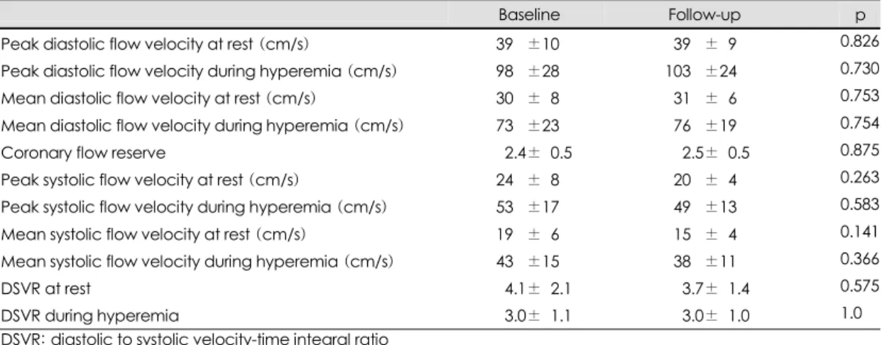
Lipid-lowering therapy caused no significant changes in the mean diastolic and systolic velocities or the dias- tolic to systolic velocity-time integral ratio (DSVR) dur- ing hyperemia or at rest (Table 3). The coronary flow reserve in all the patients showed no significant change after the lipid-lowering therapy (from 2.4±0.5 to 2.5±
0.5, p= 0.875)(Figure 2).
Table 1. Baseline patients characteristics
Age (years) 59±04
Sex (male : female) 1 : 13
Height (cm) 159±07
Body weight (Kg) 59±07
Body mass index (Kg/m2) 24±02
Systolic blood pressure (mmHg) 129±11 Diastolic blood pressure (mmHg) 82±06 Fasting blood glucose (mg/dl) 86±11 Interventricular septal wall thickness (mm) 10±01 Left ventricular posterior wall thickness (mm) 10±01 Left ventricular ejection fraction (%) 61±05
Table 2. Changes in lipid profile after lipid-lowering the- rapy
Baseline Follow-up p Total cholesterol (mg/dL) 273±27 199±22 0.001 LDL-cholesterol (mg/dL) 182±25 110±27 0.001 Triglyceride (mg/dL) 159±81 127±60 0.064 HDL-cholesterol (mg/dL) 059±15 062±17 0.182 HDL: high-density lipoprotein, LDL: low-density lipopro- tein
Correlation between lipid levels and coronary flow reserve
The baseline coronary flow reserve was found to be inversely correlated with the baseline LDL-cholesterol (p=0.012, r=-0.649)(Figure 3). However, the serum concentrations of total cholesterol (p=0.130, r=-0.425), triglyceride (p=0.201, r=0.38) and HDL-cholesterol (p=
0.951, r=-0.013) were not related to the coronary flow reserve.
Discussion
In the present study on hypercholesterolemic patients without any modifiable risk factors of coronary artery disease, the coronary flow reserve measured by trans- thoracic echocardiography did not change significantly, despite a significant decrease in the total and LDL- cholesterols after the five months of lipid-lowering the- rapy. Several factors might explain the discrepant result in the lack of significant change in the coronary flow reserve after the lipid-lowering therapy.
Table 3. Changes in coronary flow velocity and reserve after lipid-lowering therapy
Baseline Follow-up p
Peak diastolic flow velocity at rest (cm/s) 39.0±10 039.0±09 0.826
Peak diastolic flow velocity during hyperemia (cm/s) 98.0±28 103.0±24 0.730
Mean diastolic flow velocity at rest (cm/s) 30.0±08 031.0±06 0.753
Mean diastolic flow velocity during hyperemia (cm/s) 73.0±23 076.0±19 0.754
Coronary flow reserve 00.2.4±00.5 0.002.5±00.5 0.875
Peak systolic flow velocity at rest (cm/s) 24.0±08 020.0±04 0.263
Peak systolic flow velocity during hyperemia (cm/s) 53.0±17 049.0±13 0.583
Mean systolic flow velocity at rest (cm/s) 19.0±06 015.0±04 0.141
Mean systolic flow velocity during hyperemia (cm/s) 43.0±15 038.0±11 0.366
DSVR at rest .004.1±02.1 .0003.7±01.4 0.575
DSVR during hyperemia .003.0±01.1 0.003.0±01.0 1.0
DSVR: diastolic to systolic velocity-time integral ratio
4
3.5
3
2.5
2
1.5
1
Coronary flow reserve
Baseline After Tx
Pt 1 Pt 2 Pt 3 Pt 4 Pt 5 Pt 6 Pt 7 Pt 8 Pt 9 Pt 10 Pt 11 Pt 12 Pt 13 Pt 14
Figure 2. The coronary flow reserve of individual study patients at both the baseline and after lipid-lowering therapy. The coronary flow reserve in all of the patients showed significant change after lipid-lowering therapy (from 2.4±0.5 to 2.5±0.5, p=0.875).
3.5
3
2.5
2
1.5
1
Baseline coronary flow reserve
100 150 200 250 Baseline LDL-cholesterol (mg/dL)
y=-0.01x+4.7 r=-0.65 p=0.012
Figure 3. Relationship between the baseline LDL-chole- sterol levels and coronary flow reserve. A significant negative correlation was found between the baseline LDL-cholesterol and CFR. CFR: coronary flow reserve, LDL: low-density lipoprotein.
Firstly, differences in the study population are an im- portant factor. In previous studies that reported improved coronary flow reserve after lipid-lowering therapy, pa- tients with coronary artery disease,3-5) hypertension,3)4) non insulin-dependent diabetes mellitus3)5) and smokers3) were included. Moreover, in such a study,3) both the systolic- and diastolic-blood pressures decreased signi- ficantly after the lipid-lowering therapy. The coronary flow reserve has been reported to be reduced in the presence of significant coronary artery stenosis,11) in patients with diabetes due to an impaired microvascular function even in the absence of obstructive coronary atherosclerosis,12)13) and in patients with hypertension.14) In the present study, all patients with traditional risk factors of coronary heart disease, such as, hypertension, diabetes and smoking, and those with overt coronary artery disease were excluded. Therefore, only hypercho- lesterolemic patients with age- and sex-related coronary risks were included in this study, thereby excluding the possible confounding effects of modifiable coronary risk factors other than hypercholesterolemia. The baseline mean total cholesterol level of the patients enrolled in the present study (273±27 mg/dL) was not significantly lower than those of the studies by Yokoyama et al (263±33.8 mg/dL), Baller et al(241±44 mg/dL) and Guethlin et al(258±24 mg/dL).3-5)
Secondly, the female predominance of the present study may explain our results. Previously, it has been shown that, in asymptomatic familial hypercholestero- lemia patients, the coronary flow reserve was signifi- cantly higher in women than in men.15) While such a significant gender-specific difference was not evident in the control subjects, it has previously been postulated that the gender-specific differences in the coronary flow reserve might be related to the lower incidence of cor- onary artery disease in women with familial hypercho- lesterolemia.15)16) However, some previous studies favored the lack of gender specific effects of hypercholesterolemia on the coronary flow reserve, and reported the lack of a significant difference between the coronary flow reserves of men and women, in patients with hypercholestero-
lemia and normal coronary arteries,14) coronary artery disease17) and in healthy volunteers.18) In addition, Yo- koyama et al.19) recently reported no difference between men and women with familial hypercholesterolemia in terms of improved myocardial flow reserve after mod- erate- to long-term simvastatin therapy.
Thirdly, the lack of a significant reduction in the triglyceride levels in our study may present another reason for the insignificant change in the coronary flow reserve observed after lipid lowering therapy. In previous reports on the significant improvement in the coronary flow reserve after lipid-lowering therapy, the plasma triglyceride, HDL-cholesterol, total cholesterol and LDL- cholesterol concentrations changed significantly.3)4) Fur- thermore, patients with lone hypertriglyceridemia have been reported to show reduced coronary vasodilatation.20) In an animal experiment, a rise in capillary resistance due to an increased blood viscosity was suggested as a key mechanism of decreased coronary blood flow reserve during hypertriglyceridemia.21) In addition, a recent study has shown that an increase in the plasma triglyceride levels after a single high-fat meal in 15 young healthy men induced a significant reduction in the coronary flow reserve measured by transthoracic Doppler echocardio- graphy.22) In our study, the plasma triglyceride levels were not significantly changed after the lipid lowering therapy, which might be one of the causes of the insig- nificant changes in the coronary flow reserve.
Fourthly, the duration of lipid-lowering therapy may also be a factor. In previous reports, which showed an increase in the coronary flow reserve after lipid-lowe- ring treatment, the duration of therapy was longer than that used in the present study.3)4)
In the present study, the baseline coronary flow res- erve was found to be inversely related with the baseline LDL-cholesterol. Past studies have also reported the correlation between LDL-cholesterol and the coronary flow reserve.1)2)14)23) An impaired endothelial function was suggested as the main mechanism that reduced the coronary flow reserve in patients with hypercholestero- lemia.1)2)14)23)
Limitations of the study
First, our study was limited by the number of patients involved. The exclusion of other modifiable risk factors of coronary heart disease was a major enrollment- limiting factor. In addition, because of the uncomfor- table experience during dipyridamole infusion, patients were reluctant to participate in the study, as they re- quired a follow-up examination once enrolled.
Second, the duration, selection and dosage of lipid- lowering agents were not standardized. However, our study was designed to evaluate the relation between hypercholesterolemia and coronary flow reserve, and our results do not seem to be affected by the diversity of lipid lowering agents used, as the cholesterol levels were adequately controlled.
Conclusion
In the present study, no significant change in the coronary flow reserve was noted after the lipid-lowering therapy for hypercholesterolemic patients with no other risk factors of coronary heart disease, although the base- line LDL-cholesterol levels were found to correlate well with the baseline coronary flow reserve. Transthoracic Doppler echocardiography was able to easily and non- invasively evaluate the changes in the coronary flow velocity, coronary flow reserve and other related para- meters. Therefore, a controlled trial using transthoracic Doppler echocardiography to assess the effect of lipid- lowering therapy on patients showing a wider range of baseline risk factors and LDL-cholesterol level is required.
■Acknoewledgments
Grant support: This study was supported in part by research grant No. 04-2001-012-0 from the Clinical Research Insti- tute, Seoul National University Hospital
REFERENCES
1) Yokoyama I, Ohtake T, Momomura S, Nishikawa J, Sasaki Y, Omata M. Reduced coronary flow reserve in hyperchole- sterolemic patients without overt coronary stenosis. Circu- lation 1996;94:3232-8.
2) Pitkanen OP, Raitakari OT, Niinikoski H, Nuutila P, Iida H, Voipio-Pulkki LM, et al. Coronary flow reserve is impaired
in young men with familial hypercholesterolemia. J Am Coll Cardiol 1996;28:1705-11.
3) Yokoyama I, Momomura S, Ohtake T, Yonekura K, Yang W, Kobayakawa N, et al. Improvement of impaired myocardial vasodilatation due to diffuse coronary atherosclerosis in hypercholesterolemics after lipid-lowering therapy. Circula- tion 1999;100:117-22.
4) Baller D, Notohamiprodjo G, Gleichmann U, Holzinger J, Weise R, Lehmann J. Improvement in coronary flow reserve determined by positron emission tomography after 6 months of cholesterol-lowering therapy in patients with early stages of coronary atherosclerosis. Circulation 1999;99:2871-5.
5) Guethlin M, Kasel AM, Coppenrath K, Ziegler S, Delius W, Schwaiger M. Delayed response of myocardial flow res- ponse to lipid-lowering therapy with fluvastatin. Circulation 1999;99:475-81.
6) Vita JA, Yeung AC, Winniford M, Hodgson JM, Treasure CB, Klein JL, et al. Effect of cholesterol-lowering therapy on coronary endothelial vasomotor function in patients with coronary artery disease. Circulation 2000;102:846-51.
7) Janatuinen T, Laaksonen R, Vesalainen R, Raitakari O, Leh- timaki T, Nuutila P, et al. Effect of lipid-lowering therapy with pravastatin on myocardial blood flow in young mildly hypercholesterolemic adults. J Cardiovasc Pharmacol 2001;
38:561-8.
8) Crowley JJ, Shapiro LM. Transthoracic echocardiographic measurement of coronary blood flow and reserve. J Am Soc Echocardiogr 1997;10:337-43.
9) Kuriki S, Nasu M, Fukumi Ki K, Hiramori K. Noninvasive measurement of left coronary blood flow reserve by tran- sthoracic Doppler echocardiography. Echocardiography 1999;16:547-57.
10) Saraste M, Koskenvuo JW, Knuuti J, Toikka JO, Laine H, Niemi P, et al. Coronary flow reserve: measurement with transthoracic Doppler echocardiography is reproducible and comparable with positron emission tomography. Clin Physiol 2001;21:114-22.
11) Gould KL, Lipscomb K. Effects of coronary stenoses on cor- onary flow reserve and resistance. Am J Cardiol 1974;34:
48-55.
12) Nitenberg A, Valensi P, Sachs R, Dali M, Aptecar E, Attali JR. Impairment of coronary vascular reserve and Ach-in- duced coronary vasodilation in diabetic patients with angio- graphically normal coronary arteries and normal left vent- ricular systolic function. Diabetes 1993;42:1017-25.
13) Nahser PJ Jr, Brown RE, Oskarsson H, Winniford MD, Rossen JD. Maximal coronary flow reserve and metabolic coronary vasodilation in patients with diabetes mellitus.
Circulation 1995;91:635-40.
14) Antony I, Nitenberg A. Coronary vascular reserve is simi- larly reduced in hypertensive patients without any other coronary risk factors and in normotensive smokers and hypercholesterolemic patients with angiographically normal coronary arteries. Am J Hypertens 1997;10:181-8.
15) Yokoyama I, Murakami T, Ohtake T, Momomura S, Nishi- kawa J, Sasaki Y, et al. Reduced coronary flow reserve in familial hypercholesterolemia. J Nucl Med 1996;37:1937-42.
16) Mabuchi H, Koizumi J, Shimizu M, Takeda R. Development
of coronary heart disease in familial hypercholesterolemia.
Circulation 1989;79:225-32.
17) Kern MJ, Bach RG, Mechem CJ, Caracciolo EA, Aguirre FV, Miller LW, et al. Variations in normal coronary vasodil- atory reserve stratified by artery, gender, heart transplanta- tion and coronary artery disease. J Am Coll Cardiol 1996;
28:1154-60.
18) Hirata K, Shimada K, Watanabe H, Muro T, Yoshiyama M, Takeuchi K, et al. Modulation of coronary flow velocity reserve by gender, menstrual cycle and hormone replace- ment therapy. J Am Coll Cardiol 2001;38:1879-84.
19) Yokoyama I, Yonekura K, Inoue Y, Ohtomo K, Nagai R.
Long-term effect of simvastatin on the improvement of im- paired myocardial flow reserve in patients with familial hyp- ercholesterolemia without gender variance. J Nucl Cardiol 2001;8:445-51.
20) Yokoyama I, Ohtake T, Momomura S, Yonekura K, Koba-
yakawa N, Aoyagi T, et al. Altered myocardial vasodilatation in patients with hypertriglyceridemia in anatomically nor- mal coronary arteries. Arterioscler Thromb Vasc Biol 1998;
18:294-9.
21) Rim SJ, Leong-Poi H, Lindner JR, Wei K, Fisher NG, Kaul S. Decrease in coronary blood flow reserve during hyperli- pidemia is secondary to an increase in blood viscosity.
Circulation 2001;104:2704-9.
22) Hozumi T, Eisenberg M, Sugioka K, Kokkirala AR, Wa- tanabe H, Teragaki M, et al. Changes in coronary flow res- erve on transthoracic Doppler echocardiography after a single high-fat meal in young healthy men. Ann Intern Med 2002;136:523-8.
23) Kaufmann PA, Gnecchi-Ruscone T, Schafers K, Luscher TF, Camici PG. Low density lipoprotein cholesterol and cor- onary microvascular dysfunction in hypercholesterolemia. J Am Coll Cardiol 2000;36:103-9.