Hepatocyte and mesenchymal stem cell co- transplantation in rats with acute liver failure
Cheng-Maw Ho
1,2,3, Ya-Hui Chen
3, Chin-Sung Chien
3, Shu-Li Ho
1,2,*, Hui-Ling Chen
3, Rey-Heng Hu
1,2, Po-Huang Lee
1,2,41Department of Surgery, National Taiwan University Hospital, Taipei, Taiwan
2College of Medicine, National Taiwan University, Taipei, Taiwan
3Hepatitis Research Center, National Taiwan University Hospital, Taipei, Taiwan
4Department of Surgery, E-Da Hospital, I-Shou University, Kaohsiung, Taiwan
Background: Cell therapy is considered a potential alternative to liver transplantation in acute liver failure (ALF). We aimed to evaluate the add-on therapeutic benefit of he- patocyte and mesenchymal stem cell (MSC) cotransplantation over hepatocyte-only transplantations in a rat model of ALF.
Methods: ALF was induced by D-galactosamine in Sprague-Dawley rats. Freshly isolat- ed donor hepatocytes were derived from Tg (UBC-emGFP) rats and MSCs were collect- ed from the bone marrow cells of DsRed rats. Donor hepatocytes (1×10
7/mL) were intra- portally transplanted 24 hours after treatment with D-galactosamine over a 70-second interval, and donor MSCs (0.5, 1, or 2×10
6/0.5 mL) were intraportally transplanted 1 hour after the hepatocyte transplantation was complete. Animals were sacrificed after 7 and 14 days and subjected to donor cell identification, liver histology, serologic testing, and immunohistopathological examination.
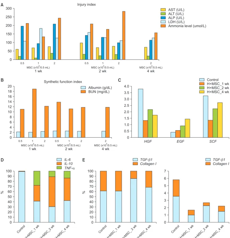
Results: MSCs were observed in the periportal area, 1 and 2 weeks after transplan- tation. Transplanted hepatocytes did not actively proliferate when compared to he- patocyte-only transplantation. Morphologically, transplanted MSCs did not appear to differentiate into hepatocytes even 2 weeks after transplantation. Cotransplantation of MSCs was associated with lower macrophage infiltration, and reduced type I collagen, hepatocyte growth factor, tumor necrosis factor-α, and interleukin 10 expression, with similar gene expression profiles for epidermal growth factor and interleukin 6, when compared to hepatocyte-only transplantation.
Conclusions: Hepatocyte and MSC cotransplantation is feasible and safe in rat mod- els of ALF. MSCs were found to survive the process and could be located within the periportal niches 2 weeks after treatment, without enhancing transplanted hepatocyte proliferation or differentiating into hepatocytes, while ameliorating the inflammatory response.
Keywords: Acute liver failure; Cell transplantation; Mesenchymal stem cells
Received February 9, 2020 Revised March 26, 2020 Accepted March 27, 2020
Corresponding author: Hui-Ling Chen Hepatitis Research Center, National Taiwan University Hospital, 7 Chung- Shan South Road, Taipei 100, Taiwan Tel: +886-2-2312-3456-67505 Fax: +886-2-2382-5962 E-mail: [email protected]
*Current affiliation: Department of Anatomy and Cell Biology, National Yang-Ming University, Taipei, Taiwan
© The Korean Society for Transplantation This is an Open Access article distributed under the terms of the Creative Commons Attribution Non-Commercial License (http://creativecommons.org/licenses/
by-nc/4.0/) which permits unrestricted non-commercial use, distribution, and reproduction in any medium, provided the original work is properly cited.
pISSN 2671-8790
eISSN 2671-8804
INTRODUCTION
Hepatocyte transplantation is a promising alternative to liver transplantation in patients with acute liver failure (ALF) and metabolic liver disease [1]. Hepatocyte trans- plantation is a safer and less invasive procedure for pa- tients when compared to whole organ transplantation.
Animal studies have clearly shown the efficacy of hepato- cyte transplantation; however, this has not translated into clinical practice where there is a limited benefit [2]. Suc- cessful translation demands high-quality cell preparation, adequate cell numbers, viability, and efficient delivery [3].
In the case of ALF, the cell dose needed to rescue/reverse the outcome is far more than that for metabolic liver dis- eases [4]. Our results demonstrate that adequate rates of cellular infusion can increase engraftment of transplanted hepatocytes in a D-galactosamine-induced acute liver injury rat model [5]. However, the engraftment efficiency was only about 2.5% 1 week after hepatocyte transplanta- tion. In addition, it is difficult for hepatocytes to proliferate in the hostile microenvironment associated with ALF [6].
Considering the urgent need for large amounts of hepato- cytes to treat ALF, there still much room for improvement in the cell based therapeutics.
In a retrorsine-treated liver injury rat model, immediate transplantation with either hepatocyte or bone marrow derived cells can ameliorate liver injury via a number of different mechanisms including hepatocyte proliferation in the former, and paracrine effects in the later [7]. Mes- enchymal stem cells (MSCs), a major component of bone marrow derived cell mixtures, have been shown to exhibit multiple beneficial effects in vitro relevant to the liver injury therapeutic context, including (1) hepatocellular functional support (improved albumin secretion, ureagen- esis, hepato-specific gene expression, cytochrome P450 activity) [8], (2) secretion of molecules that inhibit hepato-
cyte apoptosis (such as stromal-cell-derived factor-1 and vascular endothelial growth factor) [9-12] and stimulation of hepatocyte proliferation (via secretion of hepatocyte growth factor [HGF], epidermal growth factor, interleukin 6 [IL-6], and tumor necrosis factor-α [TNF-α]), (3) modula- tion of the acute phase response and suppression of the inflammatory responses including IL-1 receptor antag- onists and upregulation of anti-inflammatory cytokines like IL-10 [11], and finally (4) secrete several extracellular matrix molecules, including collagen, fibronectin and laminin necessary for liver reconstruction [13,14]. These observations suggest that MSC-derived cytokines could potentially protect the liver during injury.
In vivo, MSC or MSC-conditioned media can attenuate inflammation and augment cytokine and growth factor concentrations improving cell proliferation and providing an avenue for preventing fulminant hepatocyte failure [9,15-19]. MSC transplantation following solid organ transplantation, both clinically and experimentally, can also reduce the rate of acute rejection [19,20]. MSC trans- plantation alone, however, is not expected to exert any effect on AFL because the hostile microenvironment of ALF is not a good niche for MSCs and long-term engraft- ment rates are low [15]. Transplanted hepatocytes were unable to function, or even survive well, without stromal cell support. Thus, the addition of bone marrow-derived mesenchymal stromal cells (MSCs) during transplanta- tion could support the proliferation and functionality of the transplanted hepatocytes [13].
There are over 280 registered clinical trials examining the application of MSC, 28 of which focus on the treat- ment of liver disease [15]. While, no severe side-effects have been reported so far, the long-term benefits of these treatments remain uncertain [15]. Li et al. [18] evaluated the transplantation of human bone-marrow-derived MSCs in a porcine model of acute liver failure (ALF induced with D-galactosamine) without the use of immunosup- pressants. Most (13/15) achieved long-term survival (>6 months) while all of the animals that did not have MSC treatment died [18]. Up to 30% of the hepatocytes, in this study, were shown to be derived from the bone marrow MSCs [18]. The underlying mechanisms for this response remain unknown, and are the subject of further investiga- tion [16,21].
We have previously described the optimal rate for he- patocyte transplantation in an acute liver injury rat model to ensure optimal engraftment and repopulation [5]. If transplanted hepatocytes can proliferate properly, cell HIGHLIGHTS
• Hepatocyte and mesenchymal stem cell (MSC) cotrans- plantation was feasible and safe in rats of acute liver failure.
• Survived MSCs located within the periportal niches
2 weeks after treatment, without signs of enhancing
transplanted hepatocyte proliferation or differentiating
into hepatocytes but inflammation was ameliorated.
populations will double overtime improving the likelihood that there will be enough functional cells to compensate for the rapid loss of native hepatocytes and thus rescue the host’s liver function. It is, therefore, reasonable to as- sume that cotransplantation of hepatocytes and MSCs could provide enough support to facilitate improved sur- vival and proliferation of the transplanted hepatocytes, enhancing repopulation. The ability to properly repopulate the deteriorating liver is crucial in effective clinical inter- vention and thus the purpose of this study was to investi- gate the effects of MSC and hepatocyte cotransplantation in rats with acute liver injury.
METHODS Ethics Statement
All animal experiments were approved by the Institutional Laboratory Animal Care and Use Committee of the Na- tional University of Taiwan. All animals received humane care in accordance with the guidelines set out by the National Science Council of Taiwan (1997) and the Guide for the Care and Use of Laboratory Animals (National In- stitutes of Health publication 86–23, 1985 revision). All procedures were also performed in accordance with these guidelines.
Animals
Male Sprague-Dawley (SD) rats were used as recipient animals. Fluorescent SD rats (aged 8–10 weeks, 200–250 g) were purchased from the National Laboratory Animal Center in Taiwan and used as donor animals. These an- imals were bred in-house and maintained on standard laboratory chow and daily 12-hour light/dark cycles. All animal experiments were approved by the Institutional Laboratory Animal Care and Use Committee of the Na- tional Taiwan University (No. 20130523 and 20150405).
Isolation of Hepatocytes and MSCs for Transplantation In situ liver perfusion, collagenase digestion, and differen- tial centrifugation were used to purify hepatocytes from GFP transgenic SD rats as previously described [22,23].
The viability and purity of each preparation were as- sessed using trypan blue exclusion on a hemocytometer.
Isolated hepatocytes were resuspended 1×10
7cells/mL in phosphate buffered saline (PBS) without serum. Marrow cells, from both femurs and tibias were flushed from the
bones of the DsRed transgenic SD rats using a syringe with a 26-G needle. Bone marrow mononuclear cells were isolated using Percoll gradient density centrifugation.
MSCs were collected by depleting the cell suspensions of hematopoietic cells (CD45+) using anti-CD45 coated magnetic beads. Isolated MSCs were then resuspended to 1×10
6cells/mL in PBS without serum. MSCs were plated at a concentration of 10
6cells/mL in murine MesenCult medium (Stemcell Technologies, Vancouver, Canada) and incubated at 37°C in a 5% humidified CO
2atmosphere for 3 hours and the unattached cells were then removed.
Cells were put through a second round of purification if necessary [24].
Hepatic Tissue Histology and the Determination of Liver Repopulation
Fresh liver sections were fixed in formalin, embedded in paraffin, sectioned, and stained using hematoxylin and eosin to evaluate the histology of these samples. To iden- tify transplanted hepatocytes in the recipient liver, DsRed expression was determined using fluorescence or enzyme histochemistry in liver cryosections. To analyze the liver repopulation, three to four sections from multiple liver lobes per rat were stained for DsRed activity. Microphoto- graphs were obtained from consecutively adjacent areas to represent the whole section under ×100 magnification using a digital camera. The relative occupation of these sections by the transplanted hepatocytes was evaluated and quantified using J-Image software (National Cancer Institute, Bethesda, MD, USA).
Experimental Design
The D-galactosamine (Sigma, St. Louis, MO, USA) work- ing solution was prepared as previously described [22]
and used immediately after preparation. Acute hepatic injury was induced by D-galactosamine treatment (0.9 g/
kg, intraperitoneal injection [IP]) in male SD rats 24 hours before transplantation. Isolated hepatocytes (1×10
7/ mL) from GFP
+SD rats were transplanted intraportal- ly 24 hours after treatment with D-galactosamine at an infusion rate of 70 seconds. DsRed
+MSCs (0.5, 1, or 2×10
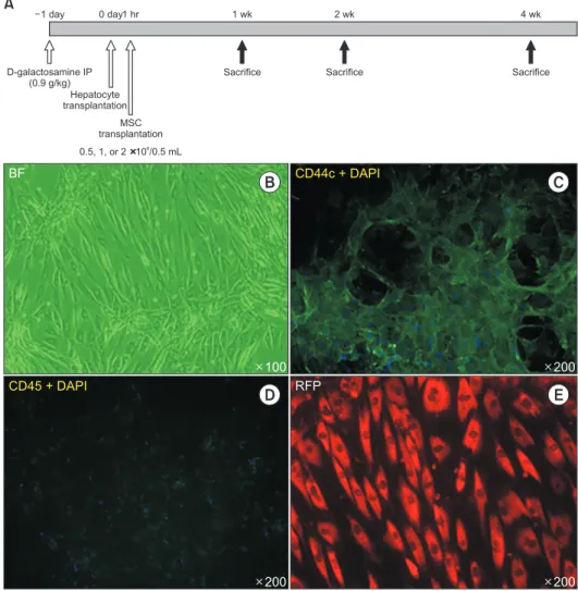
6/0.5 mL) were transplanted intraportally 1 hour after hepatocyte transplantation. The surviving rats were sac- rificed and their livers harvested at 1, 2, and 4 weeks after transplantation (Fig. 1A).
Liver Tissue Evaluations
All immunofluorescent/immunohistochemical staining
was performed according to previously described proto- cols [25]. Quantitative Reverse Transcriptase polymerase chain reaction was used to evaluate tissue specific ex- pression of HGF, EGF, SCF, IL-6, IL-10, TNFα, TGFβ1, and collagen I and was performed as previously [25].
Serological Assay
Hepatic venous blood was sampled after the recipient rats were sacrificed. Biochemical analyses (aspartate aminotransferase [AST], alanine aminotransferase [ALT], alkaline phosphatase [ALP], lactate dehydrogenase [LDH], ammonia, albumin, and blood urea nitrogen [BUN]) were performed in an animal laboratory using the standard au- tomated assays, as previously described [5].
Statistical Analysis
At least four animals per treatment were evaluated. Data are shown in a qualitative manner or presented as the
mean, as appropriate. No animal data were excluded.
RESULTS Characterization of Donor MSCs
MSCs in culture before transplantation are shown in Fig.
1B-E. They maintained the classic spindle-shape mor- phologically (Fig. 1B), expressed CD44 (Fig. 1C) and did not express hematopoietic cell marker CD45 (Fig. 1D).
They were derived from the bone marrow aspirations from the DsRed SD rats and were therefore fluoro-red positive.
Histopathological Changes after Hepatocyte and MSC Co- transplantation
One day after D-gal administration, livers of SD rats showed extensive hepatocyte necrosis, periportal focal
D-galactosamine IP (0.9 g/kg)
Hepatocyte transplantation
MSC transplantation 0.5, 1, or 2 106/0.5 mL
1 day 0 day1 hr 1 wk 2 wk
Sacrifice Sacrifice
100
BF CD44c + DAPI
CD45 + DAPI RFP
A
Sacrifice 4 wk
B C
D E
200 200
200
Fig. 1. Model of acute liver failure (ALF)
used in the cotransplantation study. (A) Ex-
perimental design for the cotransplantation
of rat hepatocytes and mesenchymal stem
cells (MSCs) in a rat model of ALF. (B-E)
Characterization of MSC. The MSCs were
spindle shaped (B) and expressed CD44c
(C) but not CD45 (D). They were fluoro-red
(+) (E) since they were derived from bone
marrow aspirates of DsRed transgenic
Sprague-Dawley rats. IP, intraperitoneal
injection; BF, blank field; DAPI, 4', 6-diamid-
ino-2-phenylindole; RFP, red fluorescent
protein.
Fig. 2. Histopathological changes after hepatocyte and mesenchymal stem cell (MSC) cotransplantation in acute liver injury. (A) Control liver. Acute
liver injury developed, 1 day after D-galactosamine treatment. Ductular reactions were noted by oval cell mark er (OV-6), expression near the portal vein.
Prominent CD163
+macrophage and low G-6-P expression were also observed. Fibrosis was limited to the periportal region. (B) One, 2 and 4 weeks after cotransplantation, γ-glutamyltransferase (γGT), was expressed in the ductular cells along with obvious signs of liver recovery. (C) Tracing of transplanted donor cells after cotransplantation. Donor hepatocytes were labelled green and MSCs were labelled red. CD163 marker expression was used to deter- mine macrophage infiltration after cotransplantation and is shown on the right. Arrows indicate the donor cells. CD31 marks the endothelial inner lining of the portal vein. GFP, green fluorescent protein; RFP, red fluorescent protein. Magnification, all ×200.
C
1wk2wk4wk
A
GTH&E
1wk2wk4wk
GT
B
OV-6 Pan-CK (green)/CD31 (red)
Pan-CK (green)/CD163 (red) G-6-P
Trichrome GT+trichrome
H&E
GFP/RFP CD31/RFP CD163
Fig. 3. Serological marker and gene expression changes. Serological changes after cotransplantation of hepatocyte and mesenchymal stem cells (MSCs)
in rats with acute liver injury. Markers of injury index (A) and synthetic function index (B). Gene expression of growth factors (C), inflammation (D) and fibrosis (E) after cotransplantation. Control: acute liver injury 1 day after D-galactosamine treatment; Baseline expression: glyceraldehyde 3-phosphate dehydrogenase; AST, aspartate aminotransferase; ALT, alanine aminotransferase; ALP, alkaline phosphatase; LDH, lactate dehydrogenase; BUN, blood urea nitrogen; H, hepatocyte; HGF, he patocyte growth factor; EGF, epidermal growth factor; SCF, stem cell factor; IL, interleukin; TNF-α, tumor necrosis factor-α; TGF-β1, transforming growth factor-β1.
C
4.0 3.5 3.0 2.5 2.0 1.5 1.0 0.5
HGF EGF SCF
0
Control H+MSC_1 wk H+MSC_2 wk H+MSC_4 wk
B A
100 90 80 70 60 50 40 30 20 10
Control 0
D
H+MSC_1 wk
H+MSC_2 wk
H+MSC_4 wk
%
IL-6 IL-10 TNF-
E
100 90 80 70 60 50 40 30 20 10
Control 0
H+MSC_1 wk
H+MSC _2wk
H+MSC_4 wk
%
TGF- 1 Collagen I
%
7 6 5 4 3 2 1
Control 0
H+MSC_1 wk
H+MSC_2 wk
H+MSC_4 wk TGF- 1 Collagen I 0.5
MSC (x10 /0.5 mL)6 300
250 200 150 100 50
1 wk 0
Injury index
1 2
2 wk 4 wk
AST (U/L) ALT (U/L) ALP (U/L) LDH (U/L)
Ammonia level (umol/L)
2
0.5 1 2
MSC (x10 /0.5 mL)6 MSC (x10 /0.5 mL)6
20 18 16 14 12 10 8 6 4 2 0
Synthetic function index
Albumin (g/dL) BUN (mg/dL)
0.5
1 wk
1 2
2 wk
2 2
1 0.5
4 wk MSC (x10 /0.5 mL)6 MSC (x10 /0.5 mL)6 MSC (x10 /0.5 mL)6