Lipopolysaccharide 로 자극한 RAW 264.7 세포에서 성체줄기세포 유래 엑소좀(exosome)의 면역 조절 효과
정수경†, 박미정†, 이지현†, 변정수, 구나연, 조인수, 차상호*
농림축산검역본부바이러스질병과
Received: April 29, 2016 / Revised: June 28, 2016 / Accepted: July 1, 2016
서 론
성체줄기세포는지방세포, 조골세포, 연골세포, 근육세포
등의세포로분화가능한다능성세포로조직재생[9], 면역
조절등다양한능력을가지고있는것으로알려져있다. 이 러한성체줄기세포는기초연구분야및심근경색[17], 이식 편대숙주병[37], 대장염[35], 간부전[36], 신부전[23], 크론병
[33], 중추신경계외상[15], 자가면역성뇌척수염[47], 류마티
스성관절염[50] 등질병과관련한임상연구분야에서면역
조절기전에대한많은연구들이진행되고있다[16].
염증반응은미생물감염, 내독소, 조직손상과같은위해 성자극을정상적으로회복하기위한필수적방어작용으로, 정상적인염증반응은시간이지남에따라염증촉진성매 개체(pro-inflammatory mediators)의생성은감소하고항염 증성매개체(anti-inflammatory mediators)는증가됨으로
써스스로염증반응조절과정을가진다[26]. 이러한염증
반응이최근암, 비만, 당뇨병등다양한질환의발병과진 행에있어중요한역할을한다고보고되면서염증반응조 절을통해염증관련질환이개선될수있는가능성을보
이게되었다[19]. 이러한염증반응에관여하여중요한역
Immunomodulatory Effect of Mesenchymal Stem Cell-Derived Exosomes in Lipopolysaccharide-Stimulated RAW 264.7 Cells
Soo-Kyung Jung†, Mi Jeong Park†, Jienny Lee†, Jeong Su Byeon, Na-Yeon Gu, In-Soo Cho, and Sang-Ho Cha* Viral Disease Division, Animal and Plant Quarantine Agency, Gimcheon 39660, Republic of Korea
Mesenchymal stem cells (MSCs) are multipotent stem cells that can be differentiated into a variety of cell types, including adipocytes, osteoblasts, chondrocytes, β-pancreatic islet cells, and neuronal cells. MSCs have been reported to exhibit immunomodulatory effects in many diseases. Many studies have reported that MSCs have distinct roles in modulating inflammatory and immune responses by releasing bioactive molecules. Exosomes are cell-derived vesicles present in biological fluids, including the blood, urine, and cultured medium of cell cultures. In this study, we investigated the immunomodulatory effects of mouse adipose tissue-derived MSCs (mAD-MSCs), cultured medium (MSC-CM) of mAD-MSCs, and mAD-MSC- derived exosomes (MSC-Exo) on lipopolysaccharide (LPS)-induced RAW 264.7 cells. We observed that the expression levels of IL-1β, TNF-α, and IL-10 were significantly increased in LPS-stimulated RAW 264.7 cells compared to those in LPS-unstimulated RAW 264.7 cells. Additionally, these values were significantly (p <
0.05) decreased in mAD-MSCs-RAW 264.7 cell co-culture groups, MSC-CM-treated groups, and MSC-Exo- treated groups. MSCs can modulate the immune system in part by secreting cytokines and growth factors.
We observed that immunomodulatory factors such as IL-1β, TNF-α, and IL-10 were secreted by mAD-MSCs under co-culturing conditions of mAD-MSCs with activated RAW 264.7 cells. In addition, mAD-MSC-derived exosomes exhibited similar immunomodulatory effects in activated RAW 264.7 cells. Therefore, our results suggest that mAD-MSCs have an immunomodulatory function through indirect contact.
Keywords: Adipose tissue, mesenchymal stem cells, exosome, indirect contact
*Corresponding author
Tel: +82-54-912-0805, Fax: +82-54-912-0812 E-mail: [email protected]
†These authors contributed equally to this work.
© 2016, The Korean Society for Microbiology and Biotechnology
할을하는것으로알려져있는대식세포(macrophages)는세 균세포막성분인 lipopolysaccharides (LPS)의자극으로활 성화된다[43]. 대식세포를 LPS로자극하면 interleukin (IL)- 1, IL-6, tumor necrosis factors-α (TNF-α) 등과같은사이 토카인및 nitric oxide (NO)의발현이증가되므로[49], 항염 증제를개발하는다른방법으로사이토카인생성을억제하 는치료제개발이연구되고있다.
엑소좀(exosome)은작은지질층으로이루어진세포분비
소낭으로세포간에단백질이나 RNA를전달하여세포간 신호전달을매개하는데[11], 최근성체줄기세포가조직을 회복하는과정이엑소좀을통한다고알려졌으나[2, 22, 27], 성체줄기세포유래엑소좀의조직회복에관한메커니즘은 명확하지않다. 본연구는쥐지방조직으로부터성체줄기세 포(mouse adipose tissue derived-mesenchymal stem cells, mAD-MSCs)를분리하여그특성을분석하였으며, 대 식세포를이용한염증유사세포모델에서성체줄기세포의 면역조절능력을평가하고자하였다. 이를위해대식세포와 성체줄기세포를공배양하고성체줄기세포상층배양액및 성체줄기세포유래엑소좀을활성화된대식세포에처리하여 성체줄기세포및성체줄기세포유래 bioactive molecules의 면역조절능력을고찰하고자하였다.
재료 및 방법
세포의 분리 및 배양
성체줄기세포는쥐의복부지방조직으로부터분리한다. 분리한 지방 조직은 300 U/ml penicillin (Gibco, USA)와 300 μg/ml streptomycin (Gibco, USA)이포함된 dulbecco’s phosphate buffered saline (DPBS; Gibco, USA)으로 2회 세척하여가위로잘게 자른다음 0.1% collagenase type Ι (Gibco, USA)과 1% bovine serum albumin (Bioworld, USA)이포함된 DPBS를넣고 37℃에서 40분간반응한다. 100 μm의 cell strainer (BD, USA)를사용하여조직부유물
을거르고 400 × g에서 5분간원심분리하여상층액을제거
한다. 원심분리된 pellet에 10% fetal bovine serum (FBS;
Gibco, USA), 100 U/ml penicillin, 100 μg/ml streptomycin이 포함된 low glucose dulbeco’s modified eagle’s media
(DMEM; Gibco, USA)를 넣고 잘 섞은 다음 세포 배양용
175 T-flask에 1 × 10⁶개의세포를분주하여 5% CO2, 37℃
세포배양기에서배양한다. 다음날세포배양액을교체하며,
3일간격으로세포배양액을교체하여세포가 80% 밀집하
면 0.25% trypsin-EDTA (Gibco, USA)를처리하여세포를 계대배양한다. 대식세포(ATCC, USA)는 5% CO2, 37℃의 세포배양기에서 DMEM 배지로배양한다. 2일마다세포배 양액을 교체하며, 세포가 80% 밀집하면 0.25% trypsin-
EDTA를처리하여세포를계대배양한다.
유세포 분석(flow cytometric analysis)을 통한 세포 표면 특 이 단백질 발현 검사
FACS CaliburTM flow cytometer (BD, USA)를 이용한 유세포분석을통해서지방조직으로부터분리한세포의세 포표면특이단백질의발현을조사한다. 해당세포에형광 물질 fluorescein isothiocyanate (FITC) 또는 phycoerythrin (PE)이결합된 CD34, CD45 (BD, USA) 및 CD44 (Abcam, UK) 항체를사용하였으며, Cell Quest Pro (BD, USA) 프로 그램을사용하여결과를분석한다.
중배엽 분화 유도 및 염색
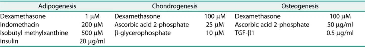
중배엽 세포로 분화능을 확인하고자 adipogenesis, chondrogenesis, osteogenesis 유도용맞춤배지(Table 1)를 사용하여분화를유도한다. 3−4일주기로분화용맞춤배지 를교체하며, 21일째에분화확인용키트를이용하여분화 여부를 관찰한다. Adipogenesis 분화는 Oil Red-O stain kit, chondrogenesis 분화는 Alcian Blue stain kit, osteogenesis 분화는 Alzarin Red stain kit를이용한특이 세포염색을통해성체줄기세포의중배엽분화능을확인한 다(IHC World, USA).
성체줄기세포와 대식세포 공배양
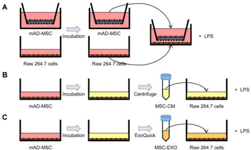
성체줄기세포와대식세포의공배양은 trans-well culture 방법을이용한다. 0.4 μm pore 크기의 insert와 lower well 에성체줄기세포와대식세포를각각 24시간배양하고, insert 와 lower well을결합한다음 1 μg/ml LPS (Sigma, USA)를 이용하여대식세포를자극하고 24시간경과후대식세포를 모아다음실험에사용한다. 또한, 성체줄기세포상층배양 액(cultured medium; CM)을얻고자성체줄기세포를 12−36 시간동안배양하여상층액을수집한다음원심분리하여세 포등을제거하고다음실험에사용한다.
Table 1. Composition of differentiation media.
Adipogenesis Chondrogenesis Osteogenesis
Dexamethasone Indomethacin
Isobutyl methylxanthine Insulin
1 μM 200 μM 500 μM 20 μg/ml
Dexamethasone
Ascorbic acid 2-phosphate β-glycerophosphate
100 μM 25 μM 10 μM
Dexamethasone
Ascorbic acid 2-phosphate TGF-β1
100 μM 50 μg/ml 0.5 μg/ml
엑소좀(exosome)의 분리
ExoQuick kit (SBI, USA)를사용하여성체줄기세포유래 엑소좀을분리한다. 성체줄기세포를배양한세포상층액을 원심 분리하여 준비한 다음, 제조사의 안내 방법에 따라 ExoQuick exosome precipitation solution을넣고 4℃에서
24시간동안반응시킨뒤 350 × g에서원심분리하여엑소
좀을분리하고다음실험에사용한다[27, 40].
정량적 중합효소연쇄반응(quantitative real-time RT-PCR) 을 이용한 면역 조절인자 분석
RNeasy mini kit (Qiagen, USA)를이용하여 RNA를추 출하고, Nano-drop 1000 (Thermo, USA)을사용하여 RNA
상태와 농도를 측정하며, cDNA 합성을 위해 GoScriptTM
Reverse Transcriptase (Promega, USA)를 사용한다. Quantitative real-time RT-PCR (qPCR)은 96 well plate 에 LightCycler® 480 SYBR Green I Master Mix (Roche Diagnostics, USA)를사용하여 predenaturation (95℃, 10분), annealing (62℃, 10초), elongation (72℃, 10초)을 45 cycle 반복한다. 그리고 2−ΔCt 계산법을이용하여 melting curve를분석하고[30], GAPDH 발현을 reference gene으로 이용하여 qPCR 결과를분석한다.
통계학적 분석
독립적인 3회(n = 3)의실험을수행하였으며, 결과는평균 과표준편차(mean ± SD)를표시하여나타내었다. 두실험 군 사이의 유의성은 Student’s t-test (JMP® 6.0; SAS Institute Inc., USA) 및 one-way ANOVA (analysis of variance)를사용하여검정하였으며, 유의성있는결과(p <
0.05)를판정한다.
결과 및 고찰
성체줄기세포의 분리 및 증식
성체줄기세포는 1970년대사람골수로부터처음분리되 었으며[13, 14], 이러한성체줄기세포는지방조직, 제대, 제 대혈등다양한조직으로부터분리, 연구되고있다[18, 24,
51]. 본연구는쥐의복부지방조직으로부터세포를분리하
여해당세포가성체줄기세포의특성중하나인섬유아세포 모양으로부착해서잘자라는것을확인하였다(Fig. 1A).
세포 표면 특이 단백질 발현 및 중배엽 분화 유도능 확인 세포표면특이단백질을확인하고자유세포분석방법을 통해 CD11b, CD31, CD34, CD44, CD45 및 CD117의발현 을확인하였다. 그결과성체줄기세포의 positive marker인 CD44의 발현을 확인하였고, negative marker인 CD11b,
CD31, CD34, CD45 및 CD117은발현되지않음을확인하였
다(Fig. 1B). 다음으로해당세포의중배엽세포분화능을확
인하고자지방세포, 골세포, 연골세포로분화를 유도하였 다. 그결과지방세포로분화를유도한성체줄기세포를 Oil
Red-O로염색하여붉은색으로염색된지방방울이축적되
어지방세포로분화됨을확인하였다. 다음으로골세포로분 화를유도한성체줄기세포를 Alizarin Red로염색하여갈색 으로염색된골기질구조가확인되어골세포로분화됨을확 인하였다. 또한, 연골세포로분화를유도한성체줄기세포를
Alcian Blue로염색하여파란색으로염색된연골기질을확
인, 연골세포로분화됨을확인하였다(Fig. 1C). 즉, 지방세 포, 골세포, 연골세포에대한특이세포염색을통해쥐의복 부지방조직으로부터분리한성체줄기세포의중배엽분화 능을확인하였다.
염증 유사 대식세포 모델에 미치는 성체줄기세포와 상층 배 양액의 영향
성체줄기세포를이용한이식치료분야에서성체줄기세포 를손상부위에이식하면기능적개선은보이나성체줄기세 포가이식부위내에서적절한세포유형으로분화되는모 습은보기드물다고보고되었으며[12, 39, 42], 성체줄기세 포이식치료에서나타난이러한기능적개선이성체줄기세 포와조직의직접적인접촉에의존하지않는다고보고되기 도하였다[5, 7, 20, 32]. 이러한결과들은성체줄기세포에의 한조직의손상감소및회복이성체줄기세포의직접적인분 화보다는성체줄기세포가분비하는 bioactive molecules을 통해이루어지고있을가능성들을제안한다[4]. 염증과관련 한다양한사이토카인중, IL-1β는외부감염이나염증이발 생하였을때국소적으로생산되는데, 과도한 IL-1β의생성 은염증성질환이나암을유발하며[41], T 세포, B 세포, NK 세포활성에직접적으로관여하여염증반응을유도하는것 으로알려져있다[8, 34]. 또한 TNF-α는염증성전구사이토 카인으로주로자극된대식세포에서분비되지만섬유아세 포, T 세포, B 세포, 내피세포및상피세포에서도분비되어
급성염증반응에서중요한역할을한다. IL-10은다양한특
성을가진사이토카인으로적응면역(adaptive immunity)에 관련된세포를조절하며 T 세포, B 세포, NK 세포와비만세 포를활성화시켜면역을자극하는특성을가지고있다[31,
46]. 또한루프스와뇌척수염과같은자가면역질환에서비정
상적인혈관형성을유도한다고알려져있으며[6], 오히려건 선을악화시키는데에중요한역할을한다고도보고되었다
[38]. 흑색종이나몇몇림프종에서 IL-10의과발현이발견되
었고이는암의진행과관련이있다고보고되었으며[1], 그 람음성균성폐렴모델에서도과발현된 IL-10이대식세포의 표현형을변형시켜사망률을증가시킨다고보고되었다[10].
본연구는 LPS를이용한대식세포염증유사세포모델에 서성체줄기세포의면역조절능력을평가하였다. 대식세포 를 LPS로자극하여대식세포에서염증관련면역인자로알 려져있는 IL-1β, IL-10 및 TNF-α등의사이토카인생산을 확인하였으며, 순수하게대식세포에미치는성체줄기세포상 층배양액의영향을관찰하고자대식세포의 mRNA에서사 이토카인생산을확인하였다[45]. IL-1β는 LPS로 12시간자 극한단독실험군에서최대로생성되었으며, 성체줄기세포
와공배양한실험군과상층배양액을처리한실험군에서통 계학적으로유의하게 IL-1β의생성이억제됨을확인하였다 (Fig. 3A). TNF-α또한 LPS로 12시간자극한단독실험군에 서최대로생성되었으며, 성체줄기세포와공배양한실험군 과상층배양액을처리한실험군에서통계학적으로유의하 게 TNF-α의생성이억제됨을확인하였다(Fig. 3B). 다음으
로면역관련저해인자로알려져있는 IL-10의생성을확인
한결과, IL-10은 LPS로 36시간자극한단독실험군에서최 Fig. 1. Isolation, characterization and differentiation potential of mouse adipose tissue-derived mesenchymal stem cells (mAD- MSCs). (A) The mAD-MSCs obtained from mouse adipose tissue were able to attach to the culture plates and expand in vitro at passage 0 and 2 (×100). (B) For further characterization of the mAD-MSCs, cell surface markers were analyzed by FACS. mAD-MSCs were strongly positive for CD44, but negative for CD11b, CD45, CD31, CD34 and CD117. (C) mAD-MSCs were differentiated to adipogenesis, osteo- genesis and chondrogenesis. mAD-MSCs were cultured in adipogenic, osteogenic or chondrogenic medium for up to 21 days. Adipo- cytes (Oil Red-O), osteocytes (Alizarin Red), and chondrocytes (Alcian Blue) were positively stained in differentiated cells.
대로생성되었으며, 성체줄기세포와공배양한실험군과상 층배양액을처리한실험군에서통계학적으로유의하게 IL- 10의생성이억제됨을확인되었다(Fig. 3C). 대조군으로는 LPS 무자극실험군을이용하여성체줄기세포와상층배양 액을처리하였다. 그결과성체줄기세포및상층배양액단 독군에의한대식세포의면역반응은관찰되지않았다. 즉, LPS 자극에의해증가된 IL-1β, TNF-α및 IL-10의생성은 성체줄기세포를공배양한실험군뿐만아니라성체줄기세포 를배양한상층배양액을처리한실험군에서도동일한효과 를나타냄을확인하여성체줄기세포가분비하는물질에의
해대식세포의증가된면역반응이조절되고있음을확인하 였다.
염증 유사 대식세포 모델에 미치는 성체줄기세포 유래 엑소 좀의 영향
최근성체줄기세포를적용한심근경색, 급성호흡부전, 심 근허혈, 이식편대숙주병등다양한질병연구모델에서성체 줄기세포의치료효과가성체줄기세포유래엑소좀에의한 새로운세포간신호전달에의한다고보고되었다[2, 22, 25,
27]. Bian 등[2]은심근경색모델에서엑소좀에의한혈류의
Fig. 3. Immunomodulatory effects of mAD-MSCs and MSC-CM on LPS-stimulated RAW 264.7 cells. mAD-MSCs and MSC-CM sig- nificantly reduced the expression levels of (A) IL-1β (12 h) (B) TNF-α (12 h), and (C) IL-10 (36 h) in stimulated RAW 264.7 cells. The mRNA levels of anti-inflammatory or pro-inflammatory cytokines were analyzed by quantitative real-time RT-PCR and normalized against the level of GAPDH. Also, each data from three independent experiments were evaluated and expressed as mean ± SD (*p < 0.05).
Fig. 2. Schematic diagram of co-culture experiment and representation of the in vitro co-culture system. Firstly, mAD-MSCs (1 × 10⁶ cells/well) were plated on the inner transwell membrane (0.4 μm pore size) and co-cultured with RAW 264.7 cells (2 × 10⁵ cells/
well) stimulated with mitogen (LPS 1 μg/ml). After coculturing for 12-36 h, culture supernatants were collected. Exosomes were isolated from mAD-MSCs supernatants using ExoQuick kit (SBI, USA) according to the manufacture’s protocol. In this study, we examined the immunomodulatory effects of mAD-MSCs in 3 groups: Group A, mAD-MSCs; Group B, MSC-CM; and Group C, MSC-Exo.
개선을보고하였고, Li 등[27]은급성호흡부전모델에서엑 소좀이기관지폐포세척액내백혈구와중성구의수치를낮 춰허혈성저산소증의전단계를막는다고보고하였다. Lai 등[25]은심근허혈모델에서엑소좀이경색부위를줄인다고 보고하였고 Kordelas 등[22]은엑소좀을이용하여처음으로 인간이식편대숙주병들에게엑소좀을투여하여이후환자 들의증상이개선됨을보고하였다. 이러한결과들은엑소좀 을이용하여다양한질병분야에치료적인적용이가능하고 또한추가연구에대한가치가있음을의미한다. 또한이러 한성체줄기세포유래엑소좀이성체줄기세포가정기적으로 분비하는성장인자보다더욱기능적인역할을한다고알려 졌다[2, 3, 21, 22, 27−29, 44, 48]. 엑소좀은내피세포의증 식과이동, 관생성능력을증가시키고[2], Akt의인산화와 Bcl-2의발현증가및 Bax의발현은감소시키며 caspage-3
의분해를감소시켜신경세포독성을완화한다[25]. 또한, 신
경교의성상세포와뉴런에 miR-133β를전달하여유전자발 현을조절함으로써뇌졸중에있어신경돌기재형성과기능
적회복에도움을준다고알려졌다[44].
본연구는 LPS를이용한대식세포염증유사세포모델에 서성체줄기세포유래엑소좀의면역조절능력을평가하였 다. 대식세포를 LPS로자극하여대식세포에서 IL-10과 TNF- α사이토카인생산을확인하였으며, 이에성체줄기세포유 래엑소좀의영향을확인하고자하였다. LPS로자극한단독 실험군에서 TNF-α는최대로생성되었으며, 성체줄기세포 유래엑소좀을처리한실험군에서통계학적으로유의하게 TNF-α의생성이억제됨을확인하였다(Fig. 4A). 또한, LPS 로자극한단독실험군에서최대로생성된 IL-10 또한성체 줄기세포유래엑소좀을처리한실험군에서통계학적으로
유의하게 IL-10의생성이억제됨을확인하였다(Fig. 4B).
요 약
본연구는대식세포에서 LPS를이용하여염증유사세포 모델을만들고염증유사대식세포모델에서성체줄기세포 의면역조절능력을평가하였다. LPS 자극에의해증가된 IL-1β, TNF-α및 IL-10의생성은성체줄기세포를공배양한 실험군뿐만아니라성체줄기세포를배양한상층배양액을 처리한실험군에서도동일한효과를나타내었으며, 또한성 체줄기세포유래엑소좀을염증유사대식세포모델에처리 하여유사한결과를관찰하였다. 이결과는성체줄기세포자 체의염증억제기능보다는성체줄기세포유래엑소좀을포 함하여성체줄기세포가분비하는 bioactive molecules에의 해세포간신호전달이이루어지고있음을의미하며, 이러 한엑소좀은염증관련질환분야에치료적적용이가능하 고또한새로운염증치료제개발의툴로사용될수있음을 시사한다.
Acknowledgments
This work was funded by the Animal and Plant Quarantine Agency (M-1543083-2014-17-01), Republic of Korea.
References
1. Asadullah K, Sterry W, Volk HD. 2003. Interleukin-10 therapy-- review of a new approach. Pharmacol. Rev. 55: 241-269.
2. Bian S, Zhang L, Duan L, Wang X, Min Y, Yu H. 2014. Extracellu- Fig. 4. Immunomodulatory effects of MSC-Exo on LPS-stimulated RAW 264.7 cells. MSC-Exo significantly reduced the expression levels of (A) TNF-α (12 h) and (B) IL-10 (36 h) in stimulated RAW 264.7 cells. The mRNA levels of anti-inflammatory or pro-inflammatory cytokines were analyzed by quantitative real-time RT-PCR and normalized against the level of GAPDH. Also, each data from three inde- pendent experiments were evaluated and expressed as mean ± SD (*p < 0.05).
lar vesicles derived from human bone marrow mesenchymal stem cells promote angiogenesis in a rat myocardial infarction model. J. Mol. Med. (Berl) 92: 387-397.
3. Bruno S, Grange C, Deregibus MC, Calogero RA, Saviozzi S, Col- lino F, et al. 2009. Mesenchymal stem cell-derived microvesi- cles protect against acute tubular injury. J. Am. Soc. Nephrol. 20:
1053-1067.
4. Caplan AI, Dennis JE. 2006. Mesenchymal stem cells as trophic mediators. J. Cell Biochem. 98: 1076-1084.
5. da Silva Meirelles L, Caplan AI, Nardi NB. 2008. In search of the in vivo identity of mesenchymal stem cells. Stem Cells 26: 2287- 2299.
6. Dace DS, Khan AA, Stark JL, Kelly J, Cross AH, Apte RS. 2009.
Interleukin-10 overexpression promotes Fas-ligand-depen- dent chronic macrophage-mediated demyelinating polyneu- ropathy. PLoS One 4: e7121.
7. Dai W, Hale SL, Martin BJ, Kuang JQ, Dow JS, Wold LE, Kloner RA. 2005. Allogeneic mesenchymal stem cell transplantation in postinfarcted rat myocardium: short- and long-term effects.
Circulation. 112: 214-223.
8. Delgado AV, McManus AT, Chambers JP. 2003. Production of tumor necrosis factor-alpha, interleukin 1-beta, interleukin 2, and interleukin 6 by rat leukocyte subpopulations after expo- sure to substance P. Neuropeptides 37: 355-361.
9. Dimarino AM, Caplan AI, Bonfield TL. 2013. Mesenchymal stem cells in tissue repair. Front. Immunol. 4: 201.
10. Dolgachev VA, Yu B, Sun L, Shanley TP, Raghavendran K, Hem- mila MR. 2014. Interleukin 10 overexpression alters survival in the setting of gram-negative pneumonia following lung con- tusion. Shock. 41: 301-310.
11. EL Andaloussi S, Mager I, Breakefield XO, Wood MJ. 2013. Extra- cellular vesicles: biology and emerging therapeutic opportuni- ties. Nat. Rev. Drug Discov. 12: 347-357.
12. Ferrand J, Noel D, Lehours P, Prochazkova-Carlotti M, Cham- bonnier L, Menard A, et al. 2011. Human bone marrow-derived stem cells acquire epithelial characteristics through fusion with gastrointestinal epithelial cells. PLoS One 6: e19569.
13. Friedenstein AJ, Chailakhjan RK, Lalykina KS. 1970. The devel- opment of fibroblast colonies in monolayer cultures of guinea- pig bone marrow and spleen cells. Cell Tissue Kinet. 3: 393-403.
14. Friedenstein AJ, Chailakhyan RK, Gerasimov UV. 1987. Bone marrow osteogenic stem cells: in vitro cultivation and trans- plantation in diffusion chambers. Cell Tissue Kinet. 20: 263-272.
15. Galindo LT, Filippo TR, Semedo P, Ariza CB, Moreira CM, Camara NO, et al. 2011. Mesenchymal stem cell therapy modulates the inflammatory response in experimental traumatic brain injury.
Neurol. Res. Int. 2011: 564089.
16. Gao F, Chiu SM, Motan DA, Zhang Z, Chen L, Ji HL, et al. 2016.
Mesenchymal stem cells and immunomodulation: current sta- tus and future prospects. Cell Death Dis. 7: e2062.
17. Gnecchi M, He H, Liang OD, Melo LG, Morello F, Mu H, et al.
2005. Paracrine action accounts for marked protection of isch- emic heart by Akt-modified mesenchymal stem cells. Nat. Med.
11: 367-368.
18. Hoogduijn MJ, Betjes MG, Baan CC. 2014. Mesenchymal stro- mal cells for organ transplantation: different sources and unique characteristics? Curr. Opin. Organ. Transplant. 19: 41-46.
19. Hotamisligil GS. 2006. Inflammation and metabolic disorders.
Nature 444: 860-867.
20. Iso Y, Spees JL, Serrano C, Bakondi B, Pochampally R, Song YH, et al. 2007. Multipotent human stromal cells improve cardiac function after myocardial infarction in mice without long-term engraftment. Biochem. Biophys. Res. Commun. 354: 700-706.
21. Katsuda T, Tsuchiya R, Kosaka N, Yoshioka Y, Takagaki K, Oki K, et al. 2013. Human adipose tissue-derived mesenchymal stem cells secrete functional neprilysin-bound exosomes. Sci. Rep. 3:
1197.
22. Kordelas L, Rebmann V, Ludwig AK, Radtke S, Ruesing J, Doep- pner TR, et al. 2014. MSC-derived exosomes: a novel tool to treat therapy-refractory graft-versus-host disease. Leukemia 28: 970-973.
23. Kunter U, Rong S, Djuric Z, Boor P, Muller-Newen G, Yu D, et al.
2006. Transplanted mesenchymal stem cells accelerate glo- merular healing in experimental glomerulonephritis. J. Am.
Soc. Nephrol. 17: 2202-2212.
24. Kuznetsov SA, Mankani MH, Gronthos S, Satomura K, Bianco P, Robey PG. 2001. Circulating skeletal stem cells. J. Cell Biol. 153:
1133-1140.
25. Lai RC, Arslan F, Lee MM, Sze NS, Choo A, Chen TS, et al. 2010.
Exosome secreted by MSC reduces myocardial ischemia/reper- fusion injury. Stem. Cell Res. 4: 214-222.
26. Lawrence T, Willoughby DA, Gilroy DW. 2002. Anti-inflamma- tory lipid mediators and insights into the resolution of inflam- mation. Nat. Rev. Immunol. 2: 787-795.
27. Li L, Jin S, Zhang Y. 2015. Ischemic preconditioning potentiates the protective effect of mesenchymal stem cells on endotoxin- induced acute lung injury in mice through secretion of exo- some. Int. J. Clin. Exp. Med. 8: 3825-3832.
28. Li T, Yan Y, Wang B, Qian H, Zhang X, Shen L, et al. 2013. Exo- somes derived from human umbilical cord mesenchymal stem cells alleviate liver fibrosis. Stem. Cells Dev. 22: 845-854.
29. Lin SS, Zhu B, Guo ZK, Huang GZ, Wang Z, Chen J, et al. 2014.
Bone marrow mesenchymal stem cell-derived microvesicles protect rat pheochromocytoma PC12 cells from glutamate- induced injury via a PI3K/Akt dependent pathway. Neurochem Res. 39: 922-931.
30. Livak KJ, Schmittgen TD. 2001. Analysis of relative gene expres- sion data using real-time quantitative PCR and the 2(-Delta Delta C(T)) Method. Methods 25: 402-408.
31. Mocellin S, Marincola F, Rossi CR, Nitti D, Lise M. 2004. The mul- tifaceted relationship between IL-10 and adaptive immunity:
putting together the pieces of a puzzle. Cytokine Growth Factor Rev. 15: 61-76.
32. Noiseux N, Gnecchi M, Lopez-Ilasaca M, Zhang L, Solomon SD, Deb A, et al. 2006. Mesenchymal stem cells overexpressing Akt dramatically repair infarcted myocardium and improve cardiac
function despite infrequent cellular fusion or differentiation.
Mol. Ther. 14: 840-850.
33. Panes J, Benitez-Ribas D, Salas A. 2011. Mesenchymal stem cell therapy of Crohn's disease: are the far-away hills getting closer? Gut. 60: 742-744.
34. Papayianni A. 1996. Cytokines, growth factors, and other inflammatory mediators in glomerulonephritis. Ren. Fail. 18:
725-740.
35. Parekkadan B, Upadhyay R, Dunham J, Iwamoto Y, Mizoguchi E, Mizoguchi A, et al. 2011. Bone marrow stromal cell trans- plants prevent experimental enterocolitis and require host CD11b+ splenocytes. Gastroenterology 140: 966-975.
36. Parekkadan B, van Poll D, Suganuma K, Carter EA, Berthiaume F, Tilles AW, et al. 2007. Mesenchymal stem cell-derived mole- cules reverse fulminant hepatic failure. PLoS One 2: e941.
37. Resnick IB, Barkats C, Shapira MY, Stepensky P, Bloom AI, Shi- moni A, et al. 2013. Treatment of severe steroid resistant acute GVHD with mesenchymal stromal cells (MSC). Am. J. Blood Res.
3: 225-238.
38. Schon MP, Boehncke WH. 2005. Psoriasis. N. Engl. J. Med. 352:
1899-1912.
39. Spees JL, Olson SD, Ylostalo J, Lynch PJ, Smith J, Perry A, et al.
2003. Differentiation, cell fusion, and nuclear fusion during ex vivo repair of epithelium by human adult stem cells from bone marrow stroma. Proc. Natl. Acad. Sci. USA 100: 2397-2402.
40. Teng X, Chen L, Chen W, Yang J, Yang Z, Shen Z. 2015. Mesen- chymal stem cell-derived exosomes improve the microenvi- ronment of infarcted myocardium contributing to angiogenesis and anti-inflammation. Cell Physiol. Biochem. 37: 2415-2424.
41. Tu S, Bhagat G, Cui G, Takaishi S, Kurt-Jones EA, Rickman B, et al.
2008. Overexpression of interleukin-1beta induces gastric inflammation and cancer and mobilizes myeloid-derived sup- pressor cells in mice. Cancer. Cell. 14: 408-419.
42. Vassilopoulos G, Wang PR, Russell DW. 2003. Transplanted bone marrow regenerates liver by cell fusion. Nature 422: 901- 904.
43. Xie QW, Whisnant R, Nathan C. 1993. Promoter of the mouse gene encoding calcium-independent nitric oxide synthase
confers inducibility by interferon gamma and bacterial lipo- polysaccharide. J. Exp. Med. 177: 1779-1784.
44. Xin H, Li Y, Liu Z, Wang X, Shang X, Cui Y, et al. 2013. MiR-133b promotes neural plasticity and functional recovery after treat- ment of stroke with multipotent mesenchymal stromal cells in rats via transfer of exosome-enriched extracellular particles.
Stem. Cells 31: 2737-2746.
45. Xu Y, Balasubramaniam B, Copland DA, Liu J, Armitage MJ, Dick AD. 2015. Activated adult microglia influence retinal progeni- tor cell proliferation and differentiation toward recoverin- expressing neuron-like cells in a co-culture model. Graefes Arch. Clin. Exp. Ophthalmol. 253: 1085-1096.
46. Yuk SS, Lim EM, Lee JY, Lee YJ, Kim YS, Lee TH, et al. 2010. Anti- inflammatory effects of Epimedium brevicornum water extract on lipopolysaccharide-activated RAW264.7 macrophages. Phy- tother. Res. 24: 1781-1787.
47. Zappia E, Casazza S, Pedemonte E, Benvenuto F, Bonanni I, Ger- doni E, et al. 2005. Mesenchymal stem cells ameliorate experi- mental autoimmune encephalomyelitis inducing T-cell anergy.
Blood 106: 1755-1761.
48. Zhang HC, Liu XB, Huang S, Bi XY, Wang HX, Xie LX, et al. 2012.
Microvesicles derived from human umbilical cord mesenchy- mal stem cells stimulated by hypoxia promote angiogenesis both in vitro and in vivo. Stem. Cells Dev. 21: 3289-3297.
49. Zhao Q, Shepherd EG, Manson ME, Nelin LD, Sorokin A, Liu Y.
2005. The role of mitogen-activated protein kinase phospha- tase-1 in the response of alveolar macrophages to lipopolysac- charide: attenuation of proinflammatory cytokine biosynthesis via feedback control of p38. J. Biol. Chem. 280: 8101-8108.
50. Zheng ZH, Li XY, Ding J, Jia JF, Zhu P. 2008. Allogeneic mesen- chymal stem cell and mesenchymal stem cell-differentiated chondrocyte suppress the responses of type II collagen-reac- tive T cells in rheumatoid arthritis. Rheumatology (Oxford) 47:
22-30.
51. Zuk PA, Zhu M, Mizuno H, Huang J, Futrell JW, Katz AJ, et al.
2001. Multilineage cells from human adipose tissue: implica- tions for cell-based therapies. Tissue Eng. 7: 211-228.