Therapeutic potentials occurring during the early differentiation process of mesenchymal stem cells in a rats model with
thioacetamide-induced liver fibrosis
Sang-Tae Choi1, Shin Hwang2, Hea-Nam Hong3, You-Jin Won3, Chul-Soo Ahn2, Tae-Yong Ha2, Gi-Won Song2, Dong-Hwan Jung2, Gil-Chun Park2, and Sung-Gyu Lee2
Department of Surgery, 1Gachon University Gil Hospital, Incheon, 2Asan Medical Center, University of Ulsan College of Medicine, 3Department of Anatomy and Cell Biology, University of
Ulsan College of Medicine, Seoul, Korea
Backgrounds/Aims: Mesenchymal stem cells (MSCs) have the capacity to differentiate into hepatocytes, The purpose of this study is to investigate the MSCs’ differentiation process and therapeutic potentials by comparing isolated MSCs with HGF-treated MSCs in rat’s model with thiacetamide (TAA)-induced cirrhosis. Methods: Male Sprague-Dawley (SD) rats, weighing 100-150 g were used in this study. To induce liver fibrosis, recipient rats were taken with 0.04% thio- acetamide (TAA) in the drinking water (400 mg TAA/L) for 8 weeks. The rats underlying liver cirrhosis were divided into 3 groups according to the transplanted materials, compared to normal saline as control (I) and isolated MSCs (II) HGF-treated MSCs. Results: Severe hepatic fibrosis and hepatocyte destruction were detected in the control group.
Less hepatic cirrhosis and collagen formation, more hepatocyte regeneration and glycogen storage were detected in isolated MSCs compared to HGF-treated MSCs group, Distribution of red autofluorescence is mainly localized near the sinusoids in isolated MSCs, scattered away the sinusoids in HGF-treated MSCs group. MSCs transdifferentiated into CK-19 postive Oval cells and then to albulmin-producing hepatocytes, HGF treated MSCs differentiated into hep- atocyte without the intermediate oval cells phase. HGF treated MSCs became the CK18-positive, MSCs became CD 90-positive. Conclusions: Significant hepatocyte differentiation occurred in not HGF-treated MSCs but isolated MSCs group unexpectedly. These results suggest that the beneficial effect of MSCs on in rat’s model with TAA-induced cir- rhosis may occur during early differentiation course of MSCs. Mature hepatocyte itself has a little effect on the accel- erated differentiation and functional capacity of hepatic lineage cell-line. (Korean J Hepatobiliary Pancreat Surg 2013;17:21-33)
Key Words: Mesenchymal stem cell; Hepatocyte; Differentiation; Liver cirrhosis; Rat model; Thioacetamide
Received: January 5, 2013; Revised: February 10, 2013; Accepted: February 15, 2013 Corresponding author: Shin Hwang
Department of Surgery, Asan Medical Center, University of Ulsan College of Medicine, 388-1, Poongnap-dong, Songpa-gu, Seoul 138-736, Korea Tel: +82-2-3010-3930, Fax: +82-2-3010-6701, E-mail: [email protected]
Copyright Ⓒ 2013 by The Korean Association of Hepato-Biliary-Pancreatic Surgery Korean Journal of Hepato-Biliary-Pancreatic Surgery ∙ ISSN: 1738-6349
INTRODUCTION
Liver cirrhosis is a long-term consequence of chronic hepatic damage.1 Following repeated injury, the liver un- dergoes regeneration and fibrosis in the process of tissue remodeling. It is characterized by excessive accumulation of extracellular matrix, with the formation of scar tissue encapsulating the area of injury.2 No effective therapy but transplantation is currently available for this disease.
Although it is effective, extensive clinical application is limited by the lack of donor organs.1,3,4 Thus, it is one of the great interests to search for an effective alternative treatment for this life-threatening disease.
Mesenchymal stem cells (MSCs) are defined as highly proliferating and adherent fibroblastic cells that have the potential to differentiated specific cells such as cho- drocytes, osteocytes, adipocytes, hematopoetic stromal cells.5,6 Recent reports have shown that the MSCs have the capacity to differentiate into hepatocytes. Therefore, MSCs have been considered as an important source of stem cells for cell therapy. Subsequently, others have shown the hepatic engraftment of transplanted MSCs us- ing sublethal animal models of liver injury and suggested the clinical potential of MSCs in the treatment of liver disease. These results indicate that MSCs are attractive cell sources for regenerative medicine.7,8 The cell-based
hepatocyte growth factor (HGF) is able to maintain the survival of MSCs and accelerate these effects.11-13 HGF has potent cytoprotective function on hepatocyte and stim- ulates the migration and proliferation of activated stem cell into parenchyma.14,15 Simultaneous implantation MSCs with HGF can improve liver regeneration, reduce the ani- mal’s mortality as well as have the potent anti-fibrotic ef- fect in an experimental study.16 Some studies suggest that simultaneous implantation MSCs with HGF may be a novel therapeutic option for the treatment of liver fibrosis.9 However, early differentiation process of MSCs to hepatic lineage such as progenitor oval cell or matrix component cells has not been elusive yet.
Initially, it would be expected that HGF has a principle role in the differentiation process of hepatocyte and result in the acceleration of hepatic regeneration. The rats under- lying liver cirrhosis were divided into 2 groups (group I:
Isolated MSCs; group II: HGF-treated MSCs) according to the transplanted materials and compared with normal saline as control. These results contribute to our under- standing of the stem cell’s differentiation process and therapeutic potentials by comparing MSCs with HGF treated MSCs, will provide a new concept for the develop- ment of cell therapy-based strategies for the treatment of difficult-to-treat liver diseases.
METHODS
Preparation of mesenchymal stem cells
MSCs were provided by InVitrogene Corporation (rat mesenchymal stem cells, Invitrogen, USA). MSCs are ex- tracted from isolated from the rat bone marrow are ex- panded using α-MEM glutamax Medium with 10% mes- enchymal FBS (Invitrogen, MA, USA) at a density of 1×10⁶ cells in T25 culture flask, which supports a much shorter cell doubling time(36+/−4 hours) than traditional medium (DMEM+10% FBS), resulting in a cell doubling time of 54+/−4 hours. The medium was changed every 3-5 days and cells were maintained in humidified in-
TAA-induced liver fibrosis
Male, Sprague-Dawley (SD) rats, 100-150 g, purchased from Nara-Biotech (Korea) were used. Rats were main- tained in an air-conditioned house with specific patho- gen-free conditions, and were subjected to a 12:12-h day- light/darkness and allowed unlimited access to food and water. The 0.04% TAA (Sigma, St. Louis, USA) was ad- ministrated in the drinking water (400 mg TAA/L) with a standard solid diet for induction of liver fibrosis. Liver cells were stained with Hematoxylin & Eosin (H&E), Sirius-red, and Periodic Acid Schiff (PAS) for histological examination to evaluate the extent of the liver fibrosis or regeneration. The protocol was approved by Animal Care Committee of Asan Institute for Life Sciences, Seoul, Korea.
Transplanted materials to evaluate the hepatic fibrosis and repair
Rats underlying liver cirrhosis were divided into 2 groups (group I: Isolated MSCs; group II: HGF-treated MSCs) according to the transplanted materials and com- pared with Normal Saline as control.
Isolated MSCs: MSCs were harvested after culture for 3 or 4 passages and prepared MSCs were incubated in culture medium with CELL STALKER-CSR dye for over- night at 37oC, 5% CO2. After administration TAA for 8 weeks, MSCs without HGF were added via trans-splenic infusion. Animals were anesthetized, a 3 cm-long midline abdominal incision was made, and the abdominal cavity was exposed. Isolated MSCs was injected directly into the inferior tip of the spleen using 50 ul Hamilton syringe (Hamilton Company, Nevada, USA). A total of 2×10⁶/30 ul cells were injected per rat. The pinhole at the injection site was pressed for hemostasis. Subsequently, 1 ml of normal sterile saline was administered into abdomen as fluid replacement. The abdominal wall was closed in two layers in a running fashion. All procedures were per- formed under sterile conditions. The animals was raised
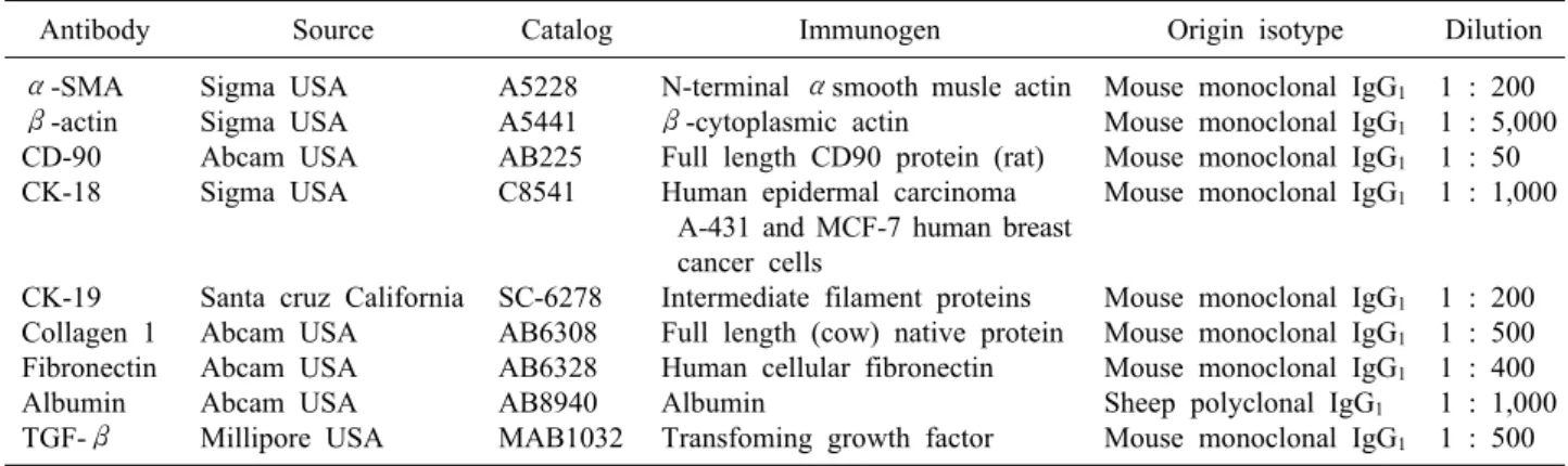
Table 1. Primary antibodies used in this study
Antibody Source Catalog Immunogen Origin isotype Dilution
α-SMA β-actin CD-90 CK-18
CK-19 Collagen 1 Fibronectin Albumin TGF-β
Sigma USA Sigma USA Abcam USA Sigma USA
Santa cruz California Abcam USA Abcam USA Abcam USA Millipore USA
A5228 A5441 AB225 C8541
SC-6278 AB6308 AB6328 AB8940 MAB1032
N-terminal αsmooth musle actin β-cytoplasmic actin
Full length CD90 protein (rat) Human epidermal carcinoma
A-431 and MCF-7 human breast cancer cells
Intermediate filament proteins Full length (cow) native protein Human cellular fibronectin Albumin
Transfoming growth factor
Mouse monoclonal IgG1
Mouse monoclonal IgG1
Mouse monoclonal IgG1
Mouse monoclonal IgG1
Mouse monoclonal IgG1
Mouse monoclonal IgG1
Mouse monoclonal IgG1
Sheep polyclonal IgG1
Mouse monoclonal IgG1
1 : 200 1 : 5,000 1 : 50 1 : 1,000
1 : 200 1 : 500 1 : 400 1 : 1,000 1 : 500
for further 2 weeks, sacrificed in 10 weeks (8 weeks after TAA administration and 2 weeks after isolated MSCs transplantation via transsplenic puncture).
HGF-treated MSCs: MSCs were harvested after cul- ture for 3 or 4 passages. Prepared MSCs were incubated in culture medium with CELL STALKER-CSR dye for overnight at 37oC, 5% CO2 and incubated with HGF (50 ng/ml) during 2 weeks extra. HGF-treated MSCs (2×10⁶/ 30 ul cells) were transplanted via transsplenic infusion.
Animals were anesthetized, a 3 cm-long midline abdomi- nal incision was made, and the abdominal cavity was exposed. HGF-treated MSCs was injected directly into the inferior tip of the spleen using 50 ul Hamilton syringe (Hamilton Company, Nevada, USA). The pinhole at the injection site was pressed for hemostasis. Subsequently, 1 ml of normal sterile saline was administered into abdomen as fluid replacement. The abdominal wall was closed in two layers in a running fashion. All procedures were per- formed under sterile circumstances. The animals were sac- rificed in 10 weeks (8 weeks after TAA administration and 2 weeks after transplantation HGF-treated MSCs via direct splenic puncture).
Histological study
The animals were perfused with 300-400 ml of fixative containing 4% paraformaldehyde in 0.1 mol/L phosphate buffer (pH 7.4), and embedded in paraffin then cut into serial 5 μm thick sections. Liver tissue slices were fixed in 4% PFA solution and embedded in paraffin. Five mi- crometer thick histological cuts from the paraffin blocks obtained from all groups. The specimens were stained with H&E, Sirius-red, and PAS according to the standard protocols.
Immunohistochemistry
Fixed sections were permeabilized for 5 min with Triton X-100 (Merck) diluted to 0.5% in PBS. Endoge- nous peroxidase activity was quenched by incubating for 5 min in a solution containing 0.3% H2O2 in 10% metha- nol. Sections were blocked with 2% normal goat serum in PBS for 1 hr and incubated with primary antibodies at the indicated dilutions (Table 1) overnight at 4oC. For negative controls, primary antibodies were omitted or re- placed with normal serum. On the following day, cells were incubated with secondary antibodies (biotinylated rabbit or mouse IgG [1 : 400; Vector, Burlingame, CA]) for 1 hour, followed by streptavidin horseradish complex (1 : 400; Vector) for 1 hour. Immunolabeling was visua- lized using a 0.05% 3,3 -diaminobenzidine/0.01% H2O2
mixture as a chromogen. For fluorescent stain, cells were incubated with secondary antibodies (FITC-, CY-3 rabbit or mouse IgG [1 : 500; Jackson Lacoratories]) for 1 hour 30 min. The results were evaluated by Leica TCS-SP2 and Laser scanner confocal microscopy.
Western blot analysis
Cells were lysed by adding sodium dodecyl sulfate-pol- yacrylamide gel electrophoresis (SDS-PAGE) sample buf- fer (62.5 mM Tris-HCl, pH 6.8, 2% SDS, 7.8% glycerol, 4.5% mercaptoethanol, and 0.1% bromphenol blue) and boiling for 5 minutes at 100oC. Lysates were clarified by centrifugation, and protein concentrations of supernatants were determined with bovine serum albumin as a standard. Proteins were separated by SDS-PAGE using the Laemmli buffering system with 4% stacking and 10%
separating gels (70 min at 130 V), and transferred to ni- trocellulose membranes with a Bio-Rad transfer unit (120
Fig. 1. Histological appearances in the TAA-induced cirrhotic livers. For comparing isolated MSCs group with normal saline group or HGF-treated MSC group, each specimen was stained with H&E stain (Original magnification: ×10, ×20); more prominent hepatocyte regeneration and decreased hepatic fibrosis were detected in isolated MSCs group, but not normal saline or HGF-treat- ed MSCs group.
min at 200 mA). Protein blots were incubated in blocking buffer [2% BSA in Tween-20/Tris-buffered saline (TBS)]
for 1 hr at room temperature on a rotating platform, then sequentially incubated with primary antibodies (overnight) and horseradish peroxidase-conjugated secondary anti- bodies (1 hr), followed by three washes. Immunoreactive bands were visualized by chemiluminescence reagents (Pierce, Rockford, IL). Optical density of bands was ana- lyzed with an Imaging Densitometer (Bio-Rad GS-670).
Statistical analysis
Adobe Photoshop 7.0 (Adobe Systems, Mountain View, CA) was used to optimize image quality and to prepare figures. The data were analyzed by a one-way ANOVA with a Tukey-Kramer multiple comparison post hoc test in GraphPad Instat 3.05 (GraphPad, San Diego, CA). Each value is expressed as mean SEM, and p<0.001 was con- sidered significant.
RESULTS
Histological differences between the TAA/
MSCs and TAA/HGF-treated MSCs
Light microscopy demonstrated typical histopathologic changes indicative of persistent liver damage. TAA in- duced liver fibrosis became more intense and severe by 8 week, with moderate bile duct proliferation and fibrotic nodules (Fig. 1), which are more or less aggravated after Normal saline infusion in control group. Less hepatic fib- rosis and more hepatocyte regeneration were detected in isolated stem cell group compared to normal saline or HGF-treated MSC group. Contrary to our expectation, HGF-treated MSCs group is not remarkable to Normal saline group in regard to the extent and degree of hepatic repair (Fig. 1).
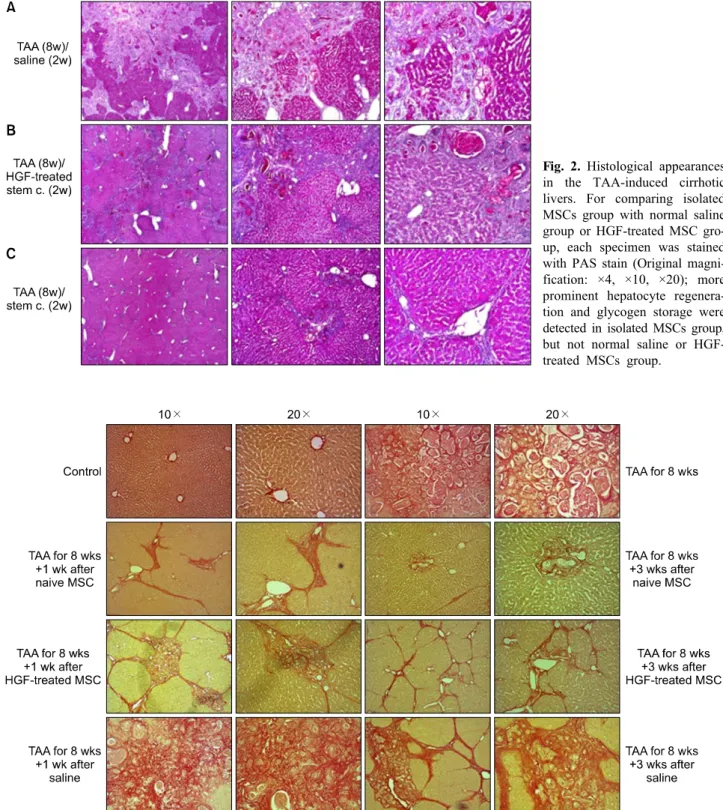
To assess fibrotic liver cells, paraffin sections were subjected to PAS staining, control group demonstrate demonstrated that glycogen-stained hepatocytes with PAS
Fig. 2. Histological appearances in the TAA-induced cirrhotic livers. For comparing isolated MSCs group with normal saline group or HGF-treated MSC gro- up, each specimen was stained with PAS stain (Original magni- fication: ×4, ×10, ×20); more prominent hepatocyte regenera- tion and glycogen storage were detected in isolated MSCs group, but not normal saline or HGF- treated MSCs group.
Fig. 3. Histological appearances in the TAA-induced cirrhotic livers. For comparing isolated MSCs group with normal saline group or HGF-treated MSC group, each specimen was stained with Sirius-red stain (Original magnification: ×10, ×20); Alleviated septal fibrosis and less collagen deposition were detected in isolated MSCs group, thickened fibrosis and more collagen deposition in normal saline or HGF-treated MSCs group.
staining was reduced and depicted a strongly time-depend- ent lack of glycogen (Fig. 2). Isolated MSCs group dem- onstrated a diffuse and homogeneous pattern of PAS
staining for glycogen (Fig. 2). Less hepatic fibrosis, more glycogen storage was detected in isolated stem cell group, compared to HGF-treated stem cell group (Fig. 2).
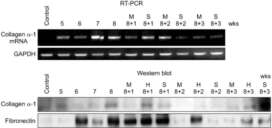
Fig. 4. Western blot analysis and RT-PCR study also showed these serial changes of fibrosis, Quantification of fibrosis induction and MSC-associated effects were assessed through antibody studies for collagen α-1 and fibronectin and RT-PCR for collagnen α-1 messanger ribonucleic andi (mRNA). The expressed amount of collagen α-1 and fibronectin were increased in 8 weeks after TAA ingestion and more decreased in additional 2 weeks after MSC infusion, contrary to control group.
Histopathologic analysis with Sirius-red staining dem- onstrated almost normalization of liver architecture in iso- lated MSCs group within 10 weeks of MSCs admin- istration (Fig. 3). Alleviated septal fibrosis and less colla- gen deposition were detected in isolated MSCs group, thickened fibrosis and more collagen deposition in normal saline or HGF-mediated MSC group. The difference be- tween HGF-treated MSC group and control group was not prominent unexpectedly (Fig. 3).
To assess the degree of hepatic fibrosis, expressed amount of collagen α-1 and fibronectin were measured using Western blot analysis and RT-PCR study. During thioacetamide ingestion, there was a time-dependent in- crease in expressed amount of collagen α-1 and fibron- ectin. After MSC implantation, however, a expressed amount of collagen and fibronection in fibrotic areas was decreased from week 1 to week 3. In contrast, expressed amount of collagen and fibronection in saline-injected rats increased still. Western blot analysis and RT-PCR study for collagen α-1 also supported these serial changes of fibrosis (Fig. 4).
The change and migration of stem cell after HGF-treatment
To evaluate differentiation of HGF-treated MSC into functional hepatocytes, we demonstrated the expression of albumin by high resolution fluorescent microscopy at 2 weeks after injection. Green autofluroresence in HGF-tr- eated stem cells indicate the differentiation to hepatocyte
and albumin expression, red autofluroresence indicated stem cell itself without differentiation (Fig. 5).
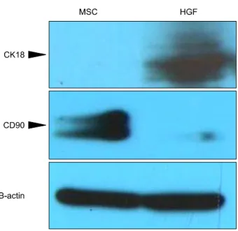
Protein expression of CK18, CD90 and β-actin in the cells was analyzed through western blot analysis (Fig. 6).
The protein bands for specific hepatic markers (CK-18) were visible in HGF-treated MSCs. The protein bands of specific stem cell marker (CD 90) were visible in Isolated MSCs. β-SMA is common in both group. Western blot analysis indicated that these specific markers were ac- tively synthesized before and after hepatic differentiation.
HGF-treated MSCs became the CK18-positive and Isolated MSCs CD90-postive (Fig. 6).
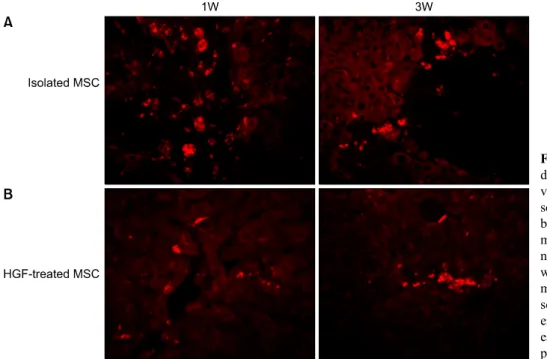
To identify Transplanted donor cell distribution to the recipient liver. We demonstrated CELL STALKER-CSR dye staining and the expression of albumin by high reso- lution fluorescent microscopy at 1 and 3 weeks after injection. Red autofluorescence is mainly localized near the sinusoids in stem cell group and scattered away the sinusoids in HGF-treated stem cells group, which indicate the migration of stem cells during the differentiation proc- ess (Fig. 7).
Comparing the serial differentiation phase between Isolated MSCs and HGF-treated MSCs
Hepatogenic differentiation of implanted MSCs was de- termined by immunohistochemistry using antibodies to al- bumin and CK19. Using CELL STALKER-CSR dye to detect MSCs, we found that these cells co-localized with
Fig. 5. Fluorescence microscope image showed that stem cells differentiate to hepatocyte and express the albumin in 2 weeks after HGF treatment. (A) Stem cells themselves without differ- entiation to hepatocyte-like cell (CELL STALKER-CSR dye sta- ining, red; Original magnifica- tion: ×10, ×20). (B) Stems cells with differentiation to hepato- cyte and albumin expression (Anti-albumin staining, green;
Original magnification: ×10, ×20).
Fig. 6. Western blot analysis indicated that these specific markers were actively synthesized before and after hepatic differentiation: HGF-treated MSCs became the CK18-pos- itive, Isolated MSCs became CD 90-postive. CD90 is a stem cell marker and CK18 is a hepatocyte marker.
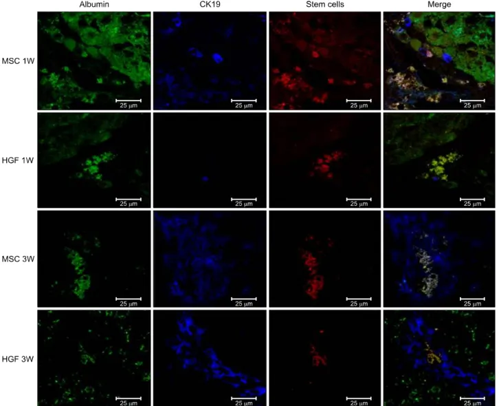
antibodies to CK19- or thy1-stained oval cells and albu- min (Fig. 8). One week after MSC implantation, CK19 stained oval cells derived from MSCs were located around the portal veins. At 3 weeks, more differentiated MSCs or hepatocyte-like cells were located toward the hepatic
parenchyma, and albumin-stained areas had expanded and overlapped the MSC-stained areas. Nearly all the CK19/thy1- and albumin-stained areas overlapped the MSC-stained areas, indicating that most MSCs had trans- differentiated into oval progenitor cells and then into functional hepatocyte-like cells after hepatic implantation.
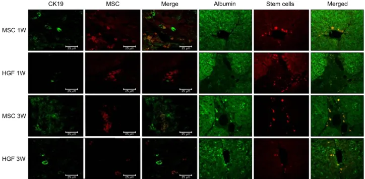
HGF-treated MSCs differentiated into hepatocyte without the oval cells phase (Fig. 9). Isolated MSCs in the merged image demonstrate co-localization of CK-19 containing vesicles and stem cell, which mean sequential different- iation (i.e., MSCs differentiate to hepatocyte-like cell via oval cell phase).
α-SMA is not only a marker for activated hepatic stel- late cells, but also for portal myofibroblasts. TUNEL rep- resents a rather specific marker with respect to hepatic stellate cells degradation and fibrosis repair. At 1 week after MSC injection, there were some TUNEL-positive non-stellate cells, but its proportion decreased after 3 weeks. This finding indicates that the proportion of stel- late cells among TUNEL-positive cells increased although total amount of TUNEL-positive cells decreased after 3 weeks (Fig. 10). To evaluate the extent of the hepatic fib- rosis and repair by High fluorescence microscope images, Isolated MSC groups express lower α-SMA, higher TUNEL and albumin expression, which have much abil-
Fig. 7. Transplanted donor cell distribution to the recipient li- vers. High fluorescence micro- scope images (×20). (A) A num- ber of Red autofluorescence is mainly localized near hepatic si- nusoids in stem cell group, which indicated better engraft- ment (B). A few red autofluore- scence is localized near periph- eral hepatic sinusoids in HGF-tr- eated stem cells group, indicated poor engraftment.
ities to increase the appotosis of hepatic stellate cells and restore the hepatic fibrosis compared with HGF-treated MSCs (Fig. 10).
DISCUSSION
The end-stage of liver disease, such as hepatic failure, end-stage cirrhosis and infections present a major health concerns. Given the shortage of organs for liver trans- plantation, an increasing emphasis is being placed on cell-based liver therapies. Some of these therapeutic ap- proaches have been moderate or short-term success.17,18 But, hepatocyte transplantation has not evolved as a viable alternative to liver transplantation, due to difficulty in a large amount of high-quality hepatocyte collection from the same donor liver. In addition, the in vitro culture of hepatocytes for transplantation is demanding and ineffi- cient.19
Stem cells are therefore considered to be a potential al- ternative for liver-directed cell therapies since it has been reported that various kinds of stem cells were able to dif- ferentiate into functioning cells of various mature tissues including hepatocytes.20,21 MSCs were shown to have mul- tiple beneficial effects in vitro that were relevant in a ther- apeutic context, including hepatocellular functional sup- port, secretion of molecules that inhibit hepatocyte apop- tosis, and modulation of an acute phase response by hep-
atocytes cultured in ALF-induced serum. BM-derived MSCs were isolated and expanded in adult rats and their multiple differentiation potential was also confirmed in vitro.8,17,18 Stem cell-derived hepatocytes may be an alter- native cell source to treat liver diseases or to be used for pharmacological purposes currently.
HGF was originally reported in 1984 to be a mitogenic protein for mature hepatocytes in primary culture. HGF is a mesenchymal-derived cytokine that acts as mitogen, motogen, and morphogen in various target cells.22-25 HGFs ares expressed primarily by a variety of mesenchymal cells, including fibroblasts, hepatic stellate cells, smooth muscle cells, and endothelia, but also by megakaryocytic and myeloic lineages.26 HGFs are classical mediator of mesenchymal epithelial interactions.27,28 The important bi- ological role of HGF is emphasized by the finding that mice that are transgenic for the HGF gene recover their original parenchymal liver cell mass twice as fast as con- trols after partial hepatectomy,29 Dong et al.30 insisted that HGFs are essential for the initiation of hepatic diffe- rentiation. HGF are the key cytokines involved in the liv- er-injury conditioned medium for the hepatic differ- entiation of BM-MSCs. Nishino et al.31 insisted that HGF gene transfer significantly improved survival after partial hepatectomy in the cirrhotic rats, HGF gene transfer to cirrhotic livers improves liver failure-associated death af- ter partial hepatectomy by upregulating expression of an
Fig. 8. Transplanted donor cell engraftment to the recipient liver and differentiation into the hepatocytes. High fluorescence microscope images (×20). Anti-CK19 staining, green. MSCs with CELL STALKER-CSR dye staining, red. Albumin positive stem cells in the merged images, yellow. Hepatogenic differentiation-related analyses showed that infusion of isolated MSCs advanced the appearance of oval cells in 1 week, leading to a higher population of hepatic progenitor cells and a lower population of MSCs at 3 weeks.
antiapoptotic protein, Bcl-xl. Yu et al.32 found that MSCs implantation with HGF significantly inhibited the for- mation of liver fibrosis in rats undergoing small for size liver transplantation, while MSCs implantation with HGF had synergistic effects in the process. Most modes have been indicated to synergistic relationships, which HGF not only stimulates hepatic regeneration but also accelerates liver function, improves fibrosis, and protects liver cells against injury.31,33-35 But, much more interesting is the dif- ference between our results and previously stated con- tents.
In this study, after rats were treated with oral admin- istration of TAA for 8 weeks, the liver exhibited a marked
increase in extracellular matrix content and displayed bun- dles of collagen surrounding the lobules leading to large fibrous septa. But In 2 week later after implantation of MSCs, Less hepatic fibrosis and more hepatocyte re- generation were detected in isolated stem cell group com- pared to normal saline or HGF-mediated MSC group (Fig.
1C). Histopathologic analysis demonstrated almost nor- malization of liver architecture in isolated MSCs group within 10 weeks of MSCs administration (Fig. 3C). Alle- viated septal fibrosis and less collagen deposition were detected in isolated MSCs group, thickened fibrosis and more collagen deposition in normal saline or HGF-medi- ated MSC group. The difference between HGF-treated
Fig. 9. Transplanted donor cell engraftment to the recipient liver and differentiation into the hepatocytes. High fluorescence microscope images (×20). Hepatogenic differentiantion of implanted MSCs was assessed by immunohistochemistry using Antibody to albunin, CK-19 and CELL STALKKER-CSR dye to assess the MSCs. One week after MSC implantation, CK19 stained oval cells derived from MSCs were located around the portal veins. At 3 weeks, more differentiated MSCs or hep- atocyte-like cells were located toward the hepatic parenchyma, and albumin-stained areas had expanded and overlapped the MSC-stained areas. Nearly all the CK19/thy1- and albumin-stained areas overlapped the MSC-stained areas, indicating that most MSCs had transdifferentiated into oval progenitor cells and then into functional hepatocyte-like cells after hepatic implantation.
MSC group and control group was not prominent (Fig.
3B and 3C).The extent and degree of hepatic repair is similar or a little better in HGF-treated MSCs group com- pared with Normal saline group (Fig. 1B), rather much decreased in HGF-treated MSCs group compared with isolated MSCs group unexpectedly. It is not clear why hepatic repair is more prominent in isolated MSCs group compared with HGF-treated MSCs group.
Recently, considerable doubt has been cast over the synergic effect between mesenchymal stem cell and HGF.
Ren et al.36 insisted that human MSC can differentiate into hepatocyte-like cells but do not accelerate the capillariza- tion and venularization of hepatic sinusoids, finally lead- ing to the partial improvement of liver function in mice with CCl4-mediated chronic liver fibrosis. Baertschiger et al.37 insisted MSC differentiated into myofibroblasts with development of fibrous tissue, when transplanted into an injured or regenerating liver, which indicate that MSC in certain circumstances might be harmful due to their fibro- genic potential.
There are several studies that emphasize about the mi- cro-milieu such as spatial configuration, cell to cell re-
action to related to cell differentiation. Qihao et al.38 in- sisted that the co-culture microenvironment plays a deci- sive role for the hepatic differentiation of MSCs and it is more efficient than HGF treatment, which emphasize on cell to cell reaction in regard to hepatic differentiation.
Lange et al.39 insisted that the rat MSCs from bone mar- row can differentiate hepatocytic cells in the presence of fetal liver cell in vitro and the presence of MSCs in co-cultures also provides a beneficial environment for ex- pansion and differentiation of fetal liver cell. Differentia- tion of stem cells might be tightly regulated by the micro- environment which is mainly composed of non-paren- chymal cells. Deng et al.40 investigated fully activated hepatic stellate cells could modulate MSCs differentiation into hepatocyte-like cells. As organogenesis is not only dependent upon soluble factors but also cell-cell inter- actions, development of 3-dimensional culture systems wherein the anatomical features of developing liver lo- bules are recreated and the use of bio-reactors making it possible to more closely control physiological parameters such as pH and glycemia, may be needed to allow the creation of hepatocytes with fully mature characteristics
Fig. 10. Hepatic fibrosis and repair by transplanted donor cell engraftment to the recipient liver. High fluorescence microscope images: Engrafted isolated MSC groups express lower α-SMA and higher TUNEL compared with HGF-treated MSCs group.
(A) Anti-α-SMA staining, red. (B) TUNEL staining, green. (C) α-SMA and TUNEL positive stem cells in the merged images, yellow. The merged images of TUNEL and α-SMA stains also revealed that apoptosis of hepatic stellate cells was less evident after HGF treatment.
and functions. The in vitro culture system still does not recreate all signals present in vivo that govern a coordi- nated maturation from pluripotent cells to terminally dif- ferentiated mature hepatocytes, despite the use of cytokine cocktails known to play a role during liver development.
To evaluate the extent of the hepatic fibrosis and repair by high fluorescence microscope images, Isolated MSC groups express lower α-SMA, higher TUNEL and albu- min expression, which have much abilities to increase the appotosis of hepatic stellate cells and restore the hepatic fibrosis compared with HGF-treated MSCs (Fig. 8). α- SMA is not only a marker for activated hepatic stellate cells, but also for portal myofibroblasts. TUNEL repre- sents a rather specific marker with respect to hepatic stel- late cells degradation and fibrosis repair. It seems that Mature hepatocyte have a less powerful potentials such as, albumin synthesis, repair of fibrosis than mesenchymal stem cell. It is interesting that MSCs have more potent capacity compared with mature hepatocyte. This suggests that there are more important things in early or inter- mediate differentiation process of stem cell before matura-
tion that have not clarified.
The mechanisms by which MSCs repair the fibrosis are still unclear. Fang et al.41 found that although albu- min-positive donor-derived cells were present at lower frequency in sections of CCL4-induced liver tissues, in- fusion of FLK1+ murine MSCs might ameliorate liver fibrosis.
Ortiz et al.42 observed that MSCs administration re- duced the degree of bleomycin-induced inflammation and collagen deposition within lung tissue, but male donor DNA accounted for 2.21×10⁻⁵% of total lung DNA in fe- male recipient mice with MSCs treatment. Some studies insisted that engrafted MSCs scattered mostly in the hep- atic connective tissue but did not differentiate into hep- atocytes expressing human albumin or alpha-fetoprotein.
Instead, these engrafted, undifferentiated MSCs secreted a variety of bioactive cytokines that may restore liver func- tion and promote regeneration.43 Although the current study did not explain how the MSCs have more potential comparing with mature hepatocytes, this study support that MSCs have more powerful anti-fibrosis, regenerative
endoderm, hepatoblasts, and finally cells with phenotypic and functional characteristics of hepatocytes, Li et al.45 have previously developed a growth-factor-free co-culture method and observed rat MSCs differentiated into hepatic progenitor cells. Co-cultured MSCs had stayed at hepatic progenitor stage until week 3, and differentiated into hep- atocytes or bile-ductal epithelial cells.
Remarkably, some studies proved the differentiation of rat multipotent adult progenitor cells to hepatocyte-like cells, even though rMAPC are isolated clonally from cul- tured rat bone marrow (BM) and have characteristics of primitive endoderm cells, which will be useful for gen- erating hepatocyte-like cells from rodent and human stem cells, and to gain insight into the early stages of liver development.44 Until recently, there has been quite un- known process to clarify the early differentiation phase of stem cell.
The results of this study demonstrated that migration and engraftment of MSCs to the injured liver area, and differentiation into functional hepatocytes from 1 to 3 weeks. Localization of differentiation cells derived from transplanted MSCs was time-dependent changed from around centrilobular hepatic veins to parenchyma. Isolated MSCs is mainly localized in sinusioids, HGF-treated stem cell is scattered away to parenchyma in 3 weeks. MSCs transdifferentiated into CK-19 postive oval cells and then to albulmin-producing hepatocytes, HGF-treated MSCs differentiated into hepatocyte without the intermediate oval cells phase. HGF treated MSCs became the CK18- positive, MSCs became CD 90-positive.
At present, hepatocyte transplantation has long been recognized as a potential treatment for life-threatening liv- er disease. The potential of hepatocyte transplantation re- mains largely doubtful yet. Authors investigated various parameters expressing more success and efficacy of using MSCs than mature hepatocyte as an alternative cell source to hepatocyte transplantation for treatment of liver disease. Unfortunately, this study leave the unanswered the question why differences occurring between Isolated
this model.
Significant heptocyte differentiation occurred in not HGF-treated MSCs but isolated MSCs groups unexpec- tedly. These results suggest that the beneficial effect of MSC on in a rat model with thiacetamide-induced cir- rhosis may occur during early differentiation course of MSCs, mature hepatocyte itself has a little effect on the acceleration of hepatic lineage cell differentiation. Further studies focused on the early process of differentiation may elucidate the new mechanism underlying MSC-associated effects.
REFERENCES
1. Alcolado R, Arthur MJ, Iredale JP. Pathogenesis of liver fibrosis.
Clin Sci (Lond) 1997;92:103-112.
2. Iredale JP. Cirrhosis: new research provides a basis for rational and targeted treatments. BMJ 2003;327:143-147.
3. Carvalho AB, Quintanilha LF, Dias JV, et al. Bone marrow mul- tipotent mesenchymal stromal cells do not reduce fibrosis or im- prove function in a rat model of severe chronic liver injury. Stem Cells 2008;26:1307-1314.
4. Mezey E, Chandross KJ, Harta G, et al. Turning blood into brain:
cells bearing neuronal antigens generated in vivo from bone marrow. Science 2000;290:1779-1782.
5. Pittenger MF, Mackay AM, Beck SC, et al. Multilineage poten- tial of adult human mesenchymal stem cells. Science 1999;284:
143-147.
6. Muraglia A, Cancedda R, Quarto R. Clonal mesenchymal progen- itors from human bone marrow differentiate in vitro according to a hierarchical model. J Cell Sci 2000;113:1161-1166.
7. Aurich I, Mueller LP, Aurich H, et al. Functional integration of hepatocytes derived from human mesenchymal stem cells into mouse livers. Gut 2007;56:405-415.
8. Sato Y, Araki H, Kato J, et al. Human mesenchymal stem cells xenografted directly to rat liver are differentiated into human hep- atocytes without fusion. Blood 2005;106:756-763.
9. Petersen BE, Bowen WC, Patrene KD, et al. Bone marrow as a potential source of hepatic oval cells. Science 1999;284:
1168-1170.
10. Zhao DC, Lei JX, Chen R, et al. Bone marrow-derived mesen- chymal stem cells protect against experimental liver fibrosis in rats. World J Gastroenterol 2005;11:3431-3440.
11. Zheng JF, Liang LJ. Intra-portal transplantation of bone marrow stromal cells ameliorates liver fibrosis in mice. Hepatobiliary Pancreat Dis Int 2008;7:264-270.
12. Seo MJ, Suh SY, Bae YC, et al. Differentiation of human adipose stromal cells into hepatic lineage in vitro and in vivo. Biochem Biophys Res Commun 2005;328:258-264.
13. Bian L, Guo ZK, Wang HX, et al. In vitro and in vivo im- munosuppressive characteristics of hepatocyte growth fac- tor-modified murine mesenchymal stem cells. In Vivo 2009;23:
21-27.
14. Constantinou MA, Theocharis SE, Mikros E. Application of me- tabonomics on an experimental model of fibrosis and cirrhosis induced by thioacetamide in rats. Toxicol Appl Pharmacol 2007;218:11-19.
15. Yu Y, Lu L, Qian X, et al. Antifibrotic effect of hepatocyte growth factor-expressing mesenchymal stem cells in small-for- size liver transplant rats. Stem Cells Dev 2010;19:903-914.
16. Roos F, Terrell TG, Godowski PJ, et al. Reduction of alpha-naph- thylisothiocyanate-induced hepatotoxicity by recombinant human hepatocyte growth factor. Endocrinology 1992;131:2540-2544.
17. Dhawan A, Mitry RR, Hughes RD. Hepatocyte transplantation for liver-based metabolic disorders. J Inherit Metab Dis 2006;29:
431-435.
18. Strom SC, Bruzzone P, Cai H, et al. Hepatocyte transplantation:
clinical experience and potential for future use. Cell Transplant 2006;15 Suppl 1:S105-110.
19. Oyagi S, Hirose M, Kojima M, et al. Therapeutic effect of trans- planting HGF-treated bone marrow mesenchymal cells into CCl4-injured rats. J Hepatol 2006;44:742-748.
20. Harris RG, Herzog EL, Bruscia EM, et al. Lack of a fusion re- quirement for development of bone marrow-derived epithelia.
Science 2004;305:90-93.
21. Krause DS, Theise ND, Collector MI, et al. Multi-organ, mul- ti-lineage engraftment by a single bone marrow-derived stem cell.
Cell 2001;105:369-377.
22. Zarnegar R, Michalopoulos GK. The many faces of hepatocyte growth factor: from hepatopoiesis to hematopoiesis. J Cell Biol 1995;129:1177-1180.
23. Matsumoto K, Nakamura T. Emerging multipotent aspects of hepatocyte growth factor. J Biochem 1996;119:591-600.
24. Rosen EM, Goldberg ID. Scatter factor and angiogenesis. Adv Cancer Res 1995;67:257-279.
25. Furlong RA, Takehara T, Taylor WG, et al. Comparison of bio- logical and immunochemical properties indicates that scatter fac- tor and hepatocyte growth factor are indistinguishable. J Cell Sci 1991;100:173-177.
26. Nusrat A, Parkos CA, Bacarra AE, et al. Hepatocyte growth fac- tor/scatter factor effects on epithelia. Regulation of intercellular junctions in transformed and nontransformed cell lines, baso- lateral polarization of c-met receptor in transformed and natural intestinal epithelia, and induction of rapid wound repair in a transformed model epithelium. J Clin Invest 1994;93:2056-2065.
27. Montesano R, Matsumoto K, Nakamura T, et al. Identification of a fibroblast-derived epithelial morphogen as hepatocyte growth factor. Cell 1991;67:901-908.
28. Rosen EM, Nigam SK, Goldberg ID. Scatter factor and the c-met receptor: a paradigm for mesenchymal/epithelial interaction. J Cell Biol 1994;127:1783-1787.
29. Shiota G, Wang TC, Nakamura T, et al. Hepatocyte growth factor in transgenic mice: effects on hepatocyte growth, liver regene- ration and gene expression. Hepatology 1994;19:962-972.
30. Dong XJ, Zhang H, Pan RL, et al. Identification of cytokines in- volved in hepatic differentiation of mBM-MSCs under liver-in- jury conditions. World J Gastroenterol 2010;16:3267-3278.
31. Nishino M, Iimuro Y, Ueki T, et al. Hepatocyte growth factor improves survival after partial hepatectomy in cirrhotic rats sup- pressing apoptosis of hepatocytes. Surgery 2008;144:374-384.
32. Yu Y, Lu L, Qian X, et al. Antifibrotic effect of hepatocyte growth factor-expressing mesenchymal stem cells in small-for- size liver transplant rats. Stem Cells Dev 2010;19:903-914.
33. Kaido T, Seto S, Yamaoka S, et al. Perioperative continuous hep- atocyte growth factor supply prevents postoperative liver failure in rats with liver cirrhosis. J Surg Res 1998;74:173-178.
34. Yagi H, Parekkadan B, Suganuma K, et al. Long-term superior performance of a stem cell/hepatocyte device for the treatment of acute liver failure. Tissue Eng Part A 2009;15:3377-3388.
35. Kisseleva T, Brenner DA. Role of hepatic stellate cells in fibro- genesis and the reversal of fibrosis. J Gastroenterol Hepatol 2007;22 Suppl 1:S73-78.
36. Ren H, Zhao Q, Cheng T, et al. No contribution of umbilical cord mesenchymal stromal cells to capillarization and venulariza- tion of hepatic sinusoids accompanied by hepatic differentiation in carbon tetrachloride-induced mouse liver fibrosis. Cytotherapy 2010;12:371-383.
37. Baertschiger RM, Serre-Beinier V, Morel P, et al. Fibrogenic po- tential of human multipotent mesenchymal stromal cells in in- jured liver. PLoS One 2009;4:e6657.
38. Qihao Z, Xigu C, Guanghui C, et al. Spheroid formation and dif- ferentiation into hepatocyte-like cells of rat mesenchymal stem cell induced by co-culture with liver cells. DNA Cell Biol 2007;26:497-503.
39. Lange C, Bruns H, Kluth D, et al. Hepatocytic differentiation of mesenchymal stem cells in cocultures with fetal liver cells. World J Gastroenterol 2006;12:2394-2397.
40. Deng X, Chen YX, Zhang X, et al. Hepatic stellate cells modu- late the differentiation of bone marrow mesenchymal stem cells into hepatocyte-like cells. J Cell Physiol 2008;217:138-144.
41. Fang B, Shi M, Liao L, et al. Systemic infusion of FLK1(+) mes- enchymal stem cells ameliorate carbon tetrachloride-induced liver fibrosis in mice. Transplantation 2004;78:83-88.
42. Ortiz LA, Gambelli F, McBride C, et al. Mesenchymal stem cell engraftment in lung is enhanced in response to bleomycin ex- posure and ameliorates its fibrotic effects. Proc Natl Acad Sci U S A 2003;100:8407-8411.
43. Tsai PC, Fu TW, Chen YM, et al. The therapeutic potential of human umbilical mesenchymal stem cells from Wharton's jelly in the treatment of rat liver fibrosis. Liver Transpl 2009;15:
484-495.
44. Roelandt P, Pauwelyn KA, Sancho-Bru P, et al. Human embry- onic and rat adult stem cells with primitive endoderm-like pheno- type can be fated to definitive endoderm, and finally hep- atocyte-like cells. PLoS One 2010;5:e12101.
45. Li TZ, Kim JH, Cho HH, et al. Therapeutic potential of bone- marrow-derived mesenchymal stem cells differentiated with growth-factor-free coculture method in liver-injured rats. Tissue Eng Part A 2010;16:2649-2659.