INTRODUCTION
Fetal cystic hygromas are congenital malformations of the lymphatic system appearing as single or multiloculated fluid- filled cavities (1). About 75% of the tumors occur in the neck, with a predilection for the left side, mainly in the posterior triangle. Ten to twenty percent of the tumors occur in the axillary region and rare locations include the mediastinum, retroperitoneal area, abdominal viscera, bones, pelvis, groin, scrotum, and the chest wall (2-7). They often progress to hy- drops fetalis and cause fetal death. There is a high prevalence of associated chromosomal abnormalities, Turner's syndrome being the most common.
Recently, we experienced a case of fetal axillary cystic hy- groma at 38 weeks' gestation in a 30-yr-old multiparous woman. Here we report this case with a brief review of the literature.
CASE REPORT
A 30-yr-old woman with gravida 3 and para 2 was referred at 35 weeks' gestation because of a mass on the left fetal chest wall detected by routine ultrasonography performed at a pri- vate clinic at 34 weeks' gestation. There was no fetal abnor- mality by ultrasonography at 28 weeks' gestation at a private
clinic. The patient's family history and previous medical history were unremarkable.
Her first normal child was delivered by cesarean section due to an arrest disorder 4 yr ago and second normal child was delivered vaginally 2 yr ago. In the current pregnancy, the maternal serum -fetoprotein checked at 16 weeks' ges- tation was normal. Ultrasound examination, performed in our hospital at 35 weeks' gestation, revealed a 6×5 cm-sized, multiseptated cystic mass in the left axillary region of the fetus (Fig. 1). Amniotic fluid amount was normal and no other structural abnormalities were found with normal echo- cardiography in the fetus. Biparietal diameter, femur length, and abdominal circumference in the fetus corresponded to 35 weeks' gestation. With the diagnosis of a fetal axillary mass, elective cesarean section was performed at 38 weeks' gestation. A 3,520 g-sized male infant was delivered with Apgar scores of 10 and 10 at 1 min and 5 min, respectively.
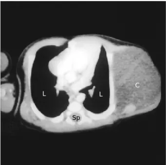
On delivery, the baby demonstrated a 6×7 cm-sized, soft cystic mass in the left axillary area (Fig. 2). No other struc- tural anomalies were identified. Umbilical cord blood taken at the delivery revealed a normal karyotype (46, XY). Chest CT was performed on the second postnatal day (Fig. 3). It showed an 8×6 cm-sized, well-marginated cystic mass with multiple septation at the left lateral chest wall without exten- sion to the upper neck or mediastinum.
Operation was performed on the 8th day after birth at the Tae-Bok Song, Cheol-Hong Kim, Seok-Mo Kim, Yoon-Ha Kim, Ji-Soo Byun, Eun-Kyung Kim*
Department of Obstetrics and Gynecology, Chonnam National University Medical School and Research Institute of Medical Sciences, Chonnam National University; Department of Diagnostic Radiology*, Chosun University Medical School, Gwangju, Korea
Address for correspondence Tae-Bok Song, M.D.
Department of Obstetrics and Gynecology Chonnam National University Medical School, 5 Hak-dong, Dong-gu, Gwangju 501-746, Korea Tel : +82-62-220-6373, Fax : +82-62-227-1637 E-mail : [email protected]
400 J Korean Med Sci 2002; 17: 400-2
ISSN 1011-8934
Copyright � The Korean Academy of Medical Sciences
Fetal Axillary Cystic Hygroma Detected by Prenatal Ultrasonography
: A Case Report
Fetal cystic hygroma is a rare developmental congenital anomaly of the lymphatic system, characterized by the formation of a multilocular, variable sized cystic mass. Most of cystic hygromas are found in the neck and other rare locations include axilla, mediastinum, and limbs. There are many papers about cystic hygro- ma colli, but there are only a few papers about fetal axillary cystic hygroma and no domestic papers. We present a case of fetal axillary cystic hygroma diagnosed antenatally followed by full-term delivery in a 30-yr-old woman. Operation was performed on the 8th day after birth and the mass was excised and confirmed as cystic hygroma.
Key Words : Lymphangioma, Cystic; Axillary; Fetal Lymphangioma, Cystic; Prenatal Diagnosis
Received : 21 May 2001 Accepted : 16 July 2001
Department of General Surgery and the mass was excised.
Microscopically, the mass showed irregularly dilated spaces lined by flattened endothelium. There was scattering of lym- phocytes in the stroma (Fig. 4). Currently, the baby grows well at the age of 1 yr and 8 months with 13.5 kg of body weight. There have been no signs of complications or sequelae.
DISCUSSION
The incidence of cystic hygroma is estimated to be 1:6,000 pregnancies but it is a relatively common anomaly in mis- carried fetuses, with a frequency of 1:875 (8). There are many papers about cystic hygroma colli, but there are only a few
Fetal Axillary Cystic Hygroma 401
Fig. 1.Transverse sonographic view of chest at 35 weeks' ges- tation demonstrating a large cystic mass with septae (arrows) in the left axillary region. Sp, fetal spine; Ht, fetal heart.
Fig. 2.A photograph demonstrating a left axillary mass (arrows) after delivery.
Fig. 4.Microscopic finding revealing dilated lymphatic vessels with lymphoid aggregates in the middle of fibrous tissue (H&E,
×100).
Fig. 3.Chest CT demonstrating a 8×6 cm-sized, well demar- cated nonenhancing cystic mass (C) in the left lateral chest.
L, fetal lung; Sp, spine.
C L L
Sp Ht Sp
papers about fetal axillary cystic hygroma (2-4) and no dome- stic papers. There is no predilection for cystic hygromas by either sex (6). Cystic hygromas are generally bilateral, thin- walled, unilocular or multilocular cysts with clear to turbid fluid. In practice the term cystic hygroma is used to refer to subcutaneous cystic spaces in the neck or to loculated cystic lesions in the noncervical regions, which have a similar ap- pearance to the nuchal cysts.
Several reviews on cystic hygroma have documented the postnatal anatomic variability (5, 10, 11). Seventy-five to ninety percent are found in the neck, 10-20% are located in the axilla (3, 7), and less than 10% are located in the extre- mities, trunk, abdomen, genitalia, etc.
Fetal nuchal cystic hygromas are thought to be etiological- ly distinct from other cystic lymphangiomas and are believed to result from inadequate drainage of the lymphatic vessels into the venous system secondary to atresia. In contradistinc- tion, cystic lymphangioma at other locations probably develop as the lymphatic anlage grow abnormally and never achieve sufficient anastomoses with the larger lymphatic channels (12). Association with other anomalies and extension to adja- cent structures leading to hydrops fetalis are frequent find- ings.
The frequency of a chromosomal abnormality associated with cystic hygroma may be as high as 78%, Turner syn- drome being the most common (13). Prognosis for cystic hygroma is grim if the karyotype is abnormal, or if ascites and pleural fluid are present, or if bilateral pleural effusions are seen. Reichler and Bronshtein (2) reported that three of five fetal axillary cystic hygromas were associated with chro- mosomal abnormalities (trisomy 21 in 2 cases and trisomy 18 in 1 case). Our case showed a normal karyotype.
The survival rate progressively improves with normal karyo- type, unilateral pleural effusions, atypical location and reso- lution of cystic hygroma (14). No single feature signifies 100% survival, however, and serial sonographic examina- tions are mandatory to evaluate any changes in the clinical manifestation. Once a cystic hygroma is detected, a careful search is indicated for fetal skin edema, ascites, pleural and pericardial effusions, and cardiac or renal anomalies.
Meticulous surgical excision is the treatment of choice when lesions are large. The operation is essentially conservative, since there is no justification for sacrificing any vital struc- tures to achieve complete removal of the benign lesion (5).
Most surgeons agree that the cystic hygroma should be excised when the diagnosis is made, because of the risk of severe com- plications. This advice should be modified if an important structure is involved and the surgery can be delayed for 6 months or 1 yr until the child has had an opportunity to grow (15). Other types of treatment have been proposed as adju- vants such as radiotherapy, injection of sclerosing agents, as- piration, and carbon dioxide laser, which are controversial
(5, 15, 16). Conservative management with observation has been recommended by some surgeons in asymptomatic pa- tients (16).
Because only small numbers of fetal cystic hygroma have been reported so far, it is uncertain if axillary cystic hygromas carry a marked risk for aneuploidy or untoward pregnancy outcome.
REFERENCES
1. Chervenak FA, Isaacson G, Blakemore KJ, Breg WR, Hobbins JC, Berkowitz RL, Tortora M, Mayden K, Mahoney MJ. Fetal cystic hygroma: cause and natural history. N Engl J Med 1983; 309: 822-5.
2. Reichler A, Bronshtein M. Early prenatal diagnosis of axillary cystic hyfroma. J Ultrasound Med 1995; 14: 581-4.
3. McCoy MC, Kuller JA, Chescheir NC, Coulson CC, Katz VL, Naka- yama DK. Prenatal diagnosis and management of massive bilateral axillary cystic lymphangioma. Obstet Gynecol 1995; 85: 853-6.
4. Hoffman-Tretin J, Koenigsberg M, Ziprkowski M. Antenatal demon- stration of axillary cystic hygroma. J Ultrasound Med 1988; 7: 233-5.
5. Tran-Ngoc-Ninh, Tran-Xuan-Ninh. Cystic hygroma in children: a report of 126 cases. J Pediatr Surg 1974; 9: 191-5.
6. Sheth S, Nussbaum AR, Hutchins GM, Sanders RC. Cystic hygromas in children: sonographic-pathologic correlation. Radiology 1987;
162: 821-4.
7. Judith E, Allanson MD. Lymphangioma. In: Stevenson RE, Hall JG, Goodman RM, editors, Human malformation and related anomalies.
New York: Oxford University Press, 1993; 288.
8. O'Brien WF, Cefalo RC, Bair DG. Ultrasonographic diagnosis of fetal cystic hygroma. Am J Obstet Gynecol 1980; 138: 464-6.
9. Cohen MM, Schwartz S, Schwartz MF, Blitzer MG, Raffel LJ, Mullins-Keene CL, Sun CC, Blakemore KJ. Antenatal detection of cystic hygroma. Obstet Gynecol Surv 1989; 44: 481-90.
10. Siegel MJ, McAlister WH, Askin FN. Lymphangiomas in children:
report of 121 cases. J Can Assoc Radiol 1979; 30: 99-102.
11. Bhattacharyya NC, Yadav K, Mitra SK, Pathak IC. Lymphangiomas in children. Aust N Z J Surg 1981; 51: 296-300.
12. Allanson JE. Lymphangioma. In: Stevenson RE, Hall JG, Goodman RM, editors, Human malformations and related anomalies. Vol. II.
Oxford: Oxford University Press, 1993: 293-305.
13. Carr RF, Ochs RH, Ritter DA, Kenny JD, Fridey JL, Ming PM. Fetal cystic hygroma and Turner's syndrome. Am J Dis Child 1986; 140:
580-3.
14. Anderson NG, Kennedy JC. Prognosis in fetal cystic hygroma. Aust N Z J Obstet Gynecol 1992; 32: 36-9.
15. Feins NR, Raffensperger JG. Cystic hygroma, lymphangioma, and lymphedema. In: Raffensperger JG, editor, Sweden's Pediatric Sur- gery. Norwalk: Appleton & Lange, 1990: 167-72.
16. Stringel G. Hemangiomas and lymphangiomas. In: Ashcraft KW, Holder TM, editors, Pediatric Surgery. Philadelphia: WB Saunders, 1993: 814-6.
402 T.-B. Song, C.-H. Kim, S.-M. Kim, et al.