29
Immune Network
서 론
말초혈액에서 2차 림프조직으로 림프구가 이동하고, 다시 림프구가 림프조직을 벗어나 혈액순환 체계를 통 하여 말초조직으로 이동하는 것은 면역반응에서 필수적 인 현상이다(1,2). 림프구는 림프조직의 high endothelial venule (HEV)을 경유하여 말초림프 조직안으로 이동하
책임저자:어성국, 전북대학교 수의과대학 미생물학교실 ꂕ 561-756, 전주시 덕진구 덕진동 1가 664-14 Tel: 063-270-3882, Fax: 063-270-3780
E-mail: vetvirus@moak.chonbuk.ac.kr
본 연구는 2002년도 전북대학교 생체안전성연구소 학술연구비의 일부지원으로 이루어졌음(CNU-BSRI, No. 2002-03).
바이러스 감염에 대한 방어효과
전북대학교 수의과대학 미생물학교실, 생체안전성 연구소
어 성 국․조 정 곤
CCR7 Ligands Induced Expansion of Memory CD4+ T Cells and Protection from Viral Infection
Seong-Kug Eo and Jeong-Gon Cho
Department of Microbiology, College of Veterinary Medicine and Bio-Safety Institute of Chonbuk National University, Chonju, Korea
ABSTRACT
Background: CC chemokine receptor (CCR) 7 and cognate CCR7 ligands, CCL21
(formerly secondary lymphoid tissue chemokine [SLC]) and CCL19 (formerly Epstein-Barr virus-induced molecule 1 ligand chemokine [ELC]), were known to establish microen- vironment for the initiation of immune responses in secondary lymphoid tissue. As described previously, coadministration of DNA vaccine with CCR7 ligand-encoding plas- mid DNA elicited enhanced humoral and cellular immunity via increasing the number of dendritic cells (DC) in secondary lymphoid tissue. The author hypothesized here that CCR7 ligand DNA could effectively expand memory CD4+ T cells to protect from viral infection likely via increasing DC number. Methods: To evaluate the effect of CCR7 ligand DNA on the expansion of memory CD4+ T cells, DO11.10.BALB/c transgenic (Tg)-mice, which have highly frequent ovalbumin (OVA)323-339 peptide-specific CD4+ T cells, were used. Tg-mice were previously injected with CCR7 ligand DNA, then immunized with OVA323-339 peptide plus complete Freund's adjuvant. Subsequently, memory CD4+ T cells in peripheral blood lymphocytes (PBL) were analyzed by FACS analysis for memory phenotype (CD44high and CD62 Llow) at memory stage. Memory CD4+ T cells recruited into inflammatory site induced with OVA-expressing virus were also analyzed. Finally, the protective efficacy against viral infection was evaluated.Results: CCR7 ligand DNA-treated Tg-mice showed more expanded CD44
high memory CD4+ T cells in PBL than control vector-treated animals. The increased number of memory CD4+ T cells recruited into inflammatory site was also observed in CCR7 ligand DNA-treated Tg-mice. Such effectively expanded memory CD4+ T cell popu- lation increased the protective immunity against virulent viral infection. Conclusion:These results document that CCR7 and its cognate ligands play an important role in intracellular infection through establishing optimal memory T cell. Moreover, CCR7 ligand could be useful as modulator in DNA vaccination against viral infection as well as cancer. (Im m une N etw ork 2003;3(1):29-37)
Key Words: CCR7 ligand, memory CD4+ T cells, DNA vaccine, viral infection
고(3), 이와 같은 이동은 selectin-mediated rolling에 의하 여 시작되며 HEV에서 integrin-mediated adhesion을 유도 할 수 있는 수 개의 신호가 필요하다. 최근 이러한 adhesion을 유도할 수 있는 수종의 chemotactic cytokine (chemokine)이 기술되었다(4). 특히, CC chemokine receptor (CCR) 7에 반응하는 두 개의 chemokine, CCL21 (SLC, secondary lymphoid tissue chemokine)과 CCL19 (ELC, Epstein-Barr virus-induced molecule 1 ligand chemokine)은 2차 림프조직에서 항상 발현되어 lymphocyte homing의 주요한 물질로 밝혀졌다(5-7). CCL21은 HEV endothelium 과 림프조직의 T 세포 영역에 존재하는 stromal cell로부 터 발현되는 반면에(8-10), CCL19은 T 세포 영역의 inter- digitating dendritic cell (DC)로부터 생성되는 것으로 밝혀 졌다(11,12). CCR7은 선택적으로 naive T 세포와 mature DC에 발현되어 있으며 일부 memory T 세포(effector- memory T cell)가 CCR7을 발현하고 있는 것으로 알려져 있다(13-15). 따라서, CCR7과 두 개의 상응 CCR7 ligand 는 면역반응을 시작하는데 필요한 미세 환경을 조성하 는 데 중요한 인자로 인식되고 있으며, 활성화된 T 세포 의 이동을 결정하는 데 중요한 역할을 하는 것으로 알려 져 있다(12-14). plt/plt 생쥐(CCR7 ligand 발현이 안 됨)가 naive lymphocyte homing과 2차 림프조직에서 DC의 위치 가 잘못 되어 있다는 보고는 이와 같은 추측을 반영한다 (16-18). 또한, CCR7 결손 생쥐는 항체반응이 신속하지 않고 contact sensitivity 및 delayed type hypersensitivity 반 응이 잘 나타나지 않는 것으로 보고되었다(19). 최근 이 와 같은 CCR7과 CCR7 ligand의 기능은 lymphocyte homing 기능을 넘어 memory T 세포의 말초조직 분포 및 naive CD4+ T 세포의 homeostatic proliferation에도 중요한 역 할을 하는 것으로 밝혀지고 있다(20,21).
각종 cytokine 또는 costimulatory molecule을 발현하도 록 고안된 plasmid DNA를 실험동물에 투여함으로써 바 이러스 감염에 대한 감수성 변화나 많은 vaccine에 의하 여 유도되는 면역반응의 특성(immune nature 또는 type) 을 원하는 방향으로 조절할 수 있게 되었다(22-24). 마찬 가지로, CCR7 ligand를 발현하는 plasmid DNA (CCR7 ligand DNA)를 DNA vaccine과 같이 투여하면 체액성 면 역과 세포성 면역이 증가되고, 이와 같은 면역증가효과 는 CCR7 ligand DNA가 DC를 2차 림프조직에 증가시킴 으로써 나타난다는 것을 알게 되었다(25). 또한, 이와 같 은 2차 림프조직에 DC의 증가는 기능장애를 갖는 CD8+
T 세포를 정상으로 회복시킬 수 있을 것으로 예상된다.
그 예로서, lymphotoxin α-deficiency (LTα-/-) 생쥐는 2 차 림프조직에 정상 생쥐보다 현저하게 적은 DC를 보여 주었고 기능장애를 갖는 CD8+ T 세포가 형성되어 바이 러스 감염에 대하여 높은 감수성을 보이는 것으로 보고 되었다(26-29). 그러나, CCR7 ligand DNA를 LTα-/- 생
쥐에 투여하면 2차 림프조직에 DC가 현저하게 증가하 고 기능장애를 갖는 CD8+ T 세포가 회복되어 바이러스 감염에 대하여 저항성이 증가됨을 보여주었다(30). 여기 서, 본 저자는 CCR7 ligand DNA를 실험동물에 투여하면 증가된 DC에 의하여 활성화된 T 세포가 증가됨으로써 항원에 대한 memory T 세포가 증가하여, 후에 바이러스 감염에 대한 저항성을 높여줄 것으로 가정하였다. 이와 같은 가정을 증명하기 위하여 높은 빈도로 ovalbumin (OVA)의 peptide 323-339에 대한 특이 CD4+ T 세포를 갖고 있는 transgenic (Tg)-생쥐 DO11.10.BALB/c를 이용 하였다. CCR7 ligand DNA로 처치된 Tg-생쥐에서 형성된 memory CD4+ T 세포를 분석한 결과, 보다 증가된 수의 memory CD4+ T 세포를 보여주었다. 더욱이 CCR7 ligand DNA의 처치는 ovalbumin발현 바이러스에 의하여 유도 된 염증 부위 내로 보다 많은 memory CD4+ T 세포가 이동된 것을 확인하였다. 이와 같이 증진된 memory CD4+
T 세포는 바이러스 감염에 대하여 저항성을 증가시켜 주는 것으로 보여진다. 따라서 본 연구논문은 CCR7 ligand DNA가 바이러스 감염에 대한 백신뿐만 아니라 암 백신 개발에 유용하게 이용될 수 있는 면역반응 조절 인자가 될 수 있음을 암시한다.
재료 및 방법
실험동물 및 바이러스. 실험동물은 4∼6주령의 BALB/
c (H-2d) 생쥐를 구입하여 사용하였으며, 높은 빈도로
OVA323-339 peptide에 대한 특이 CD4+ T 세포를 갖는 Tg-
생쥐(DO11.10)는 이전에 기술된 바와 같이 유지하여 사 용하였다(31). Herpes simplex virus type 1 (HSV-1) McKrae strain은 Vero 세포(American Type Culture Collection, Mana- ssas, VA)에서 증식시켜 사용하였으며, OVA발현 HSV-1 (HSV-ova)은 OVA유전자를 HSV-1 (KOS strain)의 glycopor- tein C유전자 안에 클로닝하여 제작되었다. HSV-ova로 부터 OVA발현은 Western blot과 Dot blot에 의하여 확인 하였다. 준비된 바이러스는 plaque assay에 의하여 정량 한 후 -70oC에 사용할 때까지 보관하였다.
Plasmid DNA 준비 및 투여. Chemokine CCL21과 CCL19
유전자는 각각 Dr. Albert Zlotnik (DNAX Research Institute, Palo Alto, CA)와 Dr. Jason G. Cyster (University of California, San Francisco, CA)로부터 공급받아 pCI-neo eukaryotic expression vector (Promega Corp., Madison, WI) 에 클로닝하였다. pCI-neo expression vector로부터 두 chemokine의 발현은 human fibroblast에 transfection한 후 RT-PCR에 의하여 확인하였다(25). Cytomegalovirus pro- moter에 의하여 HSV-1의 glycoprotein B (gB)가 발현되는 plasmid DNA (gB DNA)는 이전에 기술된 방법에 준하여 준비하였다(32,33). CCR7 ligand DNA는 5일 간격으로 두 번 100μg씩 실험동물의 양쪽 뒷다리 후부근육(biceps 또는 tibialis muscle)에 주사하였다. 일주일 후 처치된 Tg- 생쥐는 OVA323-339 peptide (생쥐당 25μg)와 complete Freund's adjuvant (CFA)를 혼합하여 면역화하였다.
FACS analysis. FACS analysis를 위하여 BD Pharmingen
(San Diego, CA)으로부터 구입한 다음과 같은 단일클론 항체들을 사용하였다. PE-labeled anti CD4, CD62L, CD44, CD25, PE-labeled IgG2a, FITC-labeled anti CD4, FITC-labeled IgG2b, PerCP-labeled streptavidin. OVA323-339peptide-specific CD4+ T 세포를 분석하기 위하여 단일클 론 항체 KJ1-26와 goat anti mouse IgG2a-FITC를 사용하였 다. 세포를 염색하기 위하여, 106∼107 세포를 1% bovine serum albumin (BSA)을 포함하는 phosphate-buffered saline (PBS) 용액(1% BSA-PBS)에 현탁 후, 단일클론 항체를 넣고 4oC에서 30분간 배양하였다. 1%BSA-PBS용액으로 세 번 씻어낸 후, 염색된 세포를 formaldehyde용액을 이 용하여 고정하였다. 고정된 세포는 다시 PBS용액에 재 현탁한 후 FACS caliber (Becton Dickinson, Mountain View, CA)를 사용하여 분석하였다.
Vaginal lymphocyte와 iliac LN 세포의 분리. Vaginal T
림프구는 이전에 기술된 방법을 조금 변형하여 분리하 였다(24,34). Vagina를 꺼내어 가로로 길게 자르고 Hank's balanced salt solution (HBSS)으로 두 번 씻어냈다. 다시 1 mM EDTA를 함유하는 HBSS로 씻고 멸균된 scalpel을 이용하여 조직을 잘게 조각 내었다. 조각난 vagina는 1 mg/ml collagenase D (Boehringer Mannheim, Indianapolis, IN)를 함유한 RPMI 배지에 넣고 가볍게 저어주면서 37oC에서 1시간 동안 배양하였다. Collagenase에 의하여 분해되어 나온 세포들은 멸균된 거즈로 걸러주고 다시 RPMI 배지로 두 번 씻어냈다. 다시 기타 조직 분해물들 은 낮은 속도(200×g, 10 min)에서 원심 분리하여 제거한 다음, 남은 세포를 RPMI 배지에 재부유시켰다. 위에서 얻은 세포로부터 T 림프구를 분리하기 위하여 nylon- wool column에 적용하였다. 일반적으로, 이와 같은 방법 에 의하여 vaginal lymphocyte를 분리할 때 7마리 실험동 물로부터 2∼3×10⁶ 세포를 얻을 수 있었다. 분리된 림프 구는 40∼60% CD4+ T 세포를 함유하는 것을 FACS analysis에 의하여 확인하였다. Iliac LN 세포는 실험동물 로부터 iliac LN를 꺼내어 주사기 plunger로 마쇄하여 준 비하였다. 적혈구는 hypotonic 0.83% ammonium chloride 용액을 이용하여 제거하였다.Vaginal challenge. 바이러스로 감염시키기 전에 실험동
물의 estrous cycle를 동시화하기 위하여 progesteron을 실 험동물에 투여하였다(35). 즉, BALB/c 생쥐에 Depo-Pro- vera (Upjohn Co., Kalamazoo, Mich) 2 mg씩 피하 주사하 였다. 5일 후, 생쥐당 5×10⁷ pfu의 virulent HSV-1 McKrae를 이용하여 vagina에 감염시켰다. 감염된 생쥐 는 매일 vaginal inflammation을 관찰하여 다음과 같은 기준에 의하여 clinical severity를 기록하였다(0, no inflam- mation; 1, mild inflammation; 2, moderate swelling and redness; 3, severe inflammation; 4, paralysis; 5, death).
결 과
CCR7 ligand DNA를 투여한 실험동물에서의 말초 혈
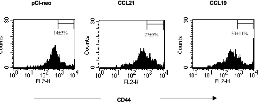
액 내의 memory CD4+ T 세포 분석. Memory CD4+T 세포 형성에 대한 CCR7 ligand의 영향을 알아보기 위 하여, OVA323-339 특이 CD4+ T 세포를 높은 빈도로 갖고 있는 Tg-생쥐(DO11.10.BALB/c)를 이용하였다. CCR7 ligand DNA를 Tg-생쥐에 5일 간격으로 두 번 양쪽 뒷다 리 후부근육에 주사한 후, 25μg OVA323-339 peptide를 CFA에 혼합하여 1주일이 지난 다음 면역화하였다. 다시 그로부터 12주가 지난 다음 말초 혈액을 채취하여 적혈 구를 hypotonic solution으로 제거하고 말초 혈액 림프구 를 분리하였다. 분리된 PBL 내에 CD44highKJ1-26+CD4 + T 세포(memory T cell)의 분포를 알아보기 위하여 FACS analysis를 하였다. CCR7 ligand DNA를 처리한 Tg- 생쥐의 경우에는 CD44highKJ1-26+CD4+ T cell이 CCL21 DNA는 27%, CCL19 DNA은 33%로 각각 나타난 반면에, 대조 plasmid DNA vector (pCI-neo)를 처리한 Tg-생쥐는 14%로 나타났다(Fig. 1). 따라서, 이와 같은 결과는 CCR7 ligand DNA을 처리하게 되면 memory stage에서 memory CD4+ T 세포의 수가 말초 혈액 내에 증가됨을 의미한다.
OVA발현 바이러스에 의하여 유도된 염증 부위 내의 memory CD4+ T 세포 분석. CCR7 ligand DNA에 의하
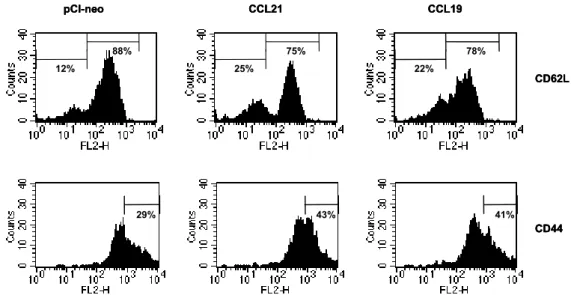
여 증가된 말초 혈액 memory CD4+ T 세포가 인위적으 로 유도된 염증 반응에 대하여 어떻게 반응하는가를 알 아보기 위하여 HSV-ova를 이용하였다. CCR7 ligand DNA로 처리된 Tg-생쥐를 OVA323-339 peptide와 CFA를 혼 합하여 면역화한 다음 12주 후, Tg-생쥐의 estrous cycle 을 맞추기 위하여 progesteron을 피하주사하고, 다시 5일 후 HSV-ova를 vaginal tract에 감염시켰다. 감염 3일 후, 첫 번째는 draining lymph node (DLN)에서 memory CD4+T 세포의 반응을 분석하기 위하여 vaginal tract의 DLN인 iliac LN을 꺼내어 림프구를 준비하였다. 두 번째는 염증 부위 내의 memory CD4+ T 세포 이동을 분석하기 위하 여 vaginal tract을 꺼내어 림프구를 분리하였다. 이와 같 이 분리된 림프구는 FACS analysis에 의하여 activation marker (CD62L, CD44, CD25) 및 KJ1-26+CD4+ T 세포 의 숫자를 분석하였다. 그 결과 CCR7 ligand DNA를 처 리한 Tg-생쥐의 iliac LN에 존재하는 KJ1-26+CD4+ T 세포의 activation marker를 분석하였을 때, CCR7 ligand DNA를 처리하게 되면 HSV-ova 감염에 대하여 보다 많 은 활성화된 KJ1-26+CD4+ T 세포가 존재하는 것을 알 수 있었다(Fig. 2). 특히 CCR7 ligand DNA를 처리한 Tg- 생쥐의 iliac LN에 IL-2 receptor인 CD25+ 세포의 경우
CCL21은 13%, CCL19은 11%인 반면에 대조 plasmid DNA를 처리한 Tg-생쥐는 단지 7%를 보여 주었다. 이와 같은 결과는 vaginal tract의 DLN인 iliac LN에 OVA323-339
peptide 특이 T 세포가 많이 존재함을 알 수 있다. 또한 memory phenotype을 보여주는 CD62LlowCD44high인 KJ1-26 +CD4+ T 세포가 CCR7 ligand DNA를 처리한 Tg-생쥐
Figure 1. CD44high memory KJ1-26+CD4+ T cells in peripheral blood of DO11.10.BALB/c mice injected with CCR7 ligand DNA.
Groups of transgenic mice preinjected with CCR7 ligand DNA or control vector (pCI-neo) were immunized with 25μg OVA323-339
peptide plus complete Freund's adjuvant. 12 weeks later, peripheral blood lymphocytes (PBL) were isolated, analyzed for CD44 expression level after gating KJ1-26+CD4+ T cells by three-color FACS analysis. The isotype controls were not represented. The graph shows average percentages±standard deviation of results obtained from four mice.
M M
14±3% 27±5% 33±11%
CD44
pCI-neo CCL21 CCL19
M M
14±3% 27±5% 33±11%
CD44
pCI-neo CCL21 CCL19
Figure 2. Activation status of KJ1-26+CD4+ T cells in iliac LN of CCR7 ligand DNA-pretreated DO11.10.BALB/c mice infected with herpes simplex virus expressing ovalbumin. Groups of transgenic mice preinjected with CCR7 ligand DNA or control vector (pCI-neo) were immunized with 25μg OVA323-339 peptide plus complete Freund's adjuvant. 12 weeks later, progesteron was subcutaneously injected into immunized transgenic mice to synchronize the estrous cycle. Five days following the administration of progesteron, the transgenic mice were intravaginally infected with herpes simplex virus expressing OVA. Iliac LNs were excised to prepare LN lymphocytes at 3 days post-infection. The activation status of KJ1-26+CD4+ T cells in iliac LN was analyzed for CD62L, CD44, and CD25 after gating KJ1-26+CD4+ T cells by three-color FACS analysis. The isotype controls were not represented. The histograph is a representative of four independent experiments.
65% 78% 74%
pCI-neo CCL21 CCL19
CD62L
21% 45% 38%
CD44
7% 13% 11% CD25
65% 78% 74%
pCI-neo CCL21 CCL19
CD62L
21% 45% 38%
CD44
21% 45% 38%
CD44
7% 13% 11% CD25
7% 13% 11% CD25
에 보다 많이 존재함을 알 수 있었다(Fig. 2).
두번째로, HSV-ova에 의하여 유도된 염증 부위 내로 이동하는 memory CD4+ T 세포를 분석하기 위하여 vagi- nal tract 림프구 중 KJ1-26+CD4+ T 세포의 숫자를 분 석하였다. CCR7 ligand DNA를 처리한 Tg-생쥐에서 HSV- ova감염에 의하여 유도된 염증 부위 내에 보다 많 은 KJ1-26+CD4+ T cell이 존재하는 것을 알 수 있다(Fig.
3). CCL21 DNA를 처리하면 7.6% KJ1-26+CD4+ T 세
포가 vaginal tract에 존재하고 CCL19 DNA를 처리하면 6.7% KJ1-26+CD4+ T 세포가 존재하는 반면에, 대조 plasmid DNA를 처리한 Tg-생쥐는 2.7% KJ1-26+CD4+
T 세포를 보여주었다(Fig. 3). 이와 같이 염증 부위인 vaginal tract에 CCR7 ligand DNA에 의하여 증가된 KJ1-26 +CD4+ T 세포의 phenotype을 분석한 결과, vaginal tract KJ1-26+CD4+ T 세포는 memory phenotype (CD62Llow CD44high)를 갖고 있는 것을 알 수 있었다(Fig. 4).
Figure 3. KJ1-26+CD4+ T cells recruited into vaginal tract of CCR7 ligand DNA-pretreated DO11.10.BALB/c mice infected with herpes simplex virus expressing ovalbumin. Groups of transgenic mice preinjected with CCR7 ligand DNA or control vector (pCI-neo) were immunized with 25μg OVA323-339 peptide plus complete Freund's adjuvant. 12 weeks later, progesteron was subcutaneously injected into immunized transgenic mice to synchronize the estrous cycle. Five days following the administration of progesteron, the transgenic mice were intravaginally infected with herpes simplex virus expressing OVA. The lymphocytes of vaginal tract were isolated as described in Material and Methods at 3 days post-infection. The number of KJ1-26+CD4+ T cells was analyzed by FACS analysis. The charts show KJ1-26+CD4+ T cells in vaginal tract pooled from four mice.
pCI-neo CCL21 CCL19
2.7% 7.6% 6.7%
CD4+
KJ1-26+
pCI-neo CCL21 CCL19
2.7% 7.6% 6.7%
CD4+
KJ1-26+
Figure 4. Activation status of KJ1-26+CD4+ T cells recruited into vaginal tract of CCR7 ligand DNA-pretreated DO11.10.BALB/c mice infected with herpes simplex virus expressing ovalbumin. Groups of transgenic mice preinjected with CCR7 ligand DNA or control vector (pCI-neo) were immunized with 25μg OVA323-339 peptide plus complete Freund's adjuvant. 12 weeks later, progesteron was subcutaneously injected into immunized transgenic mice to synchronize the estrous cycle. Five days following the administration of progesteron, the transgenic mice were intravaginally infected with herpes simplex virus expressing OVA. The lymphocytes of vaginal tract were isolated as described in Material and Methods at 3 days post-infection. The activation status of KJ1-26+CD4+ T cells was analyzed for CD62L and CD44 after gating KJ1-26+CD4+ T cells by three-color FACS analysis. The isotype controls were not represented. The histograph shows activation status of KJ1-26+CD4+ T cells in vaginal tract pooled from four mice.
12%
88%
25%
75%
22%
78%
pCI-neo CCL21 CCL19
CD62L
29% 43% 41%
CD44 12%
88%
25%
75%
22%
78%
pCI-neo CCL21 CCL19
CD62L 12%
88%
25%
75%
22%
78%
pCI-neo CCL21 CCL19
CD62L
29% 43% 41%
CD44
29% 43% 41%
CD44
바이러스 감염에 대한 방어효과. CCR7 ligand DNA에 의하여 향상된 memory CD4+ T 세포가 결국 virulent virus 감염에 대하여 방어효과를 증가시켜주는가를 알아 보기 위하여 virulent HSV-1 McKrae strain으로 vaginal challenge시험을 실행하였다. CCR7 ligand DNA로 전처 리된 BALB/c 생쥐를 7일 후, HSV-1 KOS strain의 gB를 발현하는 plasmid DNA로 면역화하였다. 그로부터 12주 후에 BALB/c 생쥐의 estrous cycle을 동시화하기 위하여 progesteron을 피하주사하고, 다시 5일 후 virulent HSV-1 McKrae 5×10⁷ pfu로 vaginal tract에 감염시켰다. gB DNA vaccine에 의한 HSV-1 감염에 대한 방어효과는 Th1-type CD4+ T cell이 주요한 방어인자로 알려져 있 기 때문에 본 vaginal challenge시험에서 gB DNA vaccine 을 선택하였다(24,32). 그 결과 CCR7 ligand DNA을 처리 한 생쥐가 대조군의 생쥐보다 virulent virus 감염에 대하 여 보다 큰 저항성을 보여주었다(Table I). Virulent HSV-1을 vaginal tract에 감염시킨 후 12일째에, CCL21 DNA 처리 군은 63%의 survival rate를 보여 주었고 CCL19 DNA 처리 군은 57%로 나타났다. 반면에 대조 plasmid DNA 처리 군의 경우에는 25%의 survival rate를 보여주었다. 한편, 아무런 처치를 하지 않은 naive 생쥐 는 감염 후 8일 안에 모두 죽었다(Table I). 또한 CCR7 ligand DNA의 처리는 virulent virus 감염에 대한 clinical severity도 대조군에 비하여 유의성 있게 감소시켰다 (Table I). 따라서, 이와 같은 결과는 CCR7 ligand DNA에 의하여 증진된 memory CD4+ T 세포는 후에 virulent virus감염에 대하여 방어효과를 향상시킨다는 것을 알 수 있다.
고 찰
본 연구논문에서는 CCR7 ligand DNA를 Tg-생쥐에 전 처리하면 항원 특이 memory CD4+ T 세포가 증가되고, 이와 같이 증가된 memory CD4+ T 세포는 바이러스 감 염에 대하여 방어효과를 향상시켜 주는 것을 보여주고 있다. 특히, CCR7 ligand DNA 처리 Tg-생쥐에서 OVA 발현 바이러스에 의하여 유도된 염증 부위 내에 보다 많 은 memory CD4+ T 세포가 존재하였다. 따라서, 이상의 결과는 CCR7과 그의 ligand는 각종 바이러스 또는 박테 리아 감염에 대한 최적의 memory T 세포를 형성하고 유 지함으로써 숙주를 보호하는 데 중요한 역할을 하는 것 으로 생각된다.
DNA vaccine이나 기타 바이러스 감염에 의하여 유도 된 memory CD4+ T 세포는 각종 병원성 바이러스의 감 염을 조절하는 데 중요한 역할을 하고 있다(36-38). 그와 같이 유도된 memory CD4+ T 세포는 바이러스 감염 시 항바이러스성 cytokine IFN-γ를 생산하거나 중화항체를 생산하도록 각종 cytokine을 이용하여 B cell의 기능을 증 진시켜준다(36-38). 더욱이, Th1-type CD4+ T 세포로부터 생성되는 IFN-γ는 최적의 cytotoxic CD8+ T 세포를 활 성화시키는 데 주요한 cytokine이다(39-41). 반대로, CD8+
T 세포는 TCR과 MHC class I molecule을 매개로 하는 세포 용해작용과 IFN-γ와 TNF-α를 생산함으로써 바이 러스를 제거한다(42,43). 물론, 본 연구논문에서는 CCR7 ligand의 memory CD8+ T 세포에 대한 영향을 고찰하지 는 않았다. 그러나, 여기서 기술하는 CCR7 ligand가 memory CD4+ T 세포를 증가시키고 바이러스 감염에 대하여 향상된 방어효과를 나타낸다는 것은 흥미로운 Table I. Summary of resistance to vaginal challenge of virulent herpes simplex virus in CCR7 ligand DNA- and control vector-pretreated animals
ꠚꠚꠚꠚꠚꠚꠚꠚꠚꠚꠚꠚꠚꠚꠚꠚꠚꠚꠚꠚꠚꠚꠚꠚꠚꠚꠚꠚꠚꠚꠚꠚꠚꠚꠚꠚꠚꠚꠚꠚꠚꠚꠚꠚꠚꠚꠚꠚꠚꠚꠚꠚꠚꠚꠚꠚꠚꠚꠚꠚꠚꠚꠚꠚꠚꠚꠚꠚꠚꠚꠚꠚꠚꠚꠚꠚꠚꠚꠚꠚꠚꠚꠚꠚꠚꠚꠚꠚꠚꠚꠚꠚꠚꠚꠚꠚꠚꠚ
No. of mice Clinical severity at day postchallenge
Survival Avg. time of
survived/no. of rateb (%) death±SD ꠏꠏꠏꠏꠏꠏꠏꠏꠏꠏꠏꠏꠏꠏꠏꠏꠏꠏꠏꠏꠏꠏꠏꠏꠏꠏꠏꠏꠏꠏꠏꠏꠏꠏꠏꠏꠏꠏꠏ
mice challengedb 6 8 10 12
ꠏꠏꠏꠏꠏꠏꠏꠏꠏꠏꠏꠏꠏꠏꠏꠏꠏꠏꠏꠏꠏꠏꠏꠏꠏꠏꠏꠏꠏꠏꠏꠏꠏꠏꠏꠏꠏꠏꠏꠏꠏꠏꠏꠏꠏꠏꠏꠏꠏꠏꠏꠏꠏꠏꠏꠏꠏꠏꠏꠏꠏꠏꠏꠏꠏꠏꠏꠏꠏꠏꠏꠏꠏꠏꠏꠏꠏꠏꠏꠏꠏꠏꠏꠏꠏꠏꠏꠏꠏꠏꠏꠏꠏꠏꠏꠏꠏꠏ
pCI-neo 2/8 25 8.3±2.6d 2.6±1.7d 3.4±2.0d 3.6±1.8d 3.8±1.8d
CCL21 5/8 63 16.2±9.8c 1.1±1.6c 1.5±2.2c 1.9±2.2 2.1±2.2
CCL19 4/7 57 15.0±8.2c 0.7±1.3c 1.3±1.6c 2.0±1.8 2.1±2.0
Naive 0/8 0 5.7±1.6 4.6±0.7 5.0±0 5.0±0 5.0±0
ꠏꠏꠏꠏꠏꠏꠏꠏꠏꠏꠏꠏꠏꠏꠏꠏꠏꠏꠏꠏꠏꠏꠏꠏꠏꠏꠏꠏꠏꠏꠏꠏꠏꠏꠏꠏꠏꠏꠏꠏꠏꠏꠏꠏꠏꠏꠏꠏꠏꠏꠏꠏꠏꠏꠏꠏꠏꠏꠏꠏꠏꠏꠏꠏꠏꠏꠏꠏꠏꠏꠏꠏꠏꠏꠏꠏꠏꠏꠏꠏꠏꠏꠏꠏꠏꠏꠏꠏꠏꠏꠏꠏꠏꠏꠏꠏꠏꠏ
aGroups of BALB/c mice preinjected with CCR7 ligand DNA or control vector (pCI-neo) were immunized with gB DNA (100μg).
Naive mice were given no treatment. 12 weeks later, the mice were subcutaneously injected with 2 mg of Depo-Provera per mouse.
Five days following the administration of Depo-Provera, the mice were challenged intravaginally with 5×10⁷ pfu of HSV-1 McKrae.
The mice were observed daily for vaginal inflammation. Moribund mice were euthanized. Clinical severity were scored as follows: 0, no inflammation; 1, mild inflammation; 2, moderate swelling and redness; 3, severe inflammation; 4, paralysis; and 5, death.
bNo. of mice survived/no. of mice challenged and survial rate (%) were determined at 12 days post-challenge.
cSignificantly different from values obtained from mice treated with control vector (P<0.5), dSignificantly different from values obtained from naive mice (P<0.5).
사실이다. 이와 같은 사실은 CCR7 ligand DNA에 의하여 2차 림프조직에 증가된 DC가 관여할 것으로 생각된다 (25,30). CCR7 ligand DNA를 DNA vaccine과 함께 투여하 면 DNA vaccine의 면역반응이 2차 림프조직에 증가된 DC에 의하여 증가된다고 보고된 바 있다(25). 또한, 현저 하게 저하된 2차 림프조직의 DC에 의하여 야기된 것으 로 보이는 기능 장애 CD8+ T 세포는 CCR7 ligand DNA 의 처치에 의하여 DC이 증가되고, 다시 정상 CD8+ T 세포로 회복된다는 사실은 위에서 기술한 추측을 반영 한다(30). 비록 본 연구에서 이용된 plasmid DNA가 2차 림프조직으로 이동하여 발현한다는 것이 증명되지 않았 지만, β-galactosidase나 green fluorescence protein (GFP) 발현 plasmid DNA를 이용한 다른 논문에서 plasmid DNA를 근육주사하면 대부분의 plasmid DNA는 인접 림 프절이나 비장에서 오랫동안 존재하는 것을 알 수 있었 다(44,45). 이와 같은 현상은 아마도 migratory cell이 주사 된 plasmid DNA를 얻어 근처의 림프절이나 비장으로 이 동시키는 것으로 생각된다(46,47). 더욱이, 본 연구논문 의 이와 같은 고찰을 지지하는 또 다른 흥미로운 보고로 는 피하 주사된 CCR7 ligand 단백질은 선택적으로 HEV 에 축적된다는 것이다(48). 이와 같은 사실은 결국, CCR7 ligand DNA로부터 발현되는 CCR7 ligand 단백질 은 HEV에 축적되어 naive T 세포와 mature DC을 2차 림 프조직에 증가시킬 것으로 생각된다. 따라서, 이와 같이 증가된 naive T cell과 mature DC은 2차 림프조직에서 접 촉의 기회가 증가할 것이고 보다 증가된 활성 T 세포는 결국 증진된 memory T 세포를 확립할 것으로 생각된다.
이러한 개념은 현재 adoptive transfer 모델을 이용하여 검 증 중에 있다.
여기서, FACS analysis를 이용하여 memory CD4+ T 세포의 말초 혈액 또는 염증 부위 내의 숫자에 초점을 맞추어 투여된 CCR7 ligand DNA의 기능을 증명하려 하 였다. 그러나, 증가된 memory CD4+ T 세포의 정량적인 기능에 대한 결과는 제시하지 않았다. 다시 말하면, 염증 부위로 이동한 memory CD4+ T 세포가 과연 항바이러 스 활성을 갖는가에 대한 척도로서 IFN-γ 생산능력이 평가되어야 할 것으로 생각된다. 이에 대한 연구는 좀 더 진행되어야 하겠지만, 그에 대한 간접적인 결과로는 CCR7 ligand DNA의 처치가 보다 활성화된 CD4+ T 세 포를 형성한다는 것이다. 더욱이, CD44는 바이러스 감 염에 의하여 형성된 염증 부위 내로 림프구가 이동하는 데 필수적인 물질로 알려져 있다(49). 본 연구논문에서 는 CCR7 ligand DNA를 Tg-생쥐에 투여하면 말초 혈액 내에 CD44high를 갖는 항원 특이 CD4+ T 세포가 증가하 였다(Fig. 1). 따라서, CCR7 ligand DNA 투여에 의하여 증가된 CD44high CD4+ T 세포가 바이러스 감염에 대하 여 방어효과를 나타낸 것으로 생각된다. 또한, 보다 많은
CD44high memory CD4+ T 세포가 염증 부위 내로 이동 하였음을 보여주었다(Fig. 3).
결론적으로 CCR7 ligand DNA의 처치는 memory CD4+
T 세포의 증가를 보여주었고 증가된 memory CD4+ T 세포는 바이러스 감염에 대하여 방어효과를 향상시킨 것으로 생각된다. 보다 염증 부위로 이동된 memory CD4 + T 세포의 기능에 대한 자세한 실험이 시도되어야 하 겠지만, 본 연구논문에서는 CCR7과 그의 CCR7 ligand는 최적의 memory T 세포를 형성하는 데 중요한 역할을 할 것으로 기대되며, 향후 CCR7 ligand는 바이러스 감염 및 암 예방과 치료에 유용하게 이용될 수 있을 것으로 생각 된다.
감사의 글
CCL21과 CCL19 유전자를 공급하여 주신 Dr. Albert Zlotnik (DNAX Research Institute)와 Dr. Jason G. Cyster (University of California)에게 감사드립니다.
참 고 문 헌
1. Von Andrian UH, Mackay CR: T-cell function and migra- tion. Two sides of the same coin. N Engl J Med 343;
1020-1034, 2000
2. Butcher EC, Picker LJ: Lymphocyte homing and home- ostasis. Science 272;60-66, 1996
3. Girard JP, Springer TA: High endothelial venules (HEVs):
specialized endothelium for lymphocyte migration. Immunol Today 16;449-457, 1995
4. Campbell JJ, Hedrick J, Zlotnik A, Siani MA, Thompson, DA, Butcher EC: Chemokines and the arrest of lymphocytes rolling under flow conditions. Science 279;381-384, 1998 5. Cyster JG: Chemokines and cell migration in secondary
lymphoid organs. Science 286;2098-2102, 1999
6. Zlotnik A, Yoshie O: Chemokines: a new classification system and their role in immunity. Immunity 12;121-127, 2000 7. Campbell JJ, Butcher EC: Chemokines in tissue-specific and
microenvironment-specific lymphocyte homing. Curr Opin Immunol 12;336-341, 2000
8. Gunn MD, Tangemann K, Tam C, Cyster JG, Rosen SD, Williams LT: A chemokine expressed in lymphoid high endo- thelial venules promotes the adhesion and chemotaxis of naive T lymphocytes. Proc Natl Acad Sci USA 95;258-263, 1998 9. Stein JV, Rot A, Luo Y, Narasimhaswamy M, Nakano H,
Gunn MD, Matsuzawa A, Quackenbush EJ, Dorf ME, von Andrian UH: The CC chemokine thymus-derived chemo- tactic agent 4 (TCA-4, secondary lymphoid tissue chemokine, 6Ckine, exodus-2) triggers lymphocyte function-associated antigen 1-mediated arrest of rolling T lymphocytes in peri- pheral lymph node high endothelial venules. J Exp Med 191;61-75, 2000
10. Warnock RA, Campbell JJ, Dorf ME, Matsuzawa A, McEvoy LM, Butcher EC: The role of chemokines in the micro- environmental control of T versus B cell arrest in Peyer's patch high endothelial venules. J Exp Med 191;77-88, 2000 11. Ngo VN, Tang HL, Cyster JG: Epstein-Barr virus-induced
molecule 1 ligand chemokine is expressed by dendritic cells in lymphoid tissues and strongly attracts naive T cells and activated B cells. J Exp Med 188;181-191, 1998
12. Dieu MC, Vanbervliet B, Vicari A, Bridon JM, Oldham E,
Ait-Yahia S, Briere F, Zlotnik A, Lebecque S, Caux C:
Selective recruitment of immature and mature dendritic cells by distinct chemokine expressed in different anatomic sites.
J Exp Med 188;373-386, 1998
13. Sallusto F, Lanzavecchia A: Mobilizing dendritic cells for tolerance, priming, and chronic inflammation. J Exp Med 189;611-614, 1999
14. Chan VW, Kothakota S, Rohan MC, Panganiban-Lustan L, Gardner JP, Wachowicz MS, Winter JA, Williams LT: Secondary lymphoid-tissue chemokine (SLC) is chemotactic for mature dendritic cells. Blood 93;3610-3616, 1999
15. Sallusto F, Lenig D, Forster R, Lipp M, Lanzavecchia A: Two subsets of memory T lymphocytes with distinct homing potentials and effector functions. Nature 401;708-712, 1999 16. Nakano H, Tamura T, Yoshimoto T, Yagita H, Miyasaka M,
Butcher EC, Nariuchi H, Kakiuchi T, Matsuzawa A: Genetic defect in T lymphocyte-specific homing into peripheral lymph nodes. Eur J Immunol 27;215-221, 1997
17. Gunn MD, Kyuwa S, Tam C, Kakiuchi T, Matsizawa A, Williams LT, Nakano H: Mice lacking expression of second- ary lymphoid organ chemokine have defect in lymphocyte homing and dendritic cell localization. J Exp Med 189;
451-460, 1999
18. Vassileva G, Soto H, Zlotnik A, Nakano H, Kakiuchi T, Hedrick JA, Lira SA: The reduced expression of 6Ckine in the plt mouse results from the deletion of one of two 6Ckine genes. J Exp Med 190;1183-1188, 1999
19. Forster R, Schubel A, Breitfeld D, Kremmer E, Renner- Muller I, Wolf E, Lipp M: CCR7 coordinates the primary immune response by establishing functional microenviron- ments in secondary lymphoid organs. Cell 99;23-33, 1999 20. Ploix C, Lo D, Carson MJ: A ligand for the chemokine rece-
ptor CCR7 can influence the homeostatic proliferation of CD4 T cells and progression of autoimmunity. J Immunol 167;
6724-6730, 2001
21. Campbell JJ, Murphy KE, Kunkel EJ, Brightling CE, Soler D, Shen Z, Boisvert J, Greenberg HB, Vierra MA, Goodman SB, Genovese MC, Wardlaw AJ, Butcher EC, Wu L: CCR7 expression and memory T cell diversity in humans. J Immunol 166;877-884, 2001
22. Xiang Z, Ertl HCJ: Manipulation of the immune response to a plasmid-encoded viral antigen by coinoculation with plasmids expressing cytokines. Immunity 2;129-135, 1995 23. Sin JI, Kim JJ, Boyer JD, Ciccarelli RB, Higgins TJ, Weiner
DB: In vivo modulation of vaccine-induced immune responses toward a Th1 phenotype increases potency and vaccine effec- tiveness in a herpes simplex virus type 2 mouse model. J Virol 73;501-509, 1999
24. Eo SK, Lee S, Chun S, Rouse BT: Modulation of immunity against herpes simplex virus infection via mucosal genetic transfer of plasmid DNA encoding chemokines. J Virol 75;
569-578, 2001.
25. Eo SK, Lee S, Kumaraguru U, Rouse BT: Immunopotenti- ation of DNA vaccine against herpes simplex virus via co-delivery of plasmid DNA expressing CCR7 ligands.
Vaccine 19;4685-4693, 2001.
26. Berger DP, Naniche D, Crowley MT, Koni PA, Flavell RA, Oldstone MA: Lymphotoxin-β-deficient mice show de- fective antiviral immunity. Virology 260;136-147, 1999 27. De Togni P, Goellner J, Ruddle NH, Streeter PR, Fick A,
Mariathasan S, Smith SC, Carlson R, Shornick LP, Strauss- Schoenberger J, Russell JH, Karr R, Chaplin DD: Abnormal development of peripheral lymphoid organs in mice deficient in lymphotoxin. Science 264;703-707, 1994
28. Wu Q, Wang Y, Wang J, Hedgeman EO, Browning JL, Fu YX: The requirement of membrane lymphotoxin for the
presence of dendritic cells in lymphoid tissue. J Exp Med 190;629-638, 1999
29. Kumaraguru U, Davis IA, Deshpande S, Tevethia SS, Rouse BT: Lymphotoxin α-/- mice develop functionally impaired CD8+ T cell responses and fail to contain virus infection of the central nervous system. J Immunol 166;1066-1074, 2001 30. Eo SK, Kumaraguru U, Rouse BT: Plasmid DNA encoding
CCR7 ligands compensate for dysfunctional CD8+ T cell responses by effects on dendritic cells. J Immunol 167;3592- 3599, 2001
31. Gangappa S, Babu JS, Thomas J, Daheshia M, Rouse BT:
Virus-induced immunoinflammatory lesions in the absence of viral antigen recognition. J Immunol 161;4289-4300, 1998 32. Manickan E, Rouse RJD, Yu Z, Wire W, Rouse BT: Genetic
immunization against herpes simplex virus: protection is medi- ated by CD4+ T lymphocytes. J Immunol 155;259-265, 1995 33. Kuklin N, Daheshia M, Karem K, Manickan E, Rouse BT:
Induction of mucosal immunity against herpes simplex virus by plasmid DNA immunization. J Virol 71;3138-3145, 1997 34. Dupuy C, Buzoni-Gatel D, Touze A, Bout D, Coursaget P:
Nasal immunization of mice human papillomavirus type 16 (HPV-16) virus-like particles or with the HPV-16 L1 gene elicits specific cytotoxic T lymphocytes in vaginal draining lymph nodes. J Virol 73;9063-9071, 2001
35. Parr MB, Kepple L, McDermott M, Drew MD, Bozzola JJ, Parr EL: A mouse model for studies of mucosal immunity to vaginal infection by herpes simplex virus type 2. Lab Investig 70;369-380, 1994
36. Eichelberger M, Allan W, Zijlstra M, Jaenisch R, Doherty PC:
Clearance of influenza virus respiratory infection in mice lacking class I major histocompatibility complex- restricted CD8+ T cells. J Exp Med 174;875-880, 1991
37. Hou S, Doherty PC, Zijlstra M, Jaenisch R, Katz JM: Delayed clearance of Sendai virus in mice lacking class I MHC- restricted CD8+ T cells. J Immunol 149;1319-1325, 1992 38. Zhong W, Roberts AD, Woodland DL: Antibody-inde-
pendent antiviral function of memory CD4+ T cells in vivo requires signals from CD8+ effector T cells. J Immunol 167;1379-1386, 2001
39. Jennings SR, Bonneau RH, Smith PM, Wolcott RM, Chervenak R: CD4-positive T lymphocytes are required for the generation of the primary but not the secondary CD8- positive cytolytic T lymphocyte response to herpes simplex virus in C57BL/6 mice. Cell Immunol 133;234-252, 1991 40. Matloubian M, Concepcion RJ, Ahmed R: CD4+ T cells are
required to sustain CD8+ cytotoxic T cell-responses during chronic viral infection. J Virol 68;8056-8063, 1994
41. Cardin RD, Brooks JW, Sarawar SR, Doherty PC: Progressive loss of CD8+ T cell-mediated control of a γ-herpesvirus in the absence of CD4+ T cells. J Exp Med 184;863-871, 1996 42. Harty JT, Tvinnereim AR, White DW: CD8+ T cell effector
mechanisms in resistance to infection. Annu Rev Immunol 18;275-308, 2000
43. Slifka MK, Whitton JL: Antigen-specific regulation of T cell-mediated cytokine production. Immunity 12;451-457, 2000 44. Osaka G, Garey K, Cuthbertson A, Godowski P, Patapott
T, Ryan A, Gadek T, Mordenti J: Pharmacokinetics, tissue distribution, and half-life expression efficiency of [32P]DNA following intravenous administration of DNA/cationic lipid complexes in mice. Use of a novel radionucleotide approach.
J Pharm Sci 85;612-618, 1996
45. Kawabata K, Takakura Y, Hashida M: The fate of plasmid DNA after intravenous injection in mice: involvement of sca- venger receptors in its hepatic uptake. Pharm Res 12;
825-830, 1995
46. Chattergoon MA, Robinson TM, Boyer JD, Weiner DB:
Systemic immune induction following DNA-based immu- nization through in vivo transfection and activation of macro- phages/antigen presenting cells. J Immunol 160;5707-5718, 1998 47. Chun S, Daheshia M, Lee S, Eo SK, Rouse BT: Distribution
fate and mechanism of immune modulation follwing mucosal delivery of plasmid DNA encoding IL-10. J Immunol 163;
2393-2402, 1999
48. Baekkevold ES, Yamanaka T, Palframan RT, Carlsen HS,
Reinholt FP, von Andrian UH, Brandtzaeg P, Haraldsen G:
The CCR7 ligand ELC (CCL19) is transcytosed in high endothelial venules and mediates T cell recruitment. J Exp Med 193;1105-1111, 2001
49. DeGrendele HC, Estess P, Siegelman MH: Requirement for CD44 in activated T cell extravasation into an inflammatory site. Science 278;672-675, 1997