- 133 -
Imaging Science in Dentistry 2015; 45: 133-5 http://dx.doi.org/10.5624/isd.2015.45.2.133
Dear Editor,
The selection of appropriate diagnostic techniques is an important step in the treatment of disease. An optimal di- agnostic method can provide essential information, while minimizing cost and harm to patients. In dental radiogra- phy, attempts have been made to reduce patient exposure to radiation. Cone-beam computed tomography (CBCT) and digital radiography were developed to achieve this goal.
CBCT is becoming the preferred cross-sectional imag- ing technique for dental practitioners
1due to its advanta- ges, such as low cost, easy accessibility, and a low radia- tion dose, compared to multi-slice computed tomography.
2Some of the applications of CBCT include the localization of supernumerary and impacted teeth,
3the examination of the temporomandibular joint,
4the detection of cysts and tumors of the jaw,
5the detection of root fractures,
6the as- sessment of root canal configurations,
7treatment planning for the placement of dental implants,
8and orthodontic dia- gnoses.
9Digital radiography is another widely popular radiogra- phic modality in dental practice, with advantages includ- ing a decreased radiation dose, easy digital storage and electronic transmission, and not requiring a darkroom.
10It is necessary to determine the level of knowledge of dental practitioners about the abovementioned imaging modali- ties and whether both conventional and novel techniques are being used efficiently in oral and maxillofacial radiol- ogy.
We assessed the level of knowledge of dental practition- ers about CBCT, digital radiography, and the correct indi- cations for referring patients to oral and maxillofacial rad- iologists. A total of 110 questionnaires were distributed among dental practitioners attending continuing education courses in the city of Isfahan, Iran. The questionnaire con- tained 19 questions addressing the demographic character- istics of the participants, their level of knowledge about CBCT, and their knowledge about appropriate indications for referring patients for digital radiography and CBCT.
Appendix 1 contains the questionnaire form, adopted from the study of Dölekoğlu et al.
11The data were analyzed using SPSS version 22 (IBM Corp., Armonk, NY, USA).
Eighty questionnaires were returned (response rate, 72.73
%). Of the respondents, 53.4% were male and 46.6% were female, with a mean age of 39.3±7.4 years (range, 25-54 years). No significant differences were observed between males and females regarding the usage of digital radiog- raphy or CBCT (P=0.990 and P=0.348, respectively).
More over, Fisher’s exact test failed to find significant dif- ferences among dentists regarding the use of digital radio- graphy or CBCT based on their time of graduation (P=
0.418 and P=0.350, respectively). The field of practice was not significantly associated with the use of digital ra- diography or CBCT (P=0.116 and P=0.135, respective- ly). The t-test revealed no significant difference among practitioners in different age groups regarding the usage of digital radiography or CBCT (P=0.218 and P=0.247, respectively). A total of 33.7% of the respondents report- ed using digital radiography. Moreover, 18.8% of dental practitioners reported referring their patients for CBCT, while 46.3% of the respondents had never referred a pa- tient for CBCT. Table 1 summarizes the reasons provided
Level of knowledge of dental practitioners in Isfahan, Iran about cone-beam computed tomography and digital radiography
Mojdeh Mehdizadeh
1, Sahar Goli Booshehri
2, Farimah Kazemzadeh
2, Parisa Soltani
2, Mahmood Reza Kalantar Motamedi
3,*
1Department of Oral and Maxillofacial Radiology, School of Dentistry, Isfahan University of Medical Sciences, Isfahan, Iran
2Dental Students Research Center, School of Dentistry, Isfahan University of Medical Sciences, Isfahan, Iran
3Department of Research, School of Dentistry, Isfahan Branch, Islamic Azad University, Isfahan, Iran
Copyright ⓒ 2015 by Korean Academy of Oral and Maxillofacial Radiology
This is an Open Access article distributed under the terms of the Creative Commons Attribution Non-Commercial License(http://creativecommons.org/licenses/by-nc/3.0) which permits unrestricted non-commercial use, distribution, and reproduction in any medium, provided the original work is properly cited.
Imaging Science in Dentistry·pISSN 2233-7822 eISSN 2233-7830 Received January 30, 2015; Revised February 26, 2015; Accepted March 3, 2015
*Correspondence to : Dr. Mahmood Reza Kalantar Motamedi
Arghavanieh Blvd., School of Dentistry, Isfahan Branch, Islamic Azad University, Postal Code: 81551-39998, Isfahan, Iran
Tel) 98-913-129-2339, Fax) 98-31-36695623, E-mail) [email protected]
*홀수페이지 시작.
*표 선 두께
=
1/0.3ptLevel of knowledge of dental practitioners in Isfahan, Iran about cone-beam computed tomography and digital radiography
- 134 -
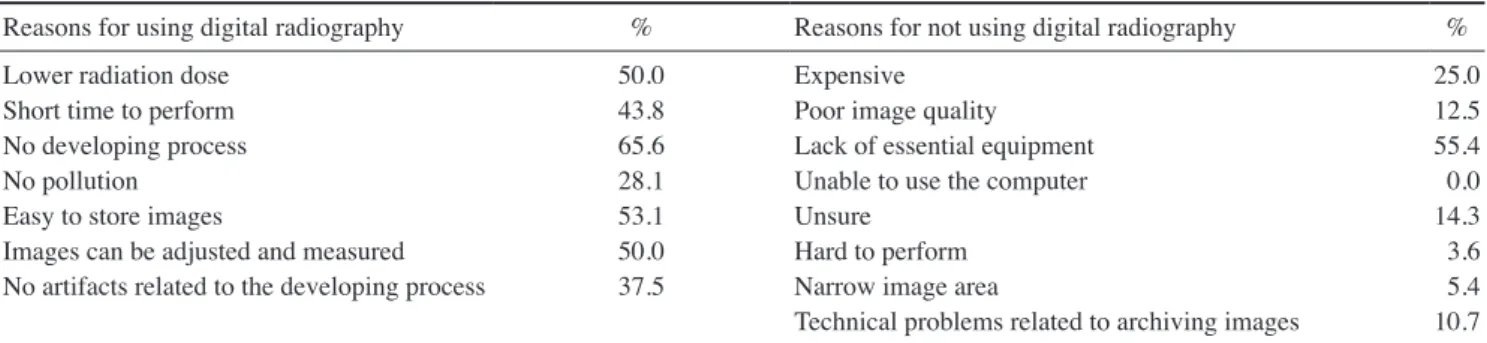
by the practitioners for using or not using digital radiogra- phy. As shown, the most important reason for using dig- ital radiography among dentists is that it eliminates the conventional film developing process. However, 55.4% of the practitioners did not use digital radiography due to lack of the necessary tools. Table 2 demonstrates the frequency of different indications for using CBCT. As shown, the most frequent indication for using CBCT is planning im- plant placement. Since the use of implants is currently growing very quickly, we can expect that CBCT will soon be used increasingly frequently in our country.
We found that dental practitioners in the city of Isfahan mostly used conventional radiography, despite their accep- table knowledge about the benefits of digital radiography.
The high cost of equipment seems to be the main obstacle in this regard. Moreover, despite an acceptable level of knowledge about the benefits of CBCT, the participants rarely referred their patients for this imaging modality.
Only five centers in our city currently possess CBCT equ- ipment. This may indicate that it is necessary to equip more oral and maxillofacial radiology centers in our city with CBCT systems.
References
1. Sudhakar KM, Hemant RD, Amit BK. Assessment of response
of dental clinicians and patients towards different imaging modalities used in diagnostic evaluation of dental implant the- rapy. Indian J Basic Appl Med Res 2012; 1: 341-50.
2. De Vos W, Casselman J, Swennen GR. Cone-beam comput- erized tomography(CBCT) imaging of the oral and maxillo- facial region: a systematic review of the literature. Int J Oral Maxillofac Surg 2009; 38: 609-25.
3. Liu DG, Zhang WL, Zhang ZY, Wu YT, Ma XC. Localization of impacted maxillary canines and observation of adjacent in- cisor resorption with cone-beam computed tomography. Oral Surg Oral Med Oral Pathol Oral Radiol Endod 2008; 105: 91- 4. Tsiklakis K, Syriopoulos K, Stamatakis HC. Radiographic 8.
examination of the temporomandibular joint using cone beam computed tomography. Dentomaxillofac Radiol 2014; 33:
196-201.
5. Closmann JJ, Schmidt BL. The use of cone beam computed tomography as an aid in evaluating and treatment planning for mandibular cancer. J Oral Maxillofac Surg 2007; 65: 766-71.
6. Hassan B, Metska ME, Ozok AR, van der Stelt P, Wesselink PR. Detection of vertical root fractures in endodontically treat- ed teeth by a cone beam computed tomography scan. J Endod 2009; 35: 719-22.
7. Patel S, Dawood A, Ford TP, Whaites E. The potential appli- cations of cone beam computed tomography in the manage- ment of endodontic problems. Int Endod J 2007; 40: 818-30.
8. Scarfe WC, Farman AG, Sukovic P. Clinical applications of cone-beam computed tomography in dental practice. J Can Dent Assoc 2006; 72: 75-80.
9. Silva MA, Wolf U, Heinicke F, Bumann A, Visser H, Hirsch E. Cone-beam computed tomography for routine orthodontic treatment planning: a radiation dose evaluation. Am J Orthod Dentofacial Orthop 2008; 133: 640.e1-5.
10. Van Der Stelt PF. Filmless imaging: the uses of digital radiog- raphy in dental practice. J Am Dent Assoc 2005; 136: 1379- 11. Dölekoğlu S, Fişekçioğlu E, İlgüy M, İlgüy D. The usage of 87.
digital radiography and cone beam computed tomography among Turkish dentists. Dentomaxillofac Radiol 2011; 40:
379-84.
Table 1. Reasons for using or not using digital radiography.
Reasons for using digital radiography % Reasons for not using digital radiography %
Lower radiation dose 50.0 Expensive 25.0
Short time to perform 43.8 Poor image quality 12.5
No developing process 65.6 Lack of essential equipment 55.4
No pollution 28.1 Unable to use the computer 0.0
Easy to store images 53.1 Unsure 14.3
Images can be adjusted and measured 50.0 Hard to perform 3.6
No artifacts related to the developing process 37.5 Narrow image area 5.4
Technical problems related to archiving images 10.7
Table 2. Frequencies of different indications for cone-beam com- puted tomography imaging.
Indications for CBCT %
Trauma 18.8
Cyst or tumor 37.5
Implant planning 62.5
Dental caries 0.0
Periodontal diseases 2.1
- 135 -
Mojdeh Mehdizadeh et al
Appendix 1. Questionnaire form.
1. Age: ... 2. Gender: ...
3. Student( ) General practitioner( ) Public dentist( ) Researcher( )
4. Location: ... 5. Year of graduation: ...
6. Do you use digital imaging techniques in your clinic?
(a)( ) Yes (b)( ) No
If you answered no to question 6, please go to question 11 7. For which kind(s) of radiography do you use digital imaging?
(a)( ) Panoramic (b)( ) Intraoral(periapical, etc.) (c)( ) Cephalometric (d)( ) All 8. Please check your reasons for using digital imaging techniques:
(a)( ) The radiation dose is much lower. (b)( ) It takes a short time to perform.
(c)( ) There is no developing process. (d)( ) There is no wastage in the developing process and it does not cause pollution.
(e)( ) It is easy to store images. (f)( ) Adjustments and measurements can be performed on images.
(g)( ) There are no artifacts related to the developing process.
9. Are you satisfied with digital imaging?
(a)( ) Not at all (b)( ) A little (c)( ) Unsure (d)( ) Satisfied (e)( ) Very satisfied 10. Does the usage of digital imaging increase the frequency of retaking radiography?
(a)( ) Yes (b)( ) No
11. Please check your reasons for not using digital imaging techniques:
(a)( ) Expensive (b)( ) Poor image quality (c)( ) I do not have essential equipment.
(d)( ) I do not know how to use the computer. (e)( ) Unsure (f)( ) Hard to perform
(g)( ) The image area is too narrow. (h)( ) Some technical problems might occur during the storage of images.
12. Which technique do you prefer when you need three-dimensional imaging of the neck and head region?
(a)( ) Computerized tomography(CT)
(b)( ) Dental volumetric tomography(DVT)/cone-beam computed tomography(CBCT) 13. Have you ever heard of CBCT/DVT?
(a)( ) Yes (b)( ) No
If you answered no to question 13, please go to question 17 14. Have you ever referred your patients for CBCT imaging?
(a)( ) Yes (b)( ) No
15. In which situations do you prefer CBCT imaging?
(a)( ) Trauma (b)( ) Cyst or tumor (c)( ) Implant planning (d)( ) Dental caries (e)( ) Periodontal diseases 16. What is the difference between CT and CBCT?
(a)( ) The radiation dose of CBCT is lower than that of CT. (b)( ) The radiation dose of CBCT is the same as that of CT.
(c)( ) The radiation dose of CBCT is higher than that of CT.
17. How long is your exposure time for intraoral digital radiography?
(a) Anterior teeth ... seconds (b) Posterior teeth ... seconds (c)( ) I do not know. (d)( ) I do not make any adjustments.
18. Do you have periodic checks and calibrations of your radiography equipment?
(a)( ) Yes (b)( ) No
19. Do you frequently attend seminars and meetings about radiology?
(a)( ) Yes (b)( ) No