http://dx.doi.org/10.12671/jkfs.2016.29.3.206
206
Copyright ⓒ 2016 The Korean Fracture Society. All rights reserved.
This is an Open Access article distributed under the terms of the Creative Commons Attribution Non-Commercial License (http://creativecommons.org/licenses/
by-nc/4.0) which permits unrestricted non-commercial use, distribution, and reproduction in any medium, provided the original work is properly cited.
Received April 15, 2016 Revised May 20, 2016 Accepted June 9, 2016
Address reprint requests to: Dongju Shin, M.D.
Department of Orthopaedic Surgery, Daegu Fatima Hospital, 99 Ayang-ro, Dong-gu, Daegu 41199, Korea
Tel: 82-53-940-7324ㆍFax: 82-53-954-7417 E-mail: [email protected]
Financial support: None. Conflict of interest: None.
원위 대퇴골 골절에서 Locking Compression Plate-Proximal Lateral Tibia를 이용한 내측 금속판 고정술
- 증례 보고 - 장세앙⋅변영수⋅한인호⋅신동주
대구파티마병원 정형외과
Medial Plating of Distal Femoral Fracture with Locking Compression Plate-Proximal Lateral Tibia
- Cases’ Report -
Se-Ang Jang, M.D., Young-Soo Byun, M.D., In-Ho Han, M.D., Dongju Shin, M.D.
Department of Orthopaedic Surgery, Daegu Fatima Hospital, Daegu, Korea
Generally, lateral plating is used for a comminuted fracture of the distal femur. However, in some cases, it has been shown that using a medial plate is necessary to achieve better outcome. Nevertheless, there are no available anatomical plates that fit either the distal medial femoral condyle or fracture fixation, except for the relatively short plate developed for distal femoral osteotomy. We found that locking compression plate-proximal lateral tibia (LCP-PLT) fits anatomically well for the contour of the ipsilateral medial femoral condyle. Moreover, LCP-PLT has less risk of breaking the thread holes since it rarely needs to be bent. We report a plastic bone model study and two cases of distal femoral fractures fixed with medial plating using LCP-PLT.
Key Words: Distal femur, Medial plating, Locking compression plate proximal lateral tibia
골다공증, 분쇄가 심한 원위 대퇴골 골절의 수술적 치료 는 여전히 어려운 도전적 대상이다. 금속판과 나사 사이가 고정되는 잠김 금속판은 강한 고정력을 제공하며 고정 소 실과 관련된 많은 문제를 해결하였으나1) 원위 대퇴골 내측
면에 비해 외측면이 더 평평하다는 해부학적 특성 및 신 경, 혈관 손상을 피할 수 있다는 장점 때문에 외측 금속판 고정술은 더 많이 이용되었고 또한 제조회사들은 외측 원 위 대퇴골 금속판만을 개발하였다. 그러나 경우에 따라서 내측 금속판 고정술이 유용하거나 필요한 경우가 있다.2) 현재 원위 대퇴골 내과에 맞는 해부학적 금속판은 원위 대 퇴골 절골술을 위해 개발된 TomoFix-medial distal femur plate (TomoFix-MDF)가 있으나 길이가 다양하지 못하고 내 과의 후방부를 고정할 수 없다. 기존의 금속판을 이용하여 대퇴골 내과를 고정할 경우 원위 대퇴골 내과는 후하방으 로 돌출되어 있어 금속판의 윤곽 성형이 힘들고 특히 잠김 금속판의 경우에는 심하게 금속판을 구부릴 경우 고정나사
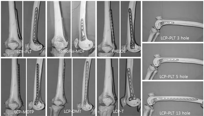
Fig. 1. Photographs of the femur bone model (3B Scientific, Hamburg, Germany) with various plates on medial condyle, locking compression plate-proximal lateral tibia (LCP-PLT), tomoFix-medial distal femur plate (TomoFix-MDF), proximal humerus inter- nal locking plate system (PHILOS), LCP-medial distal tibia plate (LCP-MDTP), LCP-distal metaphyseal tibia (LCP-DMT), and LCP-distal tibia T (LCP-T) plate, in order from upper left corner.
증례 보고
1. 플라스틱 골 모형 연구
해부학적 골 모형(3B Scientific, Hamburg, Germany)의 대퇴골 내과에 LCP-PLT (Synthes, Zuchwil, Switzerland), TomoFix-MDF (Synthes), Proximal Humerus Internal Lo- cking Plate System (PHILOS; Synthes), LCP-medial distal tibia plate (LCP-MDTP; Synthes), LCP-distal metaphyseal tibia (LCPDMT; Synthes), Locking compression plate-distal tibia T (LCP-T; Synthes) 금속판을 각각 적용하였다.
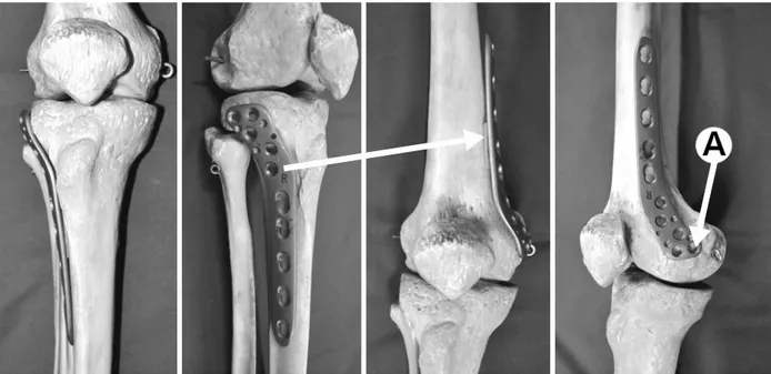
대한 금속판의 적합성을 객관적으로 측정하기는 어렵지만 LCP-PLT의 원위 후방나사 구멍(A)을 대퇴골의 내상과에 위 치시키는 것이 가장 좋은 위치로 판단되었다(Fig. 2). 나사 못 고정을 시현하기 위하여 다른 골 모형(Synbone, Malans, Switzerland)을 이용하였다. 다양한 길이의 LCP-PLT (3, 5, 7, 9, 11, 13 holes)를 원위 후방나사 구멍 (A)이 대퇴골의 내상과에 위치하도록 하고 잠김나사 가이 드를 통해 구멍을 뚫은 뒤 각각의 나사 구멍에 잠김나사들 을 고정하였다. 원위 후방나사 구멍(A)의 나사는 항상 대퇴 과간 절흔으로 향하였으며 나사 길이는 모두 40 mm였다.
나머지 구멍을 통한 나사들은 대퇴과간을 넘어 안전하게 외 과까지 고정할 수 있었다(Fig. 3).
Fig. 2. Photographs of the femur bone model (3B Scientific, Hamburg, Germany) showing the application of locking compression plate- proximal lateral tibia (LCP-PLT) on appropriate position. A: Distal posterior hole.
Fig. 3. Photographs of the fe- mur bone model (Synbone, Malans, Switzerland) with the application of locking comp- ression plate-proximal lateral ti- bia (LCP- PLT) on appropri- ate position. A: Distal post- erior screw directed to the intercondylar notch, B: 2nd row screw reached the lateral femoral condyle without pen- etration into the intercondylar notch.
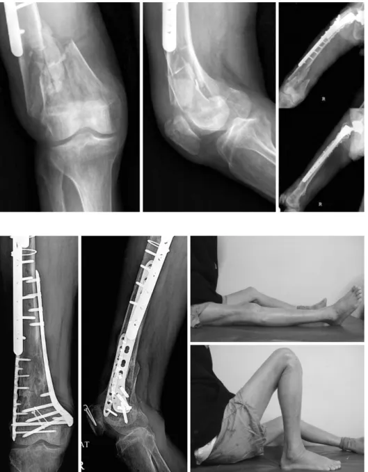
Fig. 5. Radiographs and photo- graphs of the right femur at 12 months after the surgery showing good fracture heal- ing and good range of mo- tion of the right knee.
Fig. 4. Radiographs of the right knee and the right femur showing preexisting lateral plate and comminuted fracture of the right distal femur.
내고정술을 시행한 상태였다(Fig. 4). 기존 외측 금속판으 로 인해 외측접근에 제한이 있어 내측접근을 결정하였다.
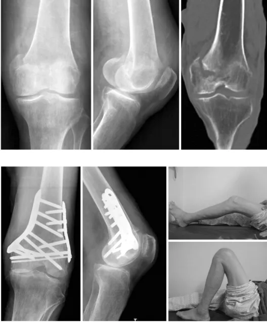
감입이 있었다(Fig. 6). 저자들은 외측 접근으로는 내측의 감입을 보다 정확히 정복하기 힘들며 외측 금속판 내고정
Fig. 6. Radiographs of the left knee and the coronal section of computed tomography scan of the left knee showing se- verely impacted medial con- dyle fracture.
Fig. 7. Radiographs and photo- graphs of the left knee at 14 weeks after surgery showing good fracture healing and good range of motion of the left knee.
술로는 내과의 견고한 고정이 힘들다고 생각하여 내측 접 근을 결정하였다. 첫 증례와 같은 접근법으로 내측 접근을 하여 금속판을 고정하였고, 외측에 작은 금속판을 최소 침 습적으로 추가하였다. 역시 술 후 14주 추시에서 순조로운 골유합과 만족스러운 임상결과를 보였다(Fig. 7).
고 찰
원위 대퇴골 골절 치료의 핵심은 일반적인 다른 관절내 골절과 같이 관혈적 정복을 통한 관절면의 정확한 해부학 적 정복 및 견고한 고정을 획득하여 조기에 관절 운동을 시행함으로써 관절 강직을 예방하는 것으로5) 현재까지 원 위 대퇴골 골절에서 관혈적정복술을 이용한 치료는 일반적
으로 외측 도달법을 통한 고정술이 시행되어 왔다.
그러나 인공구조물 주위 골절로 인해 외측에 금속판 고 정술을 시행할 수 없는 경우 및 원위 대퇴골 내과의 심한 분쇄가 동반되어 외측 내고정 시 골절부위 안정성이 떨어 져 내고정물의 손상과 골절 정복의 소실을 초래할 수 있는 경우에는 내측 금속판 단독 고정 또는 이중 금속판 고정을 하여 내측 지지대 역할과 함께 골절부위의 안정성을 높일 수 있다.2,6)
원위 대퇴골 골절에 대한 내측 접근법은 일반적으로 혈관 및 신경손상의 가능성이 높다고 알려져 있으나7) Checroun 등4)은 카데바 연구를 통하여 대퇴 내측 광근과 내전근 사 이로 접근하는 광범위 내측 접근법을 발표하였고 또한 내 전근 결절 상방 9 cm 정도에 위치하는 ‘위험 지역’을 피해
곽 성형이 힘들고 특히 잠김 금속판의 경우에는 심한 금속 판 구부림으로 인해 잠김나사 구멍의 손상이 발생하여 금 속판과 나사 사이의 고정 효과가 사라질 수 있다.3) 저자들 이 사용한 LCP-PLT 금속판은 다양한 길이를 사용할 수 있 고 동측 대퇴골 내과의 윤곽에 타 금속판에 비하여 비교적 잘 맞아 수술 중 복잡한 금속판 성형술이 필요하지 않으며 특히 내과의 특징인 후방 돌출부까지 잘 지지하는 모양을 가지고 있다.
다만 골 모형 연구를 통하여 원위 후방나사 구멍(A)에 잠김나사 고정을 시행할 경우 대퇴과간 절흔으로 향할 가 능성이 있어 이에 대한 주의가 필요하며, 이중 금속판 고 정을 시행할 시에 나사못 간의 충돌의 가능성이 존재하므 로 이에 대한 주의를 요한다.
원위 대퇴골 골절에 일반적으로 사용되는 외측 접근법이 어려운 경우 또는 내측 접근이 보다 유용한 경우에 동측 LCP-PLT를 사용한 대퇴 내측 금속판 고정은 유용한 치료 법의 하나로 고려해 볼 수 있을 것으로 생각되며 이를 임 상적으로 적용한 2예의 증례에서도 만족스러운 임상결과를 보였으나 본 금속판의 유용성과 안정성을 입증하기 위해서 는 보다 많은 증례의 적용을 통한 추가적인 연구가 필요할 것으로 생각된다.
감사의 글
본 논문의 해부학적 골 모형 실험에 도움을 주신 이태우 님께 감사드립니다.
side through medial approach. J Korean Fract Soc, 22:
246-251, 2009.
3) Boulton CL, Kim H, Shah SB, et al: Do locking screws work in plates bent at holes? J Orthop Trauma, 28: 189- 194, 2014.
4) Checroun AJ, Mekhail AO, Ebraheim NA, Jackson WT, Yeasting RA: Extensile medial approach to the femur.
J Orthop Trauma, 10: 481-486, 1996.
5) Schatzker J, Lambert DC: Supracondylar fractures of the femur. Clin Orthop Relat Res, (138): 77-83, 1979.
6) Yune SH, Rhee KJ, Park CH, Byun KY, Lee SY, Rho SK: Importance of maintenance medial buttress in treat- ment of supra-condylar and inter-condylar (T-condylar) fracture of the femur. J Korean Fracture Soc, 9: 50-58, 1996.
7) Visser J, Brinkman JM, Bleys RL, Castelein RM, van Heerwaarden RJ: The safety and feasibility of a less in- vasive distal femur closing wedge osteotomy technique: a cadaveric dissection study of the medial aspect of the dis- tal femur. Knee Surg Sports Traumatol Arthrosc, 21: 220- 227, 2013.
8) Jiamton C, Apivatthakakul T: The safety and feasibility of minimally invasive plate osteosynthesis (MIPO) on the medial side of the femur: a cadaveric injection study.
Injury, 46: 2170-2176, 2015.
9) Kim JJ, Oh HK, Bae JY, Kim JW: Radiological assess- ment of the safe zone for medial minimally invasive plate osteosynthesis in the distal femur with computed tomog- raphy angiography. Injury, 45: 1964-1969, 2014.
Copyright ⓒ 2016 The Korean Fracture Society. All rights reserved.
This is an Open Access article distributed under the terms of the Creative Commons Attribution Non-Commercial License (http://creativecommons.org/licenses/
by-nc/4.0) which permits unrestricted non-commercial use, distribution, and reproduction in any medium, provided the original work is properly cited.
http://dx.doi.org/10.12671/jkfs.2016.29.3.206
원위 대퇴골 골절에서 Locking Compression Plate-Proximal Lateral Tibia를 이용한 내측 금속판 고정술
- 증례 보고 - 장세앙⋅변영수⋅한인호⋅신동주
대구파티마병원 정형외과
일반적으로 원위 대퇴골 골절에서 외측 금속판 고정술이 흔히 사용되지만 경우에 따라서 내측 금속판 고정술을 사용해야만 하는 경우 또는 내측 금속판 고정술로 더 좋은 고정을 할 수 있는 경우가 있다. 그러나 현재 원위 대퇴골 내과에 맞는 해부학적 금속판은 원위 대퇴골 절골술을 위해 개발된 비교적 짧은 금속판밖에 없으며 골절 고정을 위한 적당한 금속판은 없는 상태이다.
Locking compression plate-proximal lateral tibia는 동측의 대퇴골 내과에 해부학적으로 잘 맞고 또한 이 금속판은 금속판 구부림이 거의 필요하지 않아 고정나사 구멍에 손상을 줄 가능성이 적었다. 저자들은 이 금속판을 이용한 플라스틱 골 모형연구 와 함께 원위 대퇴골 골절에 대하여 내측 금속판 고정술을 시행한 2예의 사례를 보고한다.
색인 단어: 원위 대퇴골, 내측 금속판 고정술, 근위 외측 경골 잠김 압박 금속판
접수일 2016. 4. 15 수정일 2016. 5. 20 게재확정 2016. 6. 9 교신저자 신동주
41199, 대구시 동구 아양로 99, 대구파티마병원 정형외과
Tel 053-940-7324, Fax 053-954-7417, E-mail [email protected]
212