DOI:10.5125/jkaoms.2010.36.5.438
438
Abstract (J Korean Assoc Oral Maxillofac Surg 2010;36:438-40)
Ⅰ. 서 론
지방종은 성숙한 지방조직의 양성 종양으로서 등, 어깨 및 복부에서 주로 발생하며 지방종의 13%는 두경부 영역 에 발생한다
1. 두경부에 발생한 지방종은 전산화단층촬영 등을 이용한 방사선적 검사에 의해 진단할 수 있다
2. 비록 양성 종양이나 천천히 성장하여 주위 조직을 변위시킬 수 있으며 그 결과 신경 압박과 기능장애를 일으킨다
3. 지방종 은 종종 노란색을 띄고 있으며 촉진 시 부드러운 특징을 보 인다. 그러나 이하선에 발생한 지방종은 발생빈도가 2.5%
정도로 낮다
4. 이하선 부위의 술전 감별진단 시에 다형성 선종이나 와르틴 종양과 혼동하기 쉬우며 액체성의 농도
때문에 낭과 감별진단하기가 어렵다
1,4. 따라서 지방종은 술전 감별진단이 어렵다. 지방종은 피막화된 종양으로서 이하선에 발생할 경우 주위 조직을 보존한 상태로 절제하 는 것을 추천하며 재발률은 1-2%로 보고되고 있다
3.
이번 증례는 이하선에 발생한 지방종을 이하선의 실질조 직을 보존하면서 종양만 제거한 증례이다.
Ⅱ. 증례보고
사십 사세의 남자 환자가 좌측 전이부의 종창으로 본원 에 내원하였다. 약 1년 전부터 좌측 볼 부위가 붓는 것을 인 지하였으나 별다른 증상이 없어 특별한 검사를 하지 않은 채로 지내고 있었다. 최근 안모에 영향을 끼칠 정도로 크기 가 커지며 압통을 느껴 치료를 위해 내원하였다. 병소는 좌 측 전이개 부위에 크기는 임상적으로 3×2 cm 정도 존재하 는 것으로 보였으며 경결감을 나타내었다. Computed tomography (CT) 상에서 병소의 경계는 명확하였으며 개구 제한, 감각이상 또는 운동이상은 보이지 않았다.(Fig. 1) 환 자의 안면신경 변화를 확인하기 위해 술전 임상사진을 채 득하였다.
김 복 주
602-715 부산광역시 서구 동대신동3가1번지 동아대학교 의료원 치과학교실 구강악안면외과 Bok-Joo Kim
Department of Oral and Maxillofacial Surgery, Department of Dentistry, Dong-A University Medical Center
3-1 Dongdaeshin-dong, Seo-gu, Busan, 602-712, Korea Tel: +82-51-240-5470 Fax: +82-51-241-5475 E-mail: samehope@naver.com
이하선 천엽에 발생한 지방종의 치험례
김정한
1∙김철훈
1∙김민구
1∙송진우
2∙정유진
2∙김복주
11
동아대학교 의료원 치과학교실 구강악안면외과,
2부산대학교 치의학전문대학원 구강악안면외과학교실
Lipoma on superficial lobe of the parotid gland: case report
Jung-Han Kim
1, Chul-Hoon Kim
1, Min-Gu Kim
1, Jin-Woo Song
2, Eu-Gene Jung
2, Bok-Joo Kim
11
Department of Oral and Maxillofacial Surgery, Department of Dentistry, Dong-A University Medical Center, Busan, Korea
2
Department of Oral and Maxillofacial Surgery, School of Dentistry, Pusan National University, Yangsan, Korea
A lipoma is a benign tumor of matured adipose tissue that usually occurs at the shoulder, back, and abdomen. 13% of lipomas occur in the head and neck area. However, the incidence of lipoma in the parotid gland is very low, approximately 2.5%. A conservational surgical excision is recommended in cases of lipoma of the parotid gland, with only 1-2% of lipomas recurring.
We report a case of a lipoma in the parotid gland that was removed by conservational surgical excision. The lesion was exposed by the pre-auricular approach and the tissue was detached. After the parotid gland envelop was exposed, a yellowish mass is observed that was easy to remove due to cap- sulation.
Most authors recommend a surgical excision of the superficial lobe of the parotid gland as the treatment for a lipoma in the parotid gland. However, enucleation only may be a sufficient treatment when a lipoma occurs in the superficial lobe or around the parotid gland. A patidectomy is not needed when a lipoma is located at the superficial lobe of the parotid gland, and a conservational surgical excision is suitable. Therefore, a clinical diagnosis is important for reducing the damage to the facial nerve.
Key words:Lipoma, Neoplasm, Parotid gland
[paper submitted 2010. 6. 7 / revised 2010. 10. 13 / accepted 2010. 10. 18 ]
*본 연구는 동아대학교 연구비 지원에 의해 이루어졌음.
이하선 천엽에 발생한 지방종의 치험례
439 초기의 임상적인 진단은 다형선종 또는 지방종으로 생각 하였다. 병소는 전이개 접근법으로 피부를 절개한 후 조직을 박리하여 이하선 피막을 노출시켰다. 이하선 피막 노출 후 노란색을 뛴 종괴가 보였고 피막으로 잘 둘러싸여 있어서 쉽 게 제거하였고(Fig. 2) 이하선 천엽은 제거하지 않았다. 층별 로 봉합을 시행하였으며 술후 감각이상이나 운동이상이 없 는 것으로 보아 안면신경의 손상은 없는 것으로 보인다.
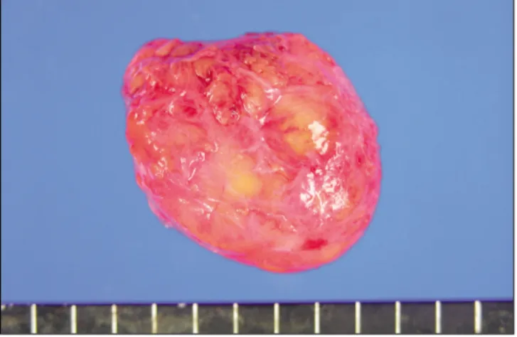
제거된 종괴는 직경이 3×2 cm으로 나타났으며 얇은 피 막으로 둘러싸인 경계가 좋고, 부드러운 난원형이었 다.(Fig. 3) 종물의 횡단면은 밝은 노란색의 균질한 지방성 단면을 보였다.(Fig. 4)
Fig. 2.Lipoma was exposed via preauricular approach.
Fig. 3. Macroscopic view (external) shows well-circum- scribed, thinly encapsulated, and oval mass with soft con- sistency.
Fig. 4. Macroscopic view (cross section) shows uniform greasy cut surface with pale yellow color.
Fig. 1.Computed tomograph (CT) scan.(enhanced view) A. Transverse view of CT scan, B. Coronal view of CT scan.
A B
J Korean Assoc Oral Maxillofac Surg 2010;36:438-40
440
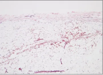
저배율의 현미경 상에서 종괴는 얇은 섬유막조직으로 잘 둘러싸여 있는 것을 볼 수 있었고(Fig. 5), 고배율의 현미경 상에서는 모양과 크기가 비슷한 성숙한 지방세포들로 구 성되어있었다.(Fig. 6)
Ⅲ. 고 찰
Kim과 Reiner
5에 의하면 지방종은 섬유성 피막으로 잘 경
계 지어지며 조직학적으로 지방조직과 비슷한 양성 종양 이다. 지방종은 전체 이하선 종양 중에 1-3%의 발생률을 보고할 만큼 드물며 주로 40대와 50대의 남성에게서 호발 하며 무통성의 연성 종물이나 전산화단층촬영이나 핵자기 공명영상 등의 도움 없이는 다형성 선종이나 와르틴 종양 또는 잔류성 낭으로 진단되기 쉽다
6,7. 전산화단층촬영사진 에서 -50에서 -150 Hounsfield units의 negative attenuation
values를 나타내는 특징을 가진다
2,8. 본 증례에서는 40대 남
성이며 CT 상에서 병소의 경계가 명확하며 negative attenu- ation values를 보이는 것을 토대로 지방종임을 의심하였고 압통이 발생하였다는 점은 지방종이 서서히 커지면서 주 위 신경을 압박하여 증상이 발생했다고 추측할 수 있겠다.
지방종은 육안검사 시 노란색을 띠며 포르말린 고정액과 같은 수용성 용액에 뜨기 때문에 특이하다. 지방세포는 정 상 지방세포와 유사하며, 핵이 한쪽으로 치우쳐 있고 둥글 고 공포화된 청명세포질을 가지고 있다. 대부분의 지방종 은 섬유격막에 의해 분리된 지방세포의 엽상을 가지며 본 증례에서도 동일한 조직소견을 보였다
9.
대부분의 저자들은 이하선 지방종의 치료를 위해 이하선 천층엽절제술을 추천한다
10,11. 하지만 이하선 주위나 천엽 에 발생할 경우에는 enucleation만으로도 충분하다. 지방종 을 적절히 제거할 경우 재발률은 1-2%이다
12. 지방종이 이
하선 심부에 발생할 경우 이하선 천층엽절제술을 시행하 는 것을 추천하나 천층에 존재할 경우 이하선 천층엽절제 술을 시행하지 않고 절제만으로 가능하므로 안면신경손상 의 가능성을 줄이기 위해서 임상진단이 중요할 것으로 판 단한다
8,11.
References
1. Williams TP, Stewart JC. Surgical pathology. In: Fonseca RJ, ed.
Oral and Maxillofacial surgery. 1st ed. Philadelphia: W.B.
Saunders; 2000:137-8.
2. Som PM, Scherl MP, Rao VM, Biller HF. Rare presentations of ordinary lipomas of the head and neck: a review. AJNR Am J Neuroradiol 1986;7:657-64.
3. Malave DA, Ziccardi VB, Greco R, Patterson GT. Lipoma of the parotid gland: report of a case. J Oral Maxillofac Surg 1994;52:
408-11.
4. Walts AE, Perzik SL. Lipomatous lesions of the parotid area.
Arch Otolaryngol 1976;102:230-2.
5. Kim YH, Reiner L. Ultrastructure of lipoma. Cancer 1982;50:
102-6.
6. Houston GD, Brannon RB. Lipoma of the parotid gland. Oral Surg Oral Med Oral Pathol 1985;60:72-4.
7. Grage TB, Lober PH, Shahon DB. Benign tumors of the major salivary glands. Surgery 1961;50:625-33.
8. Korentager R, Noyek AM, Chapnik JS, Steinhardt M, Luk SC, Cooter N. Lipoma and liposarcoma of the parotid gland: high res- olution preoperative imaging diagnosis. Laryngoscope 1988;
98:967-71.
9. Sapp JP, Eversole LR, Wysocki GP. Contemporary oral and maxillofacial pathology. 2nd ed. St. Louis, Mo: Mosby; 2004.
10. Peel RL, Gnepp DR. Diseases of the salivary glands. In: Barnes L, ed. Surgical pathology of the head and neck. Vol. 1. 1st ed.
New York, NY: Marcel Dekker; 1985:533-645.
11. Janecka IP, Conley J, Perzin KH, Pitman G. Lipoma presenting as parotid tumors. Laryngoscope 1977;87:1007-10.
12. Enzinger FM, Weiss SW. Soft tissue tumors. 2nd ed. St. Louis, Mo: Mosby; 1988.
Fig. 5.The mass is well encapsulated by thin fibromembra- nous tissue.(H&E staining, original magnification ×40)
Fig. 6. The mass is composed of mature fat cells having only a slight variation in cellular size and shape.(H&E staining, original magnification ×200)