Introduction
Coronary atherosclerosis is a chronic life-long progressive disease. Most clinical presentations of the disease are as a result of a critical narrowing of arterial lumen or a sudden thrombot- ic occlusion caused by the rupture of atherosclerotic plaque.
Unfortunately prediction of such clinically adverse events is difficult by clinical judgement or symptoms alone.1-3) Compre- hensive approach of coronary atherosclerotic plaque including qualitative and quantitative analysis is necessary for the accu- rate lesion assessment and treatment planning as well as for the prediction of future outcome (Fig. 1).4)5)
Conventional invasive coronary angiography (CAG) has been regarded as the gold standard for coronary artery disease but has limitation for assessing the extent of mild to moderate cor- onary atherosclerosis.6) Intravascular ultrasound (IVUS) or op- tical coherence tomography (OCT) are needed to quantitate the burden of atherosclerosis. Coronary computed tomography
www.kse-jcu.org http://dx.doi.org/10.4250/jcu.2015.23.4.204
REVIEW J Cardiovasc Ultrasound 2015;23(4):204-208
angiography (CCTA) can be considered as a non-invasive meth- od that may substitute CAG and IVUS.7)8) CCTA is still limited by spatial and temporal resolution but enables non-invasive patient-friendly assessment of the extent of coronary athero- sclerosis.9) Therefore it is expected that CCTA will be useful in overcoming limitations of the current diagnosis and treatment of coronary heart disease as shown below.
Demonstrating the Presence of Coronary Artery Disease
CCTA has emerged as a new noninvasive tool for screening of coronary artery disease in place of conventional stress imag- ing or electrocardiography.10)11) Therapeutic strategy of coro- nary artery disease depends on the extent and severity of coro- nary artery disease. CCTA can visualize the extent of severity of coronary artery disease with good sensitivity and excellent specificity.12)13) The performance of CCTA for plaque detection
• Received: October 12, 2015 • Revised: December 6, 2015 • Accepted: December 7, 2015
• Address for Correspondence: Jin-Ho Choi, Departments of Internal Medicine, Emergency Medicine, Samsung Medical Center, Sungkyunkwan University School of Medicine, 81 Irwon-ro, Gangnam-gu, Seoul 06351, Korea Tel: +82-2-3410-3419, Fax: +82-2-3410-3849, E-mail: [email protected]
• This is an Open Access article distributed under the terms of the Creative Commons Attribution Non-Commercial License (http://creativecommons.org/licenses/by-nc/3.0) which permits unrestricted non-commercial use, distribution, and reproduction in any medium, provided the original work is properly cited.
How to Utilize Coronary Computed
Tomography Angiography in the Treatment of Coronary Artery Disease
Hyung-Yoon Kim, MD1 and Jin-Ho Choi, MD, PhD1,2
Departments of 1Internal Medicine, 2Emergency Medicine, Heart Vascular Stroke Institute, Samsung Medical Center, Sungkyunkwan University School of Medicine, Seoul, Korea
Coronary computed tomography angiography (CCTA) has high negative predictive power for detecting coronary artery disease.
However CCTA is limited by moderate positive predictive power in the detection of myocardial ischemia. This is not unexpected because the diameter of a stenosis is a poor indicator of myocardial ischemia and discrepancy between the severity of stenosis and noninvasive tests is not uncommon. The value of stenosis for predicting future development of acute coronary syndrome repre- sented by plaque rupture has been questioned. CCTA identifies the characteristics of high-risk plaque including positive remod- eling, low density plaque and spotty or micro-calcification. Also, additional evaluation of myocardial ischemia using computa- tional flow dynamics, and luminal attenuation gradient are expected to increase both diagnostic performance for hemodynamically significant stenosis and the predictive power for future cardiovascular risk. Technical advances in CCTA would enable evaluation of both coronary artery stenosis and myocardial ischemia simultaneously with high predictive performance, and would improve vastly the clinical value of CCTA.
KEY WORDS: Coronary CT angiography · Prognosis · Coronary artery disease · Atherosclerosis · Functional ischemia · Myocardial mass.
has been validated by IVUS and OCT.7)14) Unlike stress imag- ing or invasive CAG, CCTA can detect a wide range of coronary artery disease from mild to severe disease.15)16)
Prediction of Future Coronary Heart Disease Incidents
It is difficult to predict future cardiac events through symp- toms or clinical risk factors.17)18) Clinical studies have shown that one-third of acute myocardial infarctions are asymptomatic prior to the event.19-21) Most chronic total occlusions have in- farct scar which is evidence of prior myocardial infarction but only half of them had a history of angina symptoms.22)23)
A CCTA is suitable to identify anatomical coronary stenosis, including calcified and non-calcified plaques, measure the ath- erosclerotic plaque burden and its response to treatment, and differentiate stable plaques from those that tend to rupture.24) Positive remodeling, low density (< 30 Hounsfield units) plaque, napkin-ring sign and spotty or micro-calcification detected by CCTA has been shown to have vulnerable plaque characteris- tics (Fig. 2).24-27) These non-invasive findings have been vali- dated by invasive IVUS and OCT studies.1)26)27) Moreover, non- contrast coronary calcium CT scan is also useful to evaluate the risk of coronary artery disease using a coronary calcium score and it is well demonstrated to be related to long-term prognosis.28)
Accordingly, CCTA can detect the presence of a coronary ath- erosclerosis most accurately and is one of the best non-invasive imaging modality to predict the clinical outcome of coronary ar- tery disease as well.29)
Roles of CCTA on Determining the Treatment Policy of Coronary Artery Disease
The treatment goal of coronary artery disease is the improve- ment of survival as well as relief of symptom. Revasculariza- tion by percutaneous coronary intervention or bypass surgery has relieved symptom but has been questioned for improved long-term outcome.30)31) Refinement of the need of revascular- ization strategy would be required to achieve improved clini- cal outcome after revascularization procedure including percu- taneous coronary intervention or bypass surgery. The followings are proposed treatment strategy of coronary artery disease based on the CCTA.
First is to detect coronary atherosclerosis using CCTA. CCTA has the highest diagnostic power to find coronary atheroscle- rosis among non-invasive imaging modalities and hence very high negative predictive value.1)16)
Second is to assess myocardial ischemia non-invasively with CCTA. The presence of myocardial ischemia is the most im- portant factor for making a prognosis and for the decision to perform a revascularization.31) However less than half of coro- nary arteries with a diameter stenosis ≥ 50% cause myocardial ischemia in most clinical studies.31-33) It would be greatly pref- erable to find the functional significance of a stenosis as well as the presence of a stenosis by single CCTA test. Myocardial com- puted tomography perfusion imaging could be utilized in clin- ical practice.34-36) Computational fractional flow reserve based on computed fluid dynamics analysis37)38) and contrast gradient
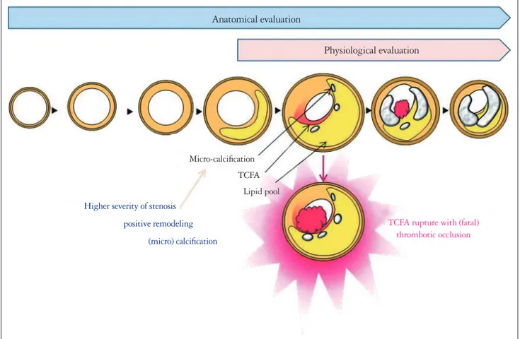
Fig. 1. Anatomical versus physiological evaluation of coronary atherosclerosis. TCFA: thin-cap fibroatheroma.
Anatomical evaluation
Physiological evaluation
TCFA rupture with (fatal) thrombotic occlusion Micro-calcification
TCFA Lipid pool Higher severity of stenosis
positive remodeling (micro) calcification
analysis39)40) are being investigated.41-43)
Third, it would be important to evaluate the amount of isch- emic myocardial mass. Several studies have investigated the relationship between the angiographic severity of stenosis and the amount of perfused myocardial tissue. Duke jeopardy score,44)45) myocardial jeopardy index,46) and Alberta Provincial Project for Outcome Assessment in Coronary Heart Disease (APPROACH) lesion score44)47) have been used to evaluate the
burden of myocardium-at-risk based on angiographic extent of stenosis. However these scoring systems are limited by semi- quantitative grading of the severity of coronary artery disease and myocardium-at-risk. Detailed anatomical variations of in- dividual vessels, myocardial mass, and location of stenosis is not taken account in these semi-quantitative scores. Nuclear perfusion studies have shown that the clinical benefit of revas- cularization became evident when the ischemic burden ex-
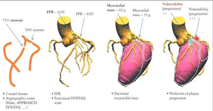
Fig. 3. Risk assessment of coronary artery disease in the future. APPROACH: Alberta Provincial Project for Outcome Assessment in Coronary Heart Disease, FFR: fractional flow reserve, PCI: percutaneous coronary intervention, SYNTAX: Synergy between PCI with Taxus and Cardiac Surgery.
75% stenosis
50% stenosis
FFR = 0.69 FFR = 0.82
Myocardial
mass = 62 g Myocardial mass = 25 g
Vulnerability (progression)
++ Vulnerability
(progression) +++
• Prediction of plaque progression
• Fractional myocardial mass
• FFR
• Functional SYNTAX score
• 2-vessel disease
• Angiographic scores (Duke, APPROACH, SYNTAX, …)
Fig. 2. Characteristics of coronary atherosclerotic plaque detected on CCTA. A: Severe stenosis. B: Positive remodeling. C: Partially calcified or
“spotty” calcification. D: Low attenuated plaque (< 30 HU). CCTA: coronary computed tomography angiography, HU: Hounsfield unit.
A B C D
ceeds 12% to 15% of total myocardial mass.48-50) CCTA enables exact quantitative assessment of the burden of myocardium- at-risk and the extent of coronary artery disease (Fig. 3).
Conclusions
CCTA can find coronary artery disease, determine the signif- icance of coronary artery disease, and guide treatment strategy including revascularization. Now it is time to validate and inves- tigate the role of CCTA in clinical practice and the impact of CCTA on the clinical outcome. Further research on this area is warranted in the future.
References
1. Sandfort V, Lima JA, Bluemke DA. Noninvasive imaging of atheroscle- rotic plaque progression: status of coronary computed tomography angiography.
Circ Cardiovasc Imaging 2015;8:e003316.
2. Laslett LJ, Alagona P Jr, Clark BA 3rd, Drozda JP Jr, Saldivar F, Wilson SR, Poe C, Hart M. The worldwide environment of cardiovascu- lar disease: prevalence, diagnosis, therapy, and policy issues: a report from the American College of Cardiology. J Am Coll Cardiol 2012;60(25 Suppl):
S1-49.
3. Fokkema ML, James SK, Albertsson P, Akerblom A, Calais F, Eriks- son P, Jensen J, Nilsson T, de Smet BJ, Sjögren I, Thorvinger B, La- gerqvist B. Population trends in percutaneous coronary intervention: 20- year results from the SCAAR (Swedish Coronary Angiography and Angioplasty Registry). J Am Coll Cardiol 2013;61:1222-30.
4. Stone GW, Maehara A, Lansky AJ, de Bruyne B, Cristea E, Mintz GS, Mehran R, McPherson J, Farhat N, Marso SP, Parise H, Tem- plin B, White R, Zhang Z, Serruys PW; PROSPECT Investigators.
A prospective natural-history study of coronary atherosclerosis. N Engl J Med 2011;364:226-35.
5. Goldstein JA, Demetriou D, Grines CL, Pica M, Shoukfeh M, O’Neill WW. Multiple complex coronary plaques in patients with acute myocardial infarction. N Engl J Med 2000;343:915-22.
6. Patel MR, Peterson ED, Dai D, Brennan JM, Redberg RF, Ander- son HV, Brindis RG, Douglas PS. Low diagnostic yield of elective coro- nary angiography. N Engl J Med 2010;362:886-95.
7. Voros S, Rinehart S, Vazquez-Figueroa JG, Kalynych A, Karmpali- otis D, Qian Z, Joshi PH, Anderson H, Murrieta L, Wilmer C, Carl- son H, Ballard W, Brown C. Prospective, head-to-head comparison of quantitative coronary angiography, quantitative computed tomography an- giography, and intravascular ultrasound for the prediction of hemodynamic significance in intermediate and severe lesions, using fractional flow reserve as reference standard (from the ATLANTA I and II Study). Am J Cardiol 2014;113:23-9.
8. Doh JH, Koo BK, Nam CW, Kim JH, Min JK, Nakazato R, Silala- hi T, Prawira H, Choi H, Lee SY, Namgung J, Kwon SU, Kwak JJ, Lee WR. Diagnostic value of coronary CT angiography in comparison with invasive coronary angiography and intravascular ultrasound in patients with intermediate coronary artery stenosis: results from the prospective multi- centre FIGURE-OUT (Functional Imaging criteria for GUiding REview of invasive coronary angiOgraphy, intravascular Ultrasound, and coronary computed Tomographic angiography) study. Eur Heart J Cardiovasc Imaging 2014;15:870-7.
9. Min JK, Shaw LJ, Berman DS. The present state of coronary computed tomography angiography a process in evolution. J Am Coll Cardiol 2010;55:
957-65.
10. Cho I, Shim J, Chang HJ, Sung JM, Hong Y, Shim H, Kim YJ, Choi BW, Min JK, Kim JY, Shim CY, Hong GR, Chung N. Prognostic val- ue of multidetector coronary computed tomography angiography in relation to
exercise electrocardiogram in patients with suspected coronary artery disease.
J Am Coll Cardiol 2012;60:2205-15.
11. Yerramasu A, Lahiri A, Venuraju S, Dumo A, Lipkin D, Underwood SR, Rakhit RD, Patel DJ. Diagnostic role of coronary calcium scoring in the rapid access chest pain clinic: prospective evaluation of NICE guidance.
Eur Heart J Cardiovasc Imaging 2014;15:886-92.
12. Heydari B, Leipsic J, Mancini GB, Min JK, Labounty T, Taylor C, Freue GV, Heilbron B. Diagnostic performance of high-definition coronary computed tomography angiography performed with multiple radiation dose reduction strategies. Can J Cardiol 2011;27:606-12.
13. Budoff MJ, Dowe D, Jollis JG, Gitter M, Sutherland J, Halamert E, Scherer M, Bellinger R, Martin A, Benton R, Delago A, Min JK. Di- agnostic performance of 64-multidetector row coronary computed tomographic angiography for evaluation of coronary artery stenosis in individuals without known coronary artery disease: results from the prospective multicenter AC- CURACY (Assessment by Coronary Computed Tomographic Angiography of Individuals Undergoing Invasive Coronary Angiography) trial. J Am Coll Cardiol 2008;52:1724-32.
14. Nakazato R, Heo R, Leipsic J, Min JK. CFR and FFR assessment with PET and CTA: strengths and limitations. Curr Cardiol Rep 2014;16:484.
15. Meijboom WB, Van Mieghem CA, van Pelt N, Weustink A, Pug- liese F, Mollet NR, Boersma E, Regar E, van Geuns RJ, de Jaegere PJ, Serruys PW, Krestin GP, de Feyter PJ. Comprehensive assessment of coronary artery stenoses: computed tomography coronary angiography versus conventional coronary angiography and correlation with fractional flow re- serve in patients with stable angina. J Am Coll Cardiol 2008;52:636-43.
16. Hoffmann U, Bamberg F, Chae CU, Nichols JH, Rogers IS, Senevi- ratne SK, Truong QA, Cury RC, Abbara S, Shapiro MD, Moloo J, Butler J, Ferencik M, Lee H, Jang IK, Parry BA, Brown DF, Udel- son JE, Achenbach S, Brady TJ, Nagurney JT. Coronary computed to- mography angiography for early triage of patients with acute chest pain: the ROMICAT (Rule Out Myocardial Infarction using Computer Assisted To- mography) trial. J Am Coll Cardiol 2009;53:1642-50.
17. Jang JJ, Krishnaswami A, Hung YY. Predictive values of Framingham risk and coronary artery calcium scores in the detection of obstructive CAD in patients with normal SPECT. Angiology 2012;63:275-81.
18. Wasfy MM, Brady TJ, Abbara S, Nasir K, Ghoshhajra BB, Truong QA, Hoffmann U, Di Carli MF, Blankstein R. Comparison of the Di- amond-Forrester method and Duke Clinical Score to predict obstructive coro- nary artery disease by computed tomographic angiography. Am J Cardiol 2012;109:998-1004.
19. Bhatia LC, Naik RH. Clinical profile of acute myocardial infarction in elderly patients. J Cardiovasc Dis Res 2013;4:107-11.
20. Sheifer SE, Gersh BJ, Yanez ND 3rd, Ades PA, Burke GL, Manolio TA. Prevalence, predisposing factors, and prognosis of clinically unrecognized myocardial infarction in the elderly. J Am Coll Cardiol 2000;35:119-26.
21. Grimm RH Jr, Tillinghast S, Daniels K, Neaton JD, Mascioli S, Crow R, Pritzker M, Prineas RJ. Unrecognized myocardial infarction:
experience in the Multiple Risk Factor Intervention Trial (MRFIT). Circu- lation 1987;75(3 Pt 2):II6-8.
22. Heyne JP, Goernig M, Feger J, Kurrat C, Werner GS, Figulla HR, Kaiser WA. Impact on adenosine stress cardiac magnetic resonance for reca- nalisation and follow up of chronic total coronary occlusions. Eur J Radiol 2007;63:384-90.
23. Choi JH, Chang SA, Choi JO, Song YB, Hahn JY, Choi SH, Lee SC, Lee SH, Oh JK, Choe Y, Gwon HC. Frequency of myocardial infarction and its relationship to angiographic collateral flow in territories supplied by chronically occluded coronary arteries. Circulation 2013;127:703-9.
24. Nakazato R, Otake H, Konishi A, Iwasaki M, Koo BK, Fukuya H, Shinke T, Hirata K, Leipsic J, Berman DS, Min JK. Atherosclerotic plaque characterization by CT angiography for identification of high-risk coronary artery lesions: a comparison to optical coherence tomography. Eur
Heart J Cardiovasc Imaging 2015;16:373-9.
25. Yamamoto H, Kitagawa T, Ohashi N, Utsunomiya H, Kunita E, Oka T, Urabe Y, Tsushima H, Awai K, Kihara Y. Noncalcified athero- sclerotic lesions with vulnerable characteristics detected by coronary CT an- giography and future coronary events. J Cardiovasc Comput Tomogr 2013;7:
192-9.
26. Arbab-Zadeh A, Fuster V. The myth of the “vulnerable plaque”: transi- tioning from a focus on individual lesions to atherosclerotic disease burden for coronary artery disease risk assessment. J Am Coll Cardiol 2015;65:846-55.
27. Puchner SB, Liu T, Mayrhofer T, Truong QA, Lee H, Fleg JL, Na- gurney JT, Udelson JE, Hoffmann U, Ferencik M. High-risk plaque detected on coronary CT angiography predicts acute coronary syndromes inde- pendent of significant stenosis in acute chest pain: results from the ROMICAT- II trial. J Am Coll Cardiol 2014;64:684-92.
28. Thomas DM, Divakaran S, Villines TC, Nasir K, Shah NR, Slim AM, Blankstein R, Cheezum MK. Management of coronary artery cal- cium and coronary cta findings. Curr Cardiovasc Imaging Rep 2015;8:18.
29. Opolski MP, Kepka C, Achenbach S, Pregowski J, Kruk M, Staruch AD, Kadziela J, Ruzyllo W, Witkowski A. Advanced computed tomo- graphic anatomical and morphometric plaque analysis for prediction of frac- tional flow reserve in intermediate coronary lesions. Eur J Radiol 2014;83:
135-41.
30. Hachamovitch R, Berman DS, Shaw LJ, Kiat H, Cohen I, Cabico JA, Friedman J, Diamond GA. Incremental prognostic value of myocardial perfusion single photon emission computed tomography for the prediction of cardiac death: differential stratification for risk of cardiac death and myocar- dial infarction. Circulation 1998;97:535-43.
31. Pijls NH, van Schaardenburgh P, Manoharan G, Boersma E, Bech JW, van’t Veer M, Bär F, Hoorntje J, Koolen J, Wijns W, de Bruyne B.
Percutaneous coronary intervention of functionally nonsignificant stenosis:
5-year follow-up of the DEFER Study. J Am Coll Cardiol 2007;49:
2105-11.
32. Douglas PS, Hoffmann U, Patel MR, Mark DB, Al-Khalidi HR, Cavanaugh B, Cole J, Dolor RJ, Fordyce CB, Huang M, Khan MA, Kosinski AS, Krucoff MW, Malhotra V, Picard MH, Udelson JE, Velazquez EJ, Yow E, Cooper LS, Lee KL; PROMISE Investigators. Out- comes of anatomical versus functional testing for coronary artery disease. N Engl J Med 2015;372:1291-300.
33. Tonino PA, De Bruyne B, Pijls NH, Siebert U, Ikeno F, van’t Veer M, Klauss V, Manoharan G, Engstrøm T, Oldroyd KG, Ver Lee PN, MacCarthy PA, Fearon WF; FAME Study Investigators. Fractional flow reserve versus angiography for guiding percutaneous coronary interven- tion. N Engl J Med 2009;360:213-24.
34. Ko BS, Cameron JD, Meredith IT, Leung M, Antonis PR, Nasis A, Crossett M, Hope SA, Lehman SJ, Troupis J, DeFrance T, Seneviratne SK. Computed tomography stress myocardial perfusion imaging in patients considered for revascularization: a comparison with fractional flow reserve.
Eur Heart J 2012;33:67-77.
35. Choo KS, Hwangbo L, Kim JH, Park YH, Kim JS, Kim J, Chun KJ, Jeong DW, Lim SJ. Adenosine-stress low-dose single-scan CT myocar- dial perfusion imaging using a 128-slice dual-source CT: a comparison with fractional flow reserve. Acta Radiol 2013;54:389-95.
36. Greif M, von Ziegler F, Bamberg F, Tittus J, Schwarz F, D’Anastasi M, Marcus RP, Schenzle J, Becker C, Nikolaou K, Becker A. CT stress perfusion imaging for detection of haemodynamically relevant coronary stenosis as defined by FFR. Heart 2013;99:1004-11.
37. Taylor CA, Fonte TA, Min JK. Computational fluid dynamics applied to cardiac computed tomography for noninvasive quantification of fractional flow reserve: scientific basis. J Am Coll Cardiol 2013;61:2233-41.
38. Kim KH, Doh JH, Koo BK, Min JK, Erglis A, Yang HM, Park KW, Lee HY, Kang HJ, Kim YJ, Lee SY, Kim HS. A novel noninva- sive technology for treatment planning using virtual coronary stenting and computed tomography-derived computed fractional flow reserve. JACC Car- diovasc Interv 2014;7:72-8.
39. Wong DT, Ko BS, Cameron JD, Nerlekar N, Leung MC, Malaia- pan Y, Crossett M, Leong DP, Worthley SG, Troupis J, Meredith IT, Seneviratne SK. Transluminal attenuation gradient in coronary com- puted tomography angiography is a novel noninvasive approach to the identi- fication of functionally significant coronary artery stenosis: a comparison with fractional flow reserve. J Am Coll Cardiol 2013;61:1271-9.
40. Stuijfzand WJ, Danad I, Raijmakers PG, Marcu CB, Heymans MW, van Kuijk CC, van Rossum AC, Nieman K, Min JK, Leipsic J, van Royen N, Knaapen P. Additional value of transluminal attenua- tion gradient in CT angiography to predict hemodynamic significance of cor- onary artery stenosis. JACC Cardiovasc Imaging 2014;7:374-86.
41. Choi AD, Joly JM, Chen MY, Weigold WG. Physiologic evaluation of ischemia using cardiac CT: current status of CT myocardial perfusion and CT fractional flow reserve. J Cardiovasc Comput Tomogr 2014;8:272-81.
42. Gonzalez JA, Lipinski MJ, Flors L, Shaw PW, Kramer CM, Salerno M. Meta-Analysis of Diagnostic Performance of Coronary Computed To- mography Angiography, Computed Tomography Perfusion, and Computed Tomography-Fractional Flow Reserve in Functional Myocardial Ischemia Assessment Versus Invasive Fractional Flow Reserve. Am J Cardiol 2015;
116:1469-78.
43. Magalhães TA, Kishi S, George RT, Arbab-Zadeh A, Vavere AL, Cox C, Matheson MB, Miller JM, Brinker J, Di Carli M, Rybicki FJ, Rochitte CE, Clouse ME, Lima JA. Combined coronary angiography and myocardial perfusion by computed tomography in the identification of flow-limiting stenosis - The CORE320 study: an integrated analysis of CT coronary angiography and myocardial perfusion. J Cardiovasc Comput To- mogr 2015;9:438-45.
44. Graham MM, Faris PD, Ghali WA, Galbraith PD, Norris CM, Badry JT, Mitchell LB, Curtis MJ, Knudtson ML; APPROACH Investigators (Alberta Provincial Project for Outcome Assessment in Coronary Heart Disease). Validation of three myocardial jeopardy scores in a population-based cardiac catheterization cohort. Am Heart J 2001;142:254-61.
45. Califf RM, Phillips HR 3rd, Hindman MC, Mark DB, Lee KL, Be- har VS, Johnson RA, Pryor DB, Rosati RA, Wagner GS. Prognostic value of a coronary artery jeopardy score. J Am Coll Cardiol 1985;5:1055- 63.
46. Ortiz-Pérez JT, Meyers SN, Lee DC, Kansal P, Klocke FJ, Holly TA, Davidson CJ, Bonow RO, Wu E. Angiographic estimates of myocardium at risk during acute myocardial infarction: validation study using cardiac magnetic resonance imaging. Eur Heart J 2007;28:1750-8.
47. Brandt PW, Partridge JB, Wattie WJ. Coronary arteriography; method of presentation of the arteriogram report and a scoring system. Clin Radiol 1977;28:361-5.
48. Cremer P, Hachamovitch R. Assessing the prognostic implications of myo- cardial perfusion studies: identification of patients at risk vs patients who may benefit from intervention? Curr Cardiol Rep 2014;16:472.
49. Iskandrian AE, Hage FG, Shaw LJ, Mahmarian JJ, Berman DS. Se- rial myocardial perfusion imaging: defining a significant change and target- ing management decisions. JACC Cardiovasc Imaging 2014;7:79-96.
50. Doukky R, Hayes K, Frogge N, Balakrishnan G, Dontaraju VS, Rangel MO, Golzar Y, Garcia-Sayan E, Hendel RC. Impact of appro- priate use on the prognostic value of single-photon emission computed tomog- raphy myocardial perfusion imaging. Circulation 2013;128:1634-43.