Introduction
Cardiac magnetic resonance imaging (CMR) is a useful di- agnostic imaging modality in patients with known or sus- pected coronary artery disease (CAD). Numerous studies have shown its efficacy in identifying the presence of CAD and its sequelae, and for risk stratifying patients for future outcome.1-3) In many conditions, such as following acute myocardial infarc- tion (MI), CMR often provides unique information not avail- able from other modalities.1) Nonetheless, CMR has limited availability and is considered by some to be a ‘boutique’ mo- dality. One possible reason is its complexity. CMR is not a sin- gle technique. Instead, it consists of multiple distinct tech- niques and a lack of understanding of which techniques to perform and how to interpret the findings in combination limits the efficacy of CMR. On the other hand, its multipara- metric nature can provide a comprehensive assessment with the potential for higher accuracy than is achievable by other modalities. In this context, this article presents a brief techni-
REVIEW J Cardiovasc Ultrasound 2016;24(2):96-103
cal overview of CMR along with a discussion of how CMR can be used in patients with known or suspected CAD. Rather than providing a detailed summary of the literature, we will emphasize a ‘real-world’ perspective in a working clinical prac- tice. We will focus on specific scenarios which highlight the unique information that CMR provides and discuss illustra- tive cases.
Technical Overview Core exam
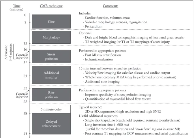
The multiplicity of techniques and the variety of informa- tion that can be obtained in a CMR exam can be exhaustive, but increasingly there has been a move towards standardiza- tion of imaging protocols tailored to specific indications, an effort spearheaded by the Society of Cardiovascular Magnetic Resonance.4) Our suggested implementation and a timeline (Fig. 1) of a CMR protocol for a standard exam is as follows:
The Use of Cardiac Magnetic Resonance in Patients with Suspected Coronary Artery Disease: A Clinical Practice Perspective
Sung-A Chang, MD, PhD1 and Raymond J. Kim, MD2,3,4
1Division of Cardiology, Department of Medicine, Heart Vascular Stroke Institute Imaging Center, Samsung Medical Center, Sungkyunkwan University School of Medicine, Seoul, Korea
2Duke Cardiovascular Magnetic Resonance Center, Departments of 3Medicine, 4Radiology, Duke University Medical Center, Durham, NC, USA
Cardiac magnetic resonance imaging (CMR) is a useful diagnostic imaging modality in patients with known or suspected coronary artery disease (CAD). It provides unique information not available from other modalities, however, it is complex. CMR is not a single technique. Instead, it consists of multiple distinct techniques and a lack of understanding of which techniques to perform and how to interpret the findings in combination limits its efficacy and widespread use. Conversely, its multiparametric nature can provide a comprehensive assessment with the potential for higher accuracy than is achievable by other modalities.
Moreover, its ability to directly assess myopathic processes often contributes insights that change patient management. In this article we provide a brief technical overview and focus on specific clinical scenarios in patients with known or suspected CAD.
We highlight the multiparametric nature of CMR and discuss cases which illustrate the unique information that CMR can contribute.
KEY WORDS: Cardiac magnetic resonance imaging · Coronary artery disease.
• Received: January 20, 2016 • Revised: February 20, 2016 • Accepted: May 10, 2016
• Address for Correspondence: Raymond J. Kim, Duke Cardiovascular Magnetic Resonance Center, Duke University Medical Center, PO Box 3934, Trent Drive, Durham, NC 27710, USA Tel: +1-919-668-3539, Fax: +1-919-668-3554, E-mail: [email protected]
• This is an Open Access article distributed under the terms of the Creative Commons Attribution Non-Commercial License (http://creativecommons.org/licenses/by-nc/3.0) which permits unrestricted non-commercial use, distribution, and reproduction in any medium, provided the original work is properly cited.
Cine images
Cine images provide comprehensive evaluation of regional and global ventricular function and overall cardiac morpholo- gy. Visual evaluation of valvular function and morphology is also performed using these images. Cine images are typically acquired in the short-axis plane from above the mitral valve through the left ventricular (LV) apex along with standard 2-, 3-, and 4-chamber long-axis views. Inter- and intra-observer reproducibility of cine CMR imaging for the quantification of LV volumes and function have been shown in multiple studies to be excellent, predominantly due to its high spatial and temporal resolution and its capacity for complete LV coverage.
The improvement in reproducibility relative to echocardiog- raphy allows a significant reduction in the sample sizes re- quired for research studies to demonstrate meaningful changes as a result of experimental therapies. This has led to increasing use of CMR in research studies that use cardiac morphology and/or function as an efficacy endpoint.5)
Stress and rest perfusion
These sequences are designed to demonstrate contrast media passage through the myocardium in a manner that reliably re- flects myocardial blood flow. The sequences are heavily T1- weighted, in order to accurately depict the passage of a T1 shortening contrast agent such as gadolinium through the myo-
cardium. It is most often used for the detection of obstructive CAD, where it is performed with pharmacological vasodilation (e.g., adenosine or regadenoson). The underlying principle is similar to that in nuclear perfusion imaging, where a vasodila- tor is used to accentuate regional differences in myocardial blood flow. However, as opposed to nuclear techniques, CMR perfusion imaging is a first pass imaging study that directly images the passage of contrast, and therefore is performed us- ing an abbreviated adenosine protocol (~3 minutes). Also, CMR perfusion imaging has higher spatial resolution (> 20 ×) than radionuclide techniques, and can depict a perfusion defect that is limited to the subendocardial layer. CMR perfusion im- aging has the additional advantage of providing a more linear depiction of myocardial blood flow in response to vasodilation, without the plateau phenomenon seen with nuclear agents.6)
Delayed enhancement
Delayed enhancement (DE) imaging allows the diagnosis and sizing of MI, assessment of viability, and other tissue char- acterization such as identification of thrombus and nonisch- emic scarring. Images are obtained 5–15 minutes after the ad- ministration of gadolinium contrast, and views are matched to the spatial location of the cine images. The DE technique ac- centuates the visibility of areas with abnormal gadolinium accu- mulation by using an inversion pulse to “null” the signal from
Comments CMR technique
Time
Includes
- Cardiac function, volumes, mass
- Valvular morphology, stenosis, regurgitation - Pericardium
Optional
- Dark and bright blood tomographic imaging of heart and great vessels - T2 weighted imaging (or T1 or T2 mapping) of acute injury Performed in appropriate patients
- Post MI risk stratification - Ischemia evaluation
15 min interval between stress/rest perfusion
- Velocity/flow imaging for valvular disease and cardiac output - Whole heart coronary MRA (may be performed prior to contrast) - Additional cine imaging
Typical sequence
- 2D or 3D, segmented (high resolution and high SNR) Useful additional sequences
- Single shot (rapid, no breath hold required, resistant to arrhythmias) - Long inversion time (~600 ms)
(useful for thrombus detection and “no-reflow” regions in acute MI) - Post contrast T1 mapping for ECV measurement and serial quantification Performed in appropriate patients
- Improves specificity of stress perfusion imaging - Quantification of myocardial blood flow reserve Cine
Morphology
Stress perfusion
Rest perfusion
Delayed enhancement 5 minute delay Additional
imaging Adenosine 3–4 minutes
(minutes)
Contrast injection
Contrast injection
45 38 33 32 25 17 16 13 5 0
Fig. 1. Timeline and potential components of a multitechnique CMR examination including stress and rest perfusion imaging. Note that the entire exam can be performed within 30–45 minutes. In cases where the patient is unable to cooperate, the cine examination can be performed with real- time imaging and delayed enhancement with single-shot techniques. This technique will reduce artifacts in patients unable to breath-hold and total exam time will be shortened further. Adapted from Kim et al. J Am Coll Cardiol 2009;55:1-16, with permission of Elsevier.1) CMR: cardiac magnetic resonance imaging, MI: myocardial infarction, MRA: magnetic resonance angiography, SNR: signal to noise ratio, ECV: extra-cellular volume fraction.
normal myocardium, resulting in abnormal areas standing out as regions of bright, “hyperenhanced” signal intensity against a background of very dark normal myocardium. The standard high-resolution DE sequence is acquired from a segmented acquisition (data from multiple heart beats), but in cases of ar- rhythmia or inability to breath hold, single-shot techniques can provide comparable data in a fraction of the imaging time with a small reduction in sensitivity.7)
The technique of DE-CMR has been extensively validated.
In animal models, DE-CMR has been shown to demonstrate acute and chronic MI with a near exact spatial match to histo- pathology specimens.8) Additionally, these studies show that DE-CMR can distinguish between reversible and irreversible injury independent of wall motion, infarct age, and reperfu- sion status. Compared with single photon emission computed tomography (SPECT) imaging, the DE-CMR technique is significantly more sensitive for the detection of subendocardial infarction, over 40% of which are missed with SPECT.9) With standard imaging parameters, DE-CMR is capable of demon- strating infarcts involving as little as one one-thousandth of to- tal LV mass, and that are undetectable by techniques that assess myocardial perfusion or contractile function. The high spatial resolution of DE-CMR has been used to visualize microinfarc- tions, involving as little as 1 g of tissue, which may occur dur- ing otherwise successful percutaneous coronary intervention.10)
Optional sequences
An overview of cardiac and great vessel anatomy is helpful in many cases and can be rapidly performed using single-shot dark blood and/or bright blood techniques. These result in a stack of still-frame images and are usually acquired in the stan- dard orthogonal imaging planes (axial, sagittal, or coronal).
T1 and T2 mapping techniques are increasingly being in- vestigated in studies of infarction as well as other myocardial disorders, and can be performed before and/or after the admin- istration of intravenous contrast. These sequences provide a quantitative assessment of regional myocardial T1 and T2 val- ues, and are less subject to surface coil sensitivity profiles that can result in variable image intensity for different regions of the heart. In the absence of gadolinium contrast, T1 and T2 values are increased in the setting of acute necrosis-related edema, and thus these sequences allow the depiction of edema and provide a metric, which may be useful for purposes of quantification and serial assessment. A parametric map of ex- tracellular volume fraction can be made by combining pre- contrast (“native”) and post-contrast T1 mapping values.11) Extracellular volume fraction will be increased both in the set- ting of acute necrosis and chronic collagenous scar. These se- quences are also being investigated for use in a variety of non- ischemic myocardial disorders, such as amyloidosis and Anderson-Fabry disease.12)13)
Multiparametric acquisitions
Each of the sequences described above provide separate and distinct pieces of information. Hence, one can get multiple data acquisitions of the same location and obtain a compre- hensive, multifaceted view of the heart. In general, if two (or more) tests provide information regarding the presence of a disease, diagnostic accuracy is not necessarily improved com- pared with one test alone. For example, if the algorithm used to determine the presence of disease requires both tests to be positive, this improves diagnostic specificity at the cost of sen- sitivity. Conversely, if the algorithm requires only one test to be positive, this generally improves sensitivity but worsens specificity. With multiparametric CMR acquisitions, it is im- portant to realize that each of the sequences described above are ‘tuned’ to highlight specific biological tissues or properties (e.g., infarction, thrombus, fat, tissue perfusion, etc.). Accord- ingly, depending on the property, the results of one test should take precedence over the results of another, and algorithms can be developed to improve diagnostic accuracy, which is not true ordinarily regarding multiple tests. Additionally, because many image artifacts are pulse-sequence specific, these are not prop- agated throughout the examination and generally do not re- duce overall scan quality.
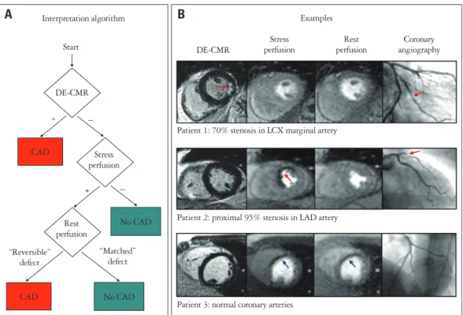
The report by Klem et al.14) demonstrates an example of an interpretation algorithm that uses multiparametric acquisi- tions to improve diagnostic accuracy. The algorithm is based on 2 principles. First, with perfusion-CMR and DE-CMR, we have independent methods to obtain information regarding the presence or absence of MI. Thus, one method could be used to confirm the results of the other. Second, DE-CMR im- age quality (e.g., signal-to-noise ratio), is far better than perfu- sion-CMR since it is less demanding in terms of scanner hard- ware (DE-CMR images can be built up over several seconds rather than in 0.1 seconds as is required for first-pass perfu- sion). Thus, DE-CMR is far more accurate for the diagnosis of MI. Conceptually, it then follows that perfusion defects that have similar intensity and extent during both stress and rest (“matched defect”) but do not have infarction on DE-CMR are artifactual and should not be considered positive for CAD.
Conversely, the presence of infarction on DE-CMR favors the diagnosis of CAD even if the results of perfusion imaging are equivocal. The algorithm and some typical images are dis- played in Fig. 2. Overall, the combination of perfusion and DE images (compared with perfusion images alone) improved the accuracy for the diagnosis of CAD from 68% to 88% in a cohort of patients with intermediate pretest probability of ob- structive CAD.
Specific Clinical Applications Unrecognized myocardial infarction
Because of late presentation, patients with MI may present without diagnostic biomarker elevation of electrocardiogram
abnormalities. Moreover, wall motion abnormalities on cardiac imaging may not occur unless the infarcted region exceeds 20% to 50% of the myocardial wall thickness. Similarly, scin- tigraphic defects may not be apparent until greater than 10 g of tissue is infarcted. Thus, because a sizable threshold of dam- age is required, echocardiography or SPECT may miss MI, particularly when it is small or subendocardial. Wall motion abnormalities may also occur in entities other than MI, such as Takotsubo cardiomyopathy, indicating a lack of specificity.
In these instances, where the diagnosis of MI is difficult, DE- CMR may prove helpful. It is notable that DE-CMR is the only imaging modality for the detection of MI that has been validated in a multicenter trial. In an international study of 282 patients with acute and 284 with chronic first-time MI, the sensitivity of DE-CMR for the detection of MI reached 99% and 94% in acute and chronic MI, respectively.15)
One might postulate that the MIs not recognized by stan- dard criteria are likely small, and of uncertain significance.
However, in the report by Kwong et al.,16) the presence of un- recognized MI detected by DE-CMR was associated with a
greater than 6-fold higher risk of major adverse cardiac events compared to the absence of such an MI. Importantly, the in- formation from DE-CMR was a stronger predictor of outcome than standard clinical risk factors and even catheterization data. Similarly, the study by Kim et al.2) reported that the presence of unrecognized MI by DE-CMR predicted an 11- fold higher risk of all-cause mortality than those without MI.
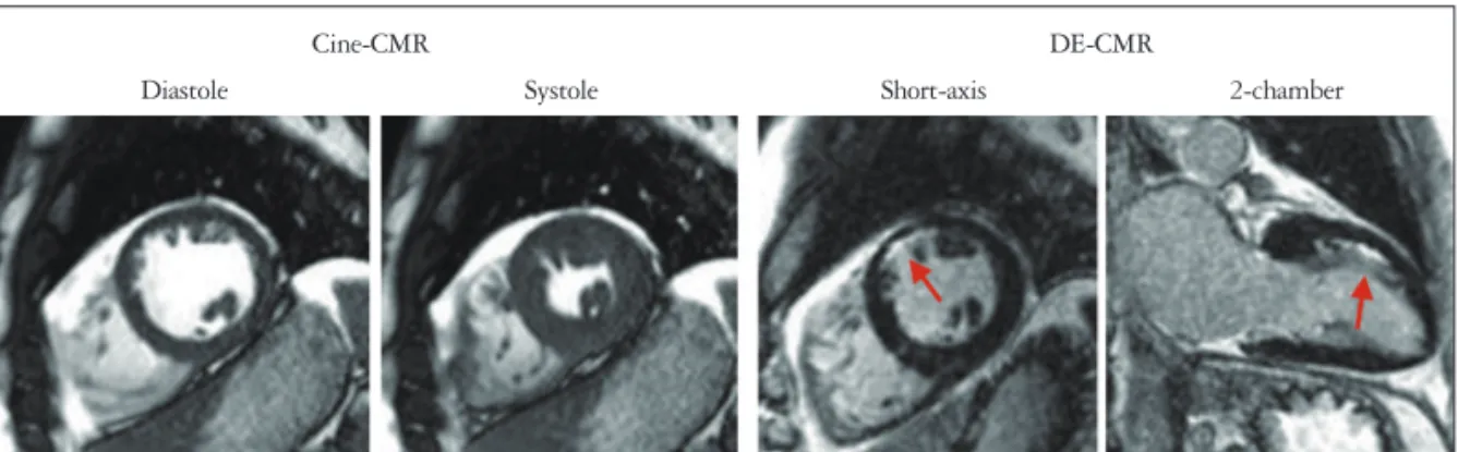
Fig. 3 demonstrates CMR images in a patient with palpita- tions who did not have any symptoms or signs of CAD. A stress echocardiogram was normal, but a Holter examination demonstrated frequent episodes of non-sustained ventricular tachyarrhythmia, which were asymptomatic. The patient was thought to have a structurally normal heart and the arrhyth- mia was thought to be right ventricular outflow tract in ori- gin. The CMR images showed normal LV size and systolic function, however, there was a focal subendocardial infarct in the mid-ventricular anterior wall, which was completely un- expected. A subsequent cardiac catheterization demonstrated occlusive coronary disease in the second diagonal branch of left anterior descending artery.
Fig. 2. Interpretation algorithm for incorporating delayed enhancement-cardiac magnetic resonance imaging (DE-CMR) with stress and rest perfusion magnetic resonance imaging (MRI) for the detection of coronary disease. A: Schema of the interpretation algorithm. 1) Positive DE-CMR study: hyperenhanced myocardium consistent with a prior myocardial infarction (MI) is detected. Does not include isolated midwall or epicardial hyperenhancement which can occur in nonischemic disorders. 2) Standard negative stress study: no perfusion defects at stress or rest. 3) Standard positive stress study: perfusion defects are present with adenosine that are absent or reduced at rest. 4) Artifactual perfusion defect: matched stress and rest perfusion defects without evidence of prior MI on DE-CMR. B: Patient examples. Top row: patient with a positive DE-CMR study demonstrating an infarct in the inferolateral wall (red arrows) although perfusion-MRI is negative. The interpretation algorithm classifies this patient as positive for coronary artery disease (CAD). Coronary angiography verified disease in a circumflex marginal artery. Middle row: patient with a negative DE-CMR study but with a prominent reversible defect in the anteroseptal wall on perfusion-MRI (red arrows). The interpretation algorithm classifies this patient as positive for CAD. Coronary angiography demonstrated a proximal 95% LAD stenosis. Bottom row: patient with a matched stress-rest perfusion defect (blue arrows) but without evidence of prior MI on DE-CMR. The interpretation algorithm classifies the perfusion defects as artifactual. Coronary angiography demonstrated normal coronary arteries. Adapted from Klem et al. J Am Coll Cardiol 2006;47:1630-8, with permission of Elsevier.14) LCX:
left circumflex artery, LAD: left anterior descending.
Examples
Patient 1: 70% stenosis in LCX marginal artery
Patient 2: proximal 95% stenosis in LAD artery
Patient 3: normal coronary arteries DE-CMR
Stress perfusion
Rest perfusion
Coronary angiography
B
+ Interpretation algorithm
Start
A
DE-CMR
Stress perfusion
No CAD No CAD Rest
perfusion
“Reversible”
defect
“Matched”
defect CAD
CAD +
+ –
–
Detection of CAD
The diagnostic performance of stress perfusion CMR has been evaluated in a number of studies in humans. A recent meta-analysis reveals that on average, the sensitivity and speci- ficity of perfusion-CMR for detecting obstructive CAD were 89% and 76%, respectively.17) Additionally, several studies have directly compared the performance of adenosine stress perfusion CMR with SPECT imaging. In the largest single- center trial to date, the Clinical evaluation of MAgnetic Reso- nance imaging in Coronary heart disease (CE-MARC) trial, 752 patients underwent SPECT and CMR, as well as invasive coronary angiography. The study found that the sensitivity of CMR for the detection of obstructive CAD exceeded that of SPECT (87% vs. 67%), with preserved specificity (both 83%).18) It is important to recognize that the CMR examina- tions in this study were multi-parametric and involved several components including cine and DE imaging in addition to stress and rest perfusion imaging. Similar to the study by Klem et al.,14) the finding of an infarct on DE images alone was sufficient to render the CMR test positive for CAD. It is likely that a key factor in the high performance of CMR was the multi-parametric information that was provided. Com- bined with the superior delineation of infarction provided by DE-CMR relative to SPECT, it is not surprising that many centers are now using stress perfusion CMR as a first-line test.
Coronary magnetic resonance angiography (MRA) may be used to directly visualize coronary anatomy and morphology.
However, coronary magnetic resonance imaging is technically demanding, leading to intermediate sensitivity and specificity values for the detection of CAD in validation studies. More re- cently, “whole-heart” free-breathing steady-state free preces- sion techniques in combination with multichannel coils and parallel acquisition has improved coronary MRA.19) The high- er inherent signal-to-noise that is available from 3T scanners is likely to improve coronary MRA further.20) That said, imaging times are still relatively long (7–10 minutes) and image quali- ty can be highly variable between patients. The CE-MARC trial also included a coronary MRA component. Of the 676 patients that had coronary MRA performed, it is notable that only 55% of coronary artery segments (15 segments for each
patient) were deemed sufficient quality to analyze. Overall, the diagnostic accuracy of CMR did not change if the coronary MRA component was excluded. Hence, we believe that re- ports that suggest that coronary MRA has similar diagnostic accuracy with cardiac computed tomography angiography are premature. From a “working” clinical practice perspective, currently we do not perform coronary MRA as part of our rou- tine CMR examination except in patients with suspected cor- onary anomalies.
Role of myocardial tissue characterization with DE-CMR
Similar to troponin, the detection of injury by DE-CMR is specific for irreversible myocardial damage but is not specific for MI. One potential advantage of DE-CMR is that the pat- tern of hyperenhancement, rather than simply the presence or extent, may offer important information regarding the etiolo- gy of myocardial damage. For this purpose, the concept that ischemic myonecrosis proceeds as a wavefront from the suben- docardium to the epicardium with increasing coronary occlu- sion time is crucial. Correspondingly, hyperenhancement pat- terns that spare the subendocardium and are limited to the middle or epicardial portion of the LV wall are usually non- ischemic in origin because significant damage in the setting of CAD almost always involves the subendocardium. Moreover, certain nonischemic disorders, such as myocarditis, amyloido- sis, hypertrophic cardiomyopathy, have characteristic hyperen- hancement patterns that suggest specific diagnoses, and a sys- tematic approach to interpreting DE-CMR images in patients with cardiomyopathy has been proposed.21)
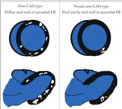
However, one caveat should be mentioned. Novices should be cautioned against overzealously interpreting non-CAD- type hyperenhancement. If there is a large area of hyperen- hancement and it is entirely midwall or epicardial throughout its course, then this is clearly a non-CAD-type pattern. Like- wise, if there are numerous islands of midwall or epicardial hyperenhancement throughout the LV and crossing into mul- tiple coronary artery territories, this is also likely nonischemic (Fig. 4). However, a single isolated ‘dot’ of hyperenhancement or a few patchy lesions of hyperenhancement in a limited focal
Diastole Systole 2-chamber
Cine-CMR
Short-axis
DE-CMR
Fig. 3. Cine and delayed-enhancement images in a patient without signs or symptoms of coronary artery disease who was believed to have a structurally normal heart. Images demonstrate a focal subendocardial infarct (arrows) with normal left ventricular size and systolic function. CMR:
cardiac magnetic resonance imaging, DE: delayed enhancement.
region of myocardium (Fig. 4) is unlikely to be nonischemic in origin, regardless of whether it spares the subendocardium or not. In our experience, the latter pattern is more likely to represent an aborted MI or atherosclerotic (or embolic) disease affecting a small secondary coronary branch, rather than a non- ischemic disorder.
DE-CMR has proven to be an excellent technique for the evaluation of patients with heart failure and/or suspected to have a cardiomyopathy. In this setting it may alleviate the need for invasive coronary angiography. However, the use of hyperenhancement patterns should not be considered merely another way to rule-in or rule-out CAD. By allowing a direct assessment of myopathic processes, tissue characterization with DE-CMR often provides a clue as to the specific etiology. Even when definitive clinical tests have already been performed, CMR can contribute information that is unique and often clinically relevant.
Fig. 5 shows representative images in 3 patients presenting with chest discomfort and ST-segment elevation. In all 3, tro- ponins were elevated, hence the diagnosis was ST-segment ele- vation MI. However, invasive coronary angiography demon- strated normal coronary arteries. CMR was performed because the diagnosis was uncertain, and in each case tissue character- ization with DE-CMR contributed unique insights to the clinical situation. In these 3 cases, CAD had already been ruled-out by coronary angiography, however, without the in- formation provided by CMR to clarify the diagnosis, patient management and potentially prognosis would have been cru- cially changed.
Finally, a more in-depth case is illustrated in Fig. 6. This is
of a 57-year-old man who underwent CMR before a scheduled radiofrequency ablation procedure for paroxysmal atrial fibril- lation. The primary purpose of the CMR was to delineate the pulmonary vein anatomy, however, at our institution, a cardiac study is routinely performed in addition to evaluate for struc- tural heart disease. The patient complained of palpitations, but otherwise was asymptomatic, and the physical examina- tion was normal. His cardiac history was significant for known single-vessel coronary disease, which had been diagnosed by invasive coronary angiography one year prior during an admis- sion for chest pain. Otherwise, he was healthy and he did not have any significant past medical history. An echocardiogram performed 2 days prior to the CMR demonstrated normal LV
Fig. 4. The cartoon schematic on the left demonstrates multiple islands of hyperenhancement in a diffuse, near global pattern, which is classic for particular types of viral myocarditis. In other words, the correct interpretation of this pattern would be “non-CAD-type”. In contrast, it would be incorrect to judge the pattern on the right, which shows only a few “dots” of hyperenhancement in a limited, focal territory, as non- CAD-type. CAD: coronary artery disease, DE: delayed enhancement.
Diffuse mid-wall or epicardial DE Non-CAD-type
Focal patchy mid-wall or epicardial DE
Pseudo non-CAD-type A
B
C
Fig. 5. Typical delayed enhancement-cardiac magnetic resonance (DE-CMR) images from 3 patients with chest discomfort, ST-segment elevation, positive troponins, and normal invasive coronary angiograms.
A: Linear, epicardial hyperenhancement (yellow arrows) is present and is indicative of myocarditis. B: Cine and DE images of a patient with sudden emotional stress and apical ballooning; the absence of hyper- enhancement is consistent with Takotsubo cardiomyopathy. C: Focal but transmural hyperenhancement (red arrow) involving the lateral apex is present and indicative of myocardial infarction (MI) because of temporary occlusion of a small diagonal branch off the distal left anterior descending coronary artery. DE-CMR with a long inversion time (600 ms) shows a thrombus (yellow arrowhead) in the left atrial appendage, suggesting that an embolus led to the MI.
size and function, normal valvular function, and only mild LV wall hypertrophy (Fig. 6A). Cine-CMR demonstrated similar findings to the echocardiogram (not shown). Remarkably, de- layed-enhancement images demonstrated widespread hyper- enhancement from the base of the LV to nearly the apex. The hyperenhancement was primarily epicardial (right ventricular side of septum), however there was also subendocardial hyper- enhancement of the basal LV lateral wall, which was less severe (Fig. 6B). The DE-CMR findings were consistent with a dif- fuse infiltrative process such as amyloidosis or sarcoidosis, hence a cardiac biopsy was performed. The biopsy demon- strated transthyretin amyloidosis. Recall that this patient pre- sented with a history of paroxysmal atrial fibrillation and sin- gle-vessel CAD and had no other known medical problems.
Now he has a biopsy-proven diagnosis of cardiac amyloidosis, which raises the question: without CMR, how long would it have taken to get the diagnosis?
CMR as a first-line test
For patients with known or suspected CAD, the conven- tional use of noninvasive imaging is to rule-in or rule-out ob- structive CAD or to assess the extent and severity of myocar- dial ischemia. While CMR can be an accurate alternative to other modalities in this regard, there are many patients that do not fit this conventional scenario. We have discussed a vari- ety of cases where invasive coronary angiography had already been performed, yet the diagnosis remained unclear. CMR is often utilized in these situations, where it is considered the fi- nal arbitrator for the diagnosis. Although well intentioned,
this can pigeonhole CMR as an important but secondary test only to be used when there are discrepancies among conven- tional tests. This is shortsighted. To paraphrase Donald Rums- feld, the former United States Secretary of Defense, the prima- ry problem is “you don’t know, what you don’t know”. In our experience when CMR is used as a first-line test, unexpected information similar to but perhaps less dramatic than that de- scribed in the last case, occurs nearly on a weekly basis. In the majority of these patients, CMR does not decide between two possible diagnoses as much as it provides an entirely new di- agnosis. In countries such as the United States, where cost to the patient for a CMR examination is no more expensive than that of SPECT,22) we believe that CMR should considered a first-line examination for the evaluation of patients with known or suspected CAD.
References
1. Kim HW, Farzaneh-Far A, Kim RJ. Cardiovascular magnetic resonance in patients with myocardial infarction: current and emerging applications. J Am Coll Cardiol 2009;55:1-16.
2. Kim HW, Klem I, Shah DJ, Wu E, Meyers SN, Parker MA, Crow- ley AL, Bonow RO, Judd RM, Kim RJ. Unrecognized non-Q-wave myocardial infarction: prevalence and prognostic significance in patients with suspected coronary disease. PLoS Med 2009;6:e1000057.
3. Shah R, Heydari B, Coelho-Filho O, Murthy VL, Abbasi S, Feng JH, Pencina M, Neilan TG, Meadows JL, Francis S, Blankstein R, Steigner M, di Carli M, Jerosch-Herold M, Kwong RY. Stress cardiac magnetic resonance imaging provides effective cardiac risk reclassification in patients with known or suspected stable coronary artery disease. Circulation 2013;128:605-14.
4. Kramer CM, Barkhausen J, Flamm SD, Kim RJ, Nagel E; Society Fig. 6. A 57-year-old patient with paroxysmal atrial fibrillation undergoing cardiac magnetic resonance imaging (CMR) prior to radiofrequency ablation procedure. A: Echocardiogram demonstrated normal left ventricular (LV) size and function and only mild LV wall hypertrophy. B: Delayed enhancement- CMR images demonstrated widespread hyperenhancement (arrows) primarily in a non-coronary artery disease-type pattern, which suggested an infiltrative process.
B
Diastole Systole
A
for Cardiovascular Magnetic Resonance Board of Trustees Task Force on Standardized Protocols. Standardized cardiovascular magnetic resonance (CMR) protocols 2013 update. J Cardiovasc Magn Reson 2013;15:91.
5. Grothues F, Braun-Dullaeus R. Serial assessment of ventricular morphol- ogy and function. Heart Fail Clin 2009;5:301-14, v.
6. Gerber BL, Raman SV, Nayak K, Epstein FH, Ferreira P, Axel L, Kraitchman DL. Myocardial first-pass perfusion cardiovascular magnetic resonance: history, theory, and current state of the art. J Cardiovasc Magn Reson 2008;10:18.
7. Sievers B, Elliott MD, Hurwitz LM, Albert TS, Klem I, Rehwald WG, Parker MA, Judd RM, Kim RJ. Rapid detection of myocardial in- farction by subsecond, free-breathing delayed contrast-enhancement cardio- vascular magnetic resonance. Circulation 2007;115:236-44.
8. Kim RJ, Fieno DS, Parrish TB, Harris K, Chen EL, Simonetti O, Bundy J, Finn JP, Klocke FJ, Judd RM. Relationship of MRI delayed contrast enhancement to irreversible injury, infarct age, and contractile func- tion. Circulation 1999;100:1992-2002.
9. Wagner A, Mahrholdt H, Holly TA, Elliott MD, Regenfus M, Parker M, Klocke FJ, Bonow RO, Kim RJ, Judd RM. Contrast-enhanced MRI and routine single photon emission computed tomography (SPECT) perfusion imaging for detection of subendocardial myocardial infarcts: an imaging study. Lancet 2003;361:374-9.
10. Ricciardi MJ, Wu E, Davidson CJ, Choi KM, Klocke FJ, Bonow RO, Judd RM, Kim RJ. Visualization of discrete microinfarction after per- cutaneous coronary intervention associated with mild creatine kinase-MB el- evation. Circulation 2001;103:2780-3.
11. White SK, Sado DM, Flett AS, Moon JC. Characterising the myocar- dial interstitial space: the clinical relevance of non-invasive imaging. Heart 2012;98:773-9.
12. Banypersad SM, Sado DM, Flett AS, Gibbs SD, Pinney JH, Maes- trini V, Cox AT, Fontana M, Whelan CJ, Wechalekar AD, Hawkins PN, Moon JC. Quantification of myocardial extracellular volume fraction in systemic AL amyloidosis: an equilibrium contrast cardiovascular magnet- ic resonance study. Circ Cardiovasc Imaging 2013;6:34-9.
13. Sado DM, White SK, Piechnik SK, Banypersad SM, Treibel T, Captur G, Fontana M, Maestrini V, Flett AS, Robson MD, Lach- mann RH, Murphy E, Mehta A, Hughes D, Neubauer S, Elliott PM, Moon JC. Identification and assessment of Anderson-Fabry disease by cardiovascular magnetic resonance noncontrast myocardial T1 mapping.
Circ Cardiovasc Imaging 2013;6:392-8.
14. Klem I, Heitner JF, Shah DJ, Sketch MH Jr, Behar V, Weinsaft J, Cawley P, Parker M, Elliott M, Judd RM, Kim RJ. Improved detection of coronary artery disease by stress perfusion cardiovascular magnetic reso- nance with the use of delayed enhancement infarction imaging. J Am Coll Cardiol 2006;47:1630-8.
15. Kim RJ, Albert TS, Wible JH, Elliott MD, Allen JC, Lee JC, Parker M, Napoli A, Judd RM; Gadoversetamide Myocardial In- farction Imaging Investigators. Performance of delayed-enhancement magnetic resonance imaging with gadoversetamide contrast for the detection and assessment of myocardial infarction: an international, multicenter, dou- ble-blinded, randomized trial. Circulation 2008;117:629-37.
16. Kwong RY, Schussheim AE, Rekhraj S, Aletras AH, Geller N, Da- vis J, Christian TF, Balaban RS, Arai AE. Detecting acute coronary syn- drome in the emergency department with cardiac magnetic resonance imaging.
Circulation 2003;107:531-7.
17. Jaarsma C, Leiner T, Bekkers SC, Crijns HJ, Wildberger JE, Nagel E, Nelemans PJ, Schalla S. Diagnostic performance of noninvasive myo- cardial perfusion imaging using single-photon emission computed tomogra- phy, cardiac magnetic resonance, and positron emission tomography imaging for the detection of obstructive coronary artery disease: a meta-analysis. J Am Coll Cardiol 2012;59:1719-28.
18. Greenwood JP, Maredia N, Younger JF, Brown JM, Nixon J, Ever- ett CC, Bijsterveld P, Ridgway JP, Radjenovic A, Dickinson CJ, Ball SG, Plein S. Cardiovascular magnetic resonance and single-photon emission computed tomography for diagnosis of coronary heart disease (CE- MARC): a prospective trial. Lancet 2012;379:453-60.
19. Kato S, Kitagawa K, Ishida N, Ishida M, Nagata M, Ichikawa Y, Katahira K, Matsumoto Y, Seo K, Ochiai R, Kobayashi Y, Sakuma H.
Assessment of coronary artery disease using magnetic resonance coronary an- giography: a national multicenter trial. J Am Coll Cardiol 2010;56:983- 91.
20. Yang Q, Li K, Liu X, Du X, Bi X, Huang F, Jerecic R, Liu Z, An J, Xu D, Zheng H, Fan Z, Li D. 3.0T whole-heart coronary magnetic reso- nance angiography performed with 32-channel cardiac coils: a single-center experience. Circ Cardiovasc Imaging 2012;5:573-9.
21. Senthilkumar A, Majmudar MD, Shenoy C, Kim HW, Kim RJ.
Identifying the etiology: a systematic approach using delayed-enhancement cardiovascular magnetic resonance. Heart Fail Clin 2009;5:349-67, vi.
22. Shah DJ, Kim HW, Kim RJ. Evaluation of ischemic heart disease.
Heart Fail Clin 2009;5:315-32, v.