서론
경구 착색은 다양한 연령대에서 잇몸, 점막, 경구개, 혀 등에서 나타나는 것으로 보고되고 있다. 잇몸착색은 질환이라기 보다는 심미적 장애에 가깝다.1 멜라닌은 갈 색색소로 가장 보편적인 천연색소이며 잇몸의 내인성 색소침착으로 잘 알려져 있다. 잇몸은 구강점막에서 착 색이 가장 두드러지게 보이는 부분이다.
기존 치은미백(gum bleaching)은 외과용 메스를 이 용하여 수술적 기법을 이용한 착색 박리술(the surgical removal of undesirable pigmentation using scalpels),2 Co2 레이저를 이용한 치료법,3 980 nm 파장 다이오드 수 술용 레이저를 이용한 치료법(A semiconductor diode surgical laser unit of wavelength 980 nm),4 Erbium, Chromium Doped Yttrium Scandium Gallium Garnet (Er-Cr:YSGG) 레이저 치료술,5,6 630 nm 다이오드 레이
*Correspondence to: Jong-Ho Lee
Professor, Department of Oral and Maxillofacial Surgery, Clinical Trial Center, Seoul National University Dental Hospital, 103 Daehak-ro, Jongno-gu, Seoul, 03080, Republic of Korea
Tel: +82-2-20722-3054, E-mail: [email protected]
Received: May 18, 2016/Last Revision: June 14, 2016/Accepted: June 14, 2016
365 nm leD laser treatment on beagle for gingival whitening without gum dermabrasion
Sung-Ho Lee
1, Ryun Kyung Kim
2, Na-Ri Seo
1, Ho-Kyung Lim
1,3, Soo-Hwan Byun
4, Young-Joon Lim
5, Soung-Min Kim
1,6, Jong-Ho Lee
1,6*
1
Department of Oral and Maxillofacial Surgery, Clinical Trial Center, Seoul National University Dental Hospital, Seoul, Republic of Korea
2
College of information and Communication Engineering, Sungkyunkwan University, Suwon, Republic of Korea
3
Department of Oral and Maxillofacial Surgery, Korea University Medical Center, Guro Hospital, Seoul, Republic of Korea
4
Department of Oral and Maxillofacial Surgery, Dongtan Sacred Heart Hospital, Hallym University Medical Center, Suwon, Republic of Korea
5
Department of Prosthodontics, Seoul National University Dental Hospital, Seoul, Republic of Korea
6
Dental Research Institute, School of Dentistry, Seoul National University, Seoul, Republic of Korea
Purpose: Gingival whitening is one of dental treatment purposes which is close to treating aesthetic disorders. Initial gingival
whitening treatment was done by dermabrasion using a high power Diode Laser. However, this treatment method cannot be free from any infection or pain after the treatment. Therefore, we have decided to progress gingival whitening treatment using a low power LED laser. Materials and Methods: The laser was irradiated on pork meat then the safety of output power, temperature change and skin denaturalization was measured. Bison 365 nm LED laser was irradiated on oral mucosal pigment of a 15 - 20 kg beagle for 15 min for 1 - 2 weeks, one or two times each. Any pigment loss was checked through Hematoxyline-Eosin staining.Results: The melanin pigments at the area of 365 nm LED Laser irradiation were decreased. Conclusion: The 365 nm LED Laser
proposed in this study is considered to compensate the bleaching effect achieved by either using Diode laser or surgical methods. (JDent Rehabil Appl Sci 2016;32(2):117-22)
Key words: gingival whitening; melanocyte; laser treatment; 365 nm laser
Copyright© 2016 The Korean Academy of Stomatognathic Function and Occlusion.
This is an Open-Access article distributed under the terms of the Creative Commons Attribution Non-Commercial License (http://creativecommons.org/
licenses/by-nc/4.0) which permits unrestricted non-commercial use, distribution, and reproduction in any medium, provided the original work is properly cited.
cc
ISSN 2384-4353 eISSN 2384-4272
저를 이용한 레이저 치료술(Laser therapy was don for gingival part by diode laser 630 nm), 808 nm 파장 연조 직 다이오드 수술용 레이저(A soft tissue diode surgical laser unit wavelength 808 nm)등7을 이용한 치료법이 존 재한다. 하지만 모든 치료법이 박피술을 이용한 시술로 서 환자의 통증이 유발되고 회복까지의 기간이 길며 수 술적 부작용이 동반 될 수도 있다는 위험성을 내재하고 있다.
이에 우리는 비손 365 nm LED 레이저를 이용하여 출 력전력과 온도 측정을 통한 안전성을 실험하고, 비글의 구강점막 착색부위에 박피술 없이 레이저를 조사하여 멜라닌 색소 감소를 통한 탈색화 효과를 확인하였다.
연구 재료 및 방법
1. 레이저 출력전력 및 온도 측정 실험
돼지고기의 표면에 비손 365nm LED 레이저를 3, 7.5, 15 조건으로 조사 시 laser power meter and laser energy meters (gentec, Quebec, Canada)를 이용하 여 출력전력을 측정하고, 200-channel expandable temperature measurement logger (ZR-RX45, OMRON corporation, Kyoto, Japan)를 이용하여 온도를 측정한 다.
2. 동물실험
생후 350 - 400일, 체중 15 - 20 kg의 비글견 수컷 4 마리(15 - 20 kg, Orient Bio, Seongnam, Korea)를 실 험 대상으로 선정하였으며, 한 마리당 두 부위의 구강내 점막 착색을 실험 대상으로 선정하였다. 비손 365 nm LED 레이저 조사 전, 체중당 10 mg의 케타민(Zoletil 50, Virbac, Carros, France)과 체중당 0.2 mg의 자일렌 (Rompen, Bayer Korea, Seoul, Korea)을 정맥 주사하여 마취상태 유지 후 실험 진행을 하였다. 비글의 구강점막 멜라닌 착색부위에 비손 365 nm LED 레이저를 조사하 며 실험 그룹은 1) Control, 2) 1주 15분간 1회 조사, 3) 1 주 15분간 2회 조사, 4) 2주 15분간 1회 조사, 5) 2주 15 분간 2회 조사로 진행하였다. 각 실험 후 염화칼륨을 정 맥 주사하여 심정지를 확인한 후, 레이저 조사부위를 채 취하였다.
3. 조직염색
레이저 조사부위를 채취한 뒤, 기존의 방법과 동일 하게 수세, 탈수, 명화 과정을 거친 뒤 파라핀 포매 후 4 µm 두께로 절단한 다음 Hematoxyline-Eosin 염색을 시 행하여 광학현미경으로 관찰하였다. 이 후 각 그룹별 멜 라닌세포 계수측정을 실시 하여 통계적 계수측정을 실 시하였다.
4. 통계분석
실험군과 대조군간의 차이에 대한 통계적인 분석은 ANOVA를 이용하였고, 유의 수준은 P < 0.05로 설정하 였다.
결과
1. 레이저 조사 시 출력전력 및 온도 변화
비손 365 nm LED 레이저를 3, 7.5, 15의 거리로 돼지 고기에 조사 시 출력전력은 각각 267 ± 8, 100 ± 5, 54
± 4.2 (mW)였다(Fig. 1A). 온도측정은 출력전력 측정과 동일한 방법으로 진행하였으며, 7.5와 15의 조사거리에 서 22.3, 54.84 (℃)로 측정되었다(Fig. 1C).
2. 비글 구강내 점막에 레이저 조사 후 착색 관찰
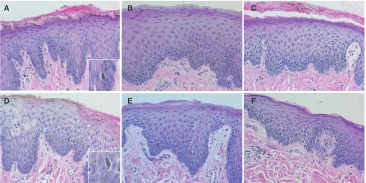
레이저를 조사하지 않은 비글 점막과 실험군 간의 멜 라닌 착색을 H/E 염색을 통해 관찰 한 결과, 레이저를 조사한 군과 Control 군간의 비교에서 멜라닌 착색의 수 가 적게 나타나는 것을 관찰하였다(Fig. 2). H/E 염색으 로 관찰한 멜라닌착색을 계수 측정한 결과 레이저 조사 후 1주 뒤 1회 레이저 조사 보다 2회 레이저 조사 시 멜 라닌 착색이 줄어드는 것으로 나타났다(Fig. 3A). 레이 저 조사 후 2주 뒤 1회 레이저 조사와 2회 레이저 조사 를 비교 관찰한 결과, 레이저를 2회 조사 시 멜라닌 착색 이 1회 레이저 조사 보다 더 줄어드는 것을 관찰하였다 (Fig. 3B). 레이저 횟수간의 비교 관찰한 결과 1회 레이 저 조사한 실험군은 2주차 실험 그룹이 1주차 실험 그룹 보다 멜라닌 착색이 더 줄어 드는 것을 관찰 할 수 있었 다(Fig. 3C). 그러나 2회 레이저 조사한 실험군 간의 비 교에서는 1주와 2주 실험그룹간에 통계적인 차이점을
Fig. 2. Melanin pigment H/E staining. (A, D) 1 weeks and 2 weeks control groups. (B, E) It was melanin pigment that
stained H/E in 1 weeks 1-time and 2 weeks 1-time irradiation. (C, F) It was melanin pigment that stained H/E in 1 weeks 2-time and 2 weeks 2-time irradiation.A B C
D E F
Fig. 1. Bison 365 nm LED laser output power & Temperature. (A, B) Change in output power when irradiated 365 nm
laser on pork, (C, D) Measurement of change in temperature when irradiated 365 nm laser. (C) 3℃ was not measured because it exceeded the 100 mW.A
Length Start 10 s 20 s 30 s 40 s 50 s 60 s 5 m 10 m 15 m
3 mW 260 273 274 275 275 275 275 275 275 275
7.5 mW 22.5 105 105 105 106 105 105 105 105 105 15 mW 58.3 58.3 55.8 55 54.7 54.4 54.3 54.2 53.5 50
C
Length Start 10 s 20 s 30 s 40 s 50 s 60 s 5 m 10 m 15 m
3 ℃ Do not measure temperature
7.5 ℃ 21.7 21.1 21.8 22 22.7 22.7 22.9 22.4 22.9 22.9 15 ℃ 21.7 22 22.6 22 21.5 20.9 20.8 22 22.9 22.9
B
Output power (mW)
300 250 200 150 100 50 0
3 7.5 15
0 10 20 30 40 50 60 300 600 900 (s)
D
Temperature (℃) 24
23
22
21
20
7.5 15
0 10 20 30 40 50 60 300 600 900 (s)
확인 할 수 없었다(Fig. 3D).
고찰
임상에서 레이저를 이용하여 잇몸의 멜라닌 착색을 제거하는 방법은 이전의 다양한 사례를 통해 보고되어 왔다. 이러한 방법들은 대부분 구강 수술, 구강 병리학, 또는 치주병리학을 기반으로 수술하였다. 또한 풍부한 수술적 경험을 기반으로 적은 출혈과 특별한 수술 기법, 그리고 레이저를 이용하여 잇몸 점막에 멜라닌 착색이 착색된 부위를 박리하여 미백 효과를 얻었다.6 그러나 이러한 방법은 시술자의 경험에 의지되며 시술 후 환자 의 통증 및 감염의 우려 유발될 수 있다. 우리는 실험 진 행을 위해 기존의 고출력 다이오드 레이저가2-7 아닌 저 출력 LED를 이용한 레이저를 사용하였다.
Yousuf et al.은 Semiconductor Diode Laser를 이용하 여 비글 잇몸의 멜라닌 착색을 탈색소 하였다.1 그러나 앞서 밝혔듯이 수술적인 치료법이 아닌 멜라닌 착색의 탈색소화 실험 및 문헌이 없어 우리 실험에서는 비글의 구강내 점막의 멜라닌 착색에 박피술 없이 비손 365 nm LED 레이저를 이용하여 탈색화 효과를 실험하였다.
비손 365 nm LED 레이저 출력전력의 기대출력은 실 험을 통해 105 - 110 mW로 설정하였다(Fig. 1A, 1B). 기 기의 조사거리 7.5에서 평균 105.5 mW의 출력전력을 확인하였으며(Fig. 1B), 이를 기반으로 15분 간의 온도 측정에서 22 ± 0.9℃의 온도 변화 폭을 관찰하였다(Fig.
1D). 다음 H/E 염색을 통해 비글 구강내 점막의 멜라닌 착색을 관찰하였으며(Fig. 2), 이것을 계수측정 한 결과 비손 365 nm LED 레이저 조사 후에 멜라닌 착색이 줄 어드는 것을 관찰하였다. 각 실험 주차에서 1회 조사와
Fig. 3. Melanin pigment counting statistical analysis. (A) Comparison of 1 weeks 1-time irradiation and 1 weeks 2-time
(*, **P < 0.05). (B) Comparison of 2 weeks 1-time irradiation and 2 weeks 2-time (* P < 0.05). (C) Comparison of 1 weeks 1-time irradiation and 2 weeks 1-time irradiation (*, ** P < 0.05). (D) Comparison of 1 weeks 2-time irradiation and 2 weeks 2-time irradiation (* P < 0.01 and ** P < 0.05).A
A number of melanonyte counting
16 14 12 10 8 6 4 2 0
Experimental groups
Control
2 weeks 15 min 1-time 2 weeks 15 min 2-time
A number of melanonyte counting
16 14 12 10 8 6 4 2 0
Experimental groups
Control
1 weeks 15 min 1-time 1 weeks 15 min 2-time
B
C
A number of melanonyte counting
16 14 12 10 8 6 4 2 0
Experimental groups
Control
1 weeks 15 min 2-time 2 weeks 15 min 2-time
A number of melanonyte counting
16 14 12 10 8 6 4 2 0
Experimental groups
Control
1 weeks 15 min 1-time 1 weeks 15 min 2-time
D
2회 조사 시 멜라닌 착색 양을 계측한 결과 2회 조사 시 멜라닌 착색이 더 많이 줄어 드는 것을 확인하였다(Fig.
3A, 3B). 주 차간의 비교 분석에서도 1주 보다 2주차에 조사한 실험군에서 많은 멜라닌 착색 감소를 확인하였 다(Fig. 3C).
이것은 기존의 다양한 파장과 종류의 다이오드 레이 저 및 다양한 기술의 수술 처치에서 보였던 탈색화 효과 를 박피술 없이 비손 365 nm LED 레이저를 이용하여 동물 실험에서 유사한 수준의 탈색화 효과를 관찰하였 다. 이것은 비손 365 nm LED 레이저가 기존 수술적 기 술 및 다이오드 레이저를 이용한 박피술을 이용한 탈색 화 효과를 대처 할 수 있을 것이라고 사료 된다. 다만 비 손 365 nm LED 레이저를 임상에 적용 하기 까지는 조 금 더 다양한 방법의 안정성 및 추가 실험을 통해 확보 해야 할 것이라고 판단 된다.
결론
본 연구에서는 비글 구강내 점막의 멜라닌 착색에 365 nm LED 레이저를 조사하여 착색된 멜라닌세포의 손실 여부를 분석하였다. 레이저 조사 후 1주와 2주차간의 멜 라닌 착색이 줄어드는 것을 확인하였으며, 각 주차간에 서도 1회와 2회간의 조사 횟수에 따라 멜라닌 착색이 줄 어드는 차이가 나타나는 것으로 관찰되었다. 다만 각 주 차간에 초기 1, 2회 간에 멜라닌 착색이 줄어드는 것은 통계적으로 차이를 보였으나, 그 이후엔 각각 차이가 없 는 것으로 나타났다.
Acknowledgements
This study was supported by a grant of Ministry for Trade, Industry, and Energy, Republic of Korea (10047615).
References
1. Yousuf A, Hossain M, Nakamura Y, Yamada Y, Kinoshita J, Matsumoto K. Removal of gingival melanin pigmentation with the semiconductor diode laser: a case report. J Clin Laser Med Surg 2000;18:263-6.
2. Patil KP, Joshi V, Waghmode V, Kanakdande V.
Gingival depigmentation : a split mouth compara- tive study between scalpel and cryosurgery. Con- temp Clin Dent 2015;6:S97-S101.
3. Monteiro LS, Costa JA, da Câmara MI, Albuquer- que R, Martins M, Pacheco JJ, Salazar F, Figueira F. Aesthetic depigmentation of gingival smoker’s melanosis using carbon dioxide lasers. Case Rep Dent 2015;2015:510589.
4. Chandna S, Kedige SD. Evaluation of pain on use of electrosurgery and diode lasers in the manage- ment of gingival hyperpigmentation: a comparative study. J Indian Soc Periodontol 2015;19:49-55.
5. Fekrazad R, Chiniforush N. One visit providing de- sirable smile by laser application. J Lasers Med Sci 2014;5:47-50.
6. Berk G, Atici K, Berk N. Treatment of gingival pig- mentation with Er, Cr:YSGG Laser. J Oral Laser Applications 2005;5:249-53.
7. Soliman MM, Al Thomali Y, Al Shammrani A, El Gazaerly H. The use of soft tissue diode laser in the treatment of oral hyper pigmentation. Int J Health Sci 2014;8:133-140.
*교신저자: 이종호
(03080)서울특별시 종로구 대학로 103 서울대학교치과병원 구강악안면외과 Tel: 02-20722-3054|E-mail: [email protected]
접수일: 2016년 5월 18일|수정일: 2016년 6월 14일|채택일: 2016년 6월 14일
잇몸 박피술 없는 치은 미백을 위한 비글에서 365 nm LED 레이저 치료
이성호
1, 김륜경
2, 서나리
1, 임호경
1,3, 변수환
4, 임영준
5, 김성민
1,6, 이종호
1,6*
1서울대학교치과병원 구강악안면외과
2성균관대학교 정보통신공학과
3고려대학교구로병원 구강악안면외과
4한림대학교동탄성심병원 구강악안면외과
5서울대학교치과병원 보철과
6서울대학교 치과대학 치학연구소
목적: 치은 미백은 치과치료의 목적으로 심미적 장애를 위한 치료에 가깝다. 기존의 치은미백술은 고출력 다이오드 레 이저를 이용하여 박피술에 의한 치료를 했왔다. 그러나 이러한 치료법은 시술 후 감염 및 통증에서 자유로울 수 없기 에, 우리는 저출력 LED 레이저를 이용하여 치은미백술을 진행하고자 하였다.
연구 재료 및 방법: 돼지고기 표면에 레이저를 조사하여 출력전력, 온도변화, 피부 변성 등의 안정성을 측정하였다. 15 - 20 kg 비글의 구강내 점막 색소에 비손 365 nm LED 레이저를 15분씩, 1 - 2주간 1, 2회씩 각각 조사하였다. 헤마토실 린-에오신 염색을 통하여 색소의 손실여부를 확인하였다.
결과: 365 nm LED 레이저 조사 부위의 멜라닌 색소가 줄어드는 것을 확인 할 수 있었다.
결론: 본 연구에서 제시하는 365 nm LED 레이저는 수술적 기술 및 다이오드 레이저를 이용한 탈색화 효과를 대처 할 수 있을 것이라고 사료 된다.
(구강회복응용과학지 2016;32(2):117-22)
주요어: 치은미백; 멜라닌색소; 레이저치료; 비손 365 nm 레이저