Corresponding author:
Shin Young ParkDepartment of Clinical Laboratory Science, Cheju Halla University, 38 Halladaehak-ro, Jeju 63092, Korea
E-mail:
[email protected]ORCID:
https://orcid.org/0000-0002-2330-5638ORIGINAL ARTICLE
Age-Related Fecal Calprotectin Concentrations in Healthy Adults
Shin Young Park
Department of Clinical Laboratory Science, Cheju Halla University, Jeju, Korea
건강한 성인의 연령별 분변 칼프로텍틴의 농도
박신영
제주한라대학교 임상병리과
ARTICLE INFO ABSTRACT
Received
August 24, 2020Revised 1
st August 30, 2020Revised 2
nd August 30, 2020Accepted
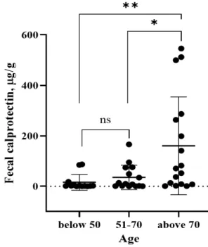
August 31, 2020Fecal calprotectin (FC) is a marker used for the differential diagnosis of inflammatory bowel disease (IBD). FC is also used to determine the effects of treatment and recurrence prediction because of its non-decomposition by bacteria, relative week stability at room temperature, and its uniform distribution within feces. Healthy male and female adults between the age of 30 and 80 living in Jeju were selected for this study. The FC concentration in the healthy control group (N=45) was distributed widely as 0∼545.9 μg/g and showed a significant difference with age in healthy adults.
The FC concentration in adults over 70 years old (80.6 years on average) was 160.3 μg/g. The result is approximately 10 times higher than in adults below 50 years (44 years on average), with FC concentrations at 15.88 μg/g. Moreover, adults over 50 years, with an average age of 59.6, had FC concentrations of 35.46 μg/g, which were two times higher than the below 50-year-old group, confirming the significant correlation between age and FC concentration. As the FC test is a non-invasive and cost-effective objective marker in IBD tests, a suitable cut-off value is required for different ages. This study provides the baseline data for differential diagnoses.
Copyright Ⓒ 2020 The Korean Society for Clinical Laboratory Science. All rights reserved.
Key words Fecal calprotectin Healthy volunteer Inflammatory bowel disease Marker
INTRODUCTION
Inflammatory bowel disease (IBD) usually affects adults in North America and Northern Europe. However, IBD’s prevalence recently expanded to ulcerative colitis and Crohn’s disease in Korea, according to recent epidemiology studies [1, 2]. IBD can be characterized by the chronic inflammation of the gastrointestinal system, requiring lifetime management and affecting
the patient’s quality of life. Diagnosis and bowel inflammation monitoring are vital in IDB diagnosis and follow up. Although diagnosis and follow-up manage- ment are closely related to its long term prognosis, the most effective monitoring methods until now are limited to colonoscopy and biopsy, which are invasive, time-consuming, and costly [3, 4]. Thus, a simpler, faster, and less invasive fecal calprotectin (FC) test has been recently reported in various studies. The chronic inflammatory disease, with its cycles of improvement and aggravation, requires a lifetime of disease activity evaluation. As a functional disease of no specific symptom and sign, it is difficult for differential diagnoses from other disorders, especially without standard diagnostic
Korean Society for Clinical Laboratory Science