Korean J Clin Lab Sci. Vol. 45, No. 1, Mar. 2013 1
www.kjcls.org ORIGINAL ARTICLE
Korean J Clin Lab Sci. 2013, 45(1):1-4 ISSN 1738-3544
Distribution of Vancomycin-resistant Enterococci Isolates Using a ChromID VRE Agar
Hyun Lee and In-Seon Yoon
Department of Laboratory Medicine, Seoul National University Hospital, Seoul 110-744, Korea
Vancomycin-resistant enterococci (VRE) have emerged as important healthcare-associated infection since last two decades. ChromID VRE agar (cIDVA) is useful for VRE rectal swab screening.
We investigated all VRE were isolated on the cIDVA. A total of 363 rectal swabs of 85 patients to test VRE screening were inoculated into bile-esculin (B-E) broth with 6 μg/mL vancomycin. After 24 hours incubation, we subcultured B-E broths were changed to black onto cIDVA. All isolates were identified by the MICROSCAN and VITEK2. The vanA gene and vancomycin minimal inhibition concentration (MIC) were detected by PCR and E-test respectively. 277 E. faecium (84.7%), 16 E.
faecalis (4.9%), 25 E. avium (7.6%), 8 E. gallinarum (2.4%) and 1 E. raffinosus (0.3%) were isolated.
10.3% of VRE detected on cIDVA were other than E. faecium and E. faecalis that presented various color from colorless to pale violet. All isolates contained vanA and vancomycin MIC were >256 μg/mL. VRE isolates other than E. faecium and E. faecalis should be objective to the contact precautions for healthcare-associated infection control if they possess vanA gene. Due to emerging enterococci carrying vanA such as E. avium, E. gallinarum, and E. raffinosus, VRE surveillance should be expanded to all isolates on chromogenic agar.
Keywords: Vancomycin-resistant entreococci, Chromogenic agar, Enterococcus avium, Entero- coccus gallinarum
Corresponding author: Hyun Lee Department of Laboratory Medicine, Seoul National University Hospital, Seoul 110-744, Korea.
Tel: 82-2-2072-2796 E-mail: [email protected]
This is an Open Access article distributed under the terms of the Creative Commons Attribution Non-Commercial License (http://creativecommons.org/licenses/by-nc/3.0) which permits unrestricted non-commercial use, distribution, and reproduction in any medium, provided the original work is properly cited.
Copyright © 2013 Korean Society of Clinical Laboratory Science. All rights reserved.
Received: September 6, 2012 Revised: January 7, 2013 Accepted: March 13, 2013
서 론
조건무산소성 그람양성알균인 장알균은 사람의 장내 정상세균 이지만 aminoglycosides와 cephalosporins에 내성이므로 장알 균에 감염되었을 경우 치료에 어려움이 있다(Cetinkaya 등, 2000;
Song 등, 2005; Delmas 등, 2007; Berktas 등, 2008). Gly- copeptide 노출에 의해 유전적 변이가 유도되는 반코마이신 내성 장알균(vancomycin- resistant enterococci, VRE)은 1986년 유 럽에서 처음 보고되었고(Kim 등, 2010) 20여년이 지난 지금도 병 원감염 감염원으로서 주목받고 있다(Harbarth 등, 2002; Cuzon 등, 2008). 이러한 VRE의 조기 검출은 신속한 병원감염 관리를 위 한 도구로서 중요하며 색소생산성 배지들은 이를 위해서 개발되었 고 Kallstrom 등(2010)의 색소생산성 배지에 대한 평가 연구에서 높은 민감도(98.6%)와 특이도(99.1%)를 보였다.
색소생산성 배지의 사용으로 환자의 직장 면봉 내지 대변에서
VRE를 분리하는데 소요되는 시간이 상당히 줄어들었으며 특히 vanA 또는 vanB 유전형 E. faecium과 E. faecalis는 ChromID VRE agar (cIDVA, bioMérieux, Marcy l’Étoile, France)에서 각 각 보라색과 청녹색으로 보이므로 VRE 선별검사에 매우 유용하다 (Cuzon 등, 2008; Kallstrom 등, 2010; Peterson 등, 2010).
하지만 최근 들어 cIDVA에서 E. faecium과 비슷한 색과 모양의 집락을 형성하는 E. faecium이 아닌 다른 장알균이 종종 분리되고 있어 저자들은 색소생산성 배지에서 다양한 색으로 집락을 형성하 는 장알균의 종류와 분리빈도 그리고 VRE 환자의 입원기간과의 연 관성을 알아보고자 하였다.
재료 및 방법 1. VRE 선별검사
본원에서 2010년 12월 1일부터 2011년 4월 18일까지 VRE 선
2 Hyun Lee and In-Seon Yoon. Distribution of Vancomycin-resistant Enterococci Isolates Using a ChromID VRE Agar
www.kjcls.org
Table 1. Demography of VRE screening
No. of patients 85
No. of rectal swabs 363
Cases of VRE 283
Cases of single VRE 245
Cases of multiple VRE 38
Cases of no VRE 80
No. of VRE isolates 327
Table 2. Results of VRE screening culture on cIDVA and genotyping
Isolates cIDVA Genotype MIC (μg/mL) Number of isolates (%)
E. faecium Violet vanA >256 277 84.7
E. faecalis Green NT NT 16 4.9
E. avium Colorless to pale violet vanA >256 25 7.7
E. gallinarum Pale violet vanA >256 8 2.4
E. raffinosus Pale violet vanA >256 1 0.3
Total 327 100.0
NT, not tested; MIC, minimum inhibitory concentration.
Fig. 1. 2% Agarose gel electrophoresis of PCR products: Lane 1.
Marker; 2-6. E. avium 7-8. E. gallinarum 9. E. raffinosus 10.
Negative control; 11. Positive control (vanA: 1032 bp).
별검사를 위해 진단검사의학과 임상미생물검사실에 의뢰된 VRE 감시환자 85명의 직장 면봉 363개를 대상으로 하였다. 각 직장 면 봉을 반코마이신(6 μg/mL)이 첨가된 bile-esculin broth (Enter- ococcosel broth, E-B, BD, Sparks, MD)에 담가서 35oC 배양기에 서 24시간 동안 배양하였다. 검정색으로 변한 E-B는 cIDVA에 접종 하였고 cIDVA에서 자란 집락은 색깔 별로 모두 그람 염색을 실시하 였다. 장알균으로 추정되는 그람 양성 사슬알균은 methyl-D- glucopyranoside (MGP) 분해능과 운동성 검사를 하였다. MGP 및 운동성 양성인 균주와 cIDVA에서 보라색과 녹색인 집락은 Microscan (Siemens, West Sacramento, California, USA)과 Vitek2 (bioMérieux, Marcy l’Étoile, France)를 이용하여 세균 동 정과 항균제 감수성 검사를 하였다. 반코마이신 최소억제농도 측정 은 분리된 장알균을 멸균생리식염수에 0.5 MacFarland로 맞춰서 Mueller-Hinton agar에 접종하고 반코마이신 E-test (AB Biodisk, Solna, Sweden) 시험지를 올려놓고 35oC에서 24시간 배 양한 다음 억제대를 측정하였다.
2. vanA 유전자 검사
cIDVA에서 분리된 VRE의 DNA는 PrepMan Ultra Sample Preparation Reagent kit (Applied Biosystem, Foster City, CA) 를 사용하여 제조사에서 제공하는 방법대로 추출하였다. VRE 집락 한 개를 200 μL PrepMan Ultra Sample Preparation Reagent (PN4322547)에 넣고 10∼30초 정도 잘 섞는다. 100oC에서 10분 간 끓인 후 16,000 g에서 3분간 원심분리하였다. 중합효소연쇄반 응의 시발체와 조건은 다음과 같다: VanA-F는 5’-CAT GAA TAG
AAT AAA AGT TGC AAT A-3’이고 VanA-R은 5’-CCC CTT TAA CGC TAA TAC GAT CAA-3’이다. 94oC 3분 predenaturation 반 응 후, denaturation은 94oC 30초, annealing은 58oC 30초, elongating은 72oC 30초, 총 30회 반복하고 72oC 5분간 post elongation을 시행하였다.
3. VRE 추적검사 기간과 다종 VRE와의 관련성
VRE 선별배양 검사의뢰일 기준으로 VRE가 분리되고 E.
faecium이 아닌 다른 반코마이신 내성 장알균이 E. faecium과 동 시에 분리되는 경우에서 30일 이상 입원한 환자와 30일 미만인 환 자에게서 분리되는 경우를 비교하여 다종 VRE와의 관련성을 분석 하였다. 통계분석은 SPSS 19.0을 이용하여 분석하였으며 통계적 유의수준은 p<0.05를 기준으로 하였다.
결 과
대상 환자 85명의 직장면봉검체 중 283개(78.0%)에서 VRE 327주가 분리되었으며 그 중 다종 VRE는 38개(10.5%)의 직장면 봉에서 분리되었다(Table 1). E. faecium 277주(84.7%), E.
Korean J Clin Lab Sci. Vol. 45, No. 1, Mar. 2013 3
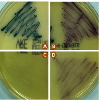
www.kjcls.org Fig. 2. Diversity of VRE color on cIDVA. A is E. faecalis and B is
E. faecium. C and D are E. avium and E. gallinarum.
Fig. 3. Multiple species VRE on cIDVA. A; E. faecalis, B; E. faecium, C; E. avium, D; E. gallinarum.
Table 3. Vancomycin-resistant enterococci with E. faecium co-infection count
P1 P2 P3 P4 P5 P6 P7 P8 P9 P10 P11 P12 P13 Total %
EFM 1 7 12 10 1 5 7 6 9 3 4 12 5 82 66.7
EFA 1 1 2 1.6
EAV 2 1 3 2.4
EFM+EAV 1 3 6 2 3 1 16 13.0
EFM+EFA 1 2 4 7 5.7
EFM+EGA 1 2 2 1 1 7 5.7
EFM+EFA+EAV 1 5 6 4.9
VRE screening period (days) 27 60 91 109 15 52 98 49 91 48 63 129 35
EFM, E. faecium; EFA, E. faecalis; EAV, E. avium; EGA, E. gallinarum.
VRE isolates were counted by patients during the VRE screening period and two of them were excluded because VRE screening tests were not requested.
faecalis 16주(4.9%)가 분리되었으며 E. avium 25주(7.6%), E.
gallinarum 8주(2.4%), E. raffinosus 1주(0.3%)가 분리되었다 (Table 2). E. faecium과 E. faecalis를 제외한 cIDVA에서 분리된 반코마이신 내성 장알균(10.3%)은 모두 vanA 유전자를 보유하였 고 반코마이신의 최소억제농도는 256 μg/mL 이상으로 고도내성 을 보였다(Table 2, Fig. 1).
cIDVA 상에서 E. avium과 E. gallinarum 그리고 E. raffinosus 의 집락은 무색이거나 연한 보라색으로 보였으며 E. avium과 E.
gallinarum은 MGP 분해능 양성이었고 E. avium은 운동성 음성, E. gallinarum은 양성으로 E. faecium과 구별할 수 있었다(Fig. 2, 3).
대상 환자의 VRE 추적검사 기간은 평균 23.3일이었지만 여러 VRE가 동시에 분리된 환자 13명은 평균 66.7일로 전체 환자의 평
균 추적검사 기간보다 2.86배 길었다. 또한 반코마이신 내성 E.
faecium과 E. avium이 동시에 분리되는 경우가 가장 많았으며(16 건, 13.0%), E. faecium과 함께 E. gallinarum이 7건(5.7%) 분리되 었다(Table 3).
반코마이신 내성 E. faecium이 분리된 환자 가운데 입원일수가 30일 이상인 환자에서 E. faecium과 다른 종류의 반코마이신 내성 장알균이 동시에 분리되는 odds ratio는 입원일수 30일 미만인 경 우보다 16.5배 더 높았다(confidence interval: 3.19∼85.25, p
<0.001).
고 찰
임상에서 분리되는 VRE 중 E. faecium은 그 빈도가 높고 vanA 또는 vanB 유전자를 보유한다(Park 등, 2010). 이러한 VRE는 병원 감염의 주요한 원인균으로 반코마이신 감수성 장알균에 비해 이환 율, 사망률이 높으며 건강관리비용 부담 또한 상당히 크다. 본원에
4 Hyun Lee and In-Seon Yoon. Distribution of Vancomycin-resistant Enterococci Isolates Using a ChromID VRE Agar
www.kjcls.org
입원한 성인 환자에게서 분리된 장알균의 반코마이신 내성률은 2006년 16.1%에서 2010년 25.6%로 해마다 꾸준히 증가하였다.
이에 VRE 선별검사는 환자격리와 감염관리 수단으로서 매우 중요 하고 조기에 실시하여 정확하게 동정해야 한다(Cha 등, 2010). 또 한 반코마이신 내성 황색포도알균(vancomycin-resistant Sta- phylococcus aureus, VRSA)의 출현을 막기 위해서도 중요하다 (Kim 등, 2010).
VRE 선별검사에 사용되는 cIDVA는 β-galactosidase (보라색) 와 β-glucosidase (청록색)를 생산하는 장알균의 특성과 반코마이 신 8 μg/mL를 함유하여 vanC 유전형 E. gallinarum과 E.
casseliflavus를 억제함으로 반코마이신 내성 E. faecium과 E.
faecalis를 신속하게 분리할 수 있다. Delmas 등(2007)의 cIDVA에 대한 평가 연구에서 cIDVA를 사용함으로서 VRE 위양성으로 인한 재검사와 분리된 장알균의 동정 및 항균제 감수성 검사 비용을 줄 일 수 있어 매우 유용하다. 하지만 일부 그람 음성 막대균과 효모균 그리고 보라색과 청록색을 제외한 다른 색을 띄는 반코마이신 내성 장알균이 cIDVA에서 자랄 수도 있다. vanA 유전형 E. gallinarum 과 E. casseliflavus는 이미 많은 연구자들에 의해서 분리되어 그들 의 존재가 널리 알려져 있으므로 눈에 보이는 집락의 색깔만으로 판단해서는 안 된다(Dutka-Malen 등, 1994).
본 연구에서 장알균이 분비하는 효소와 반응하여 색이 변하는 cIDVA의 고유 장점에 반하는 장알균이 다수 분리됨으로서 병원 내 감염을 관리할 때 주의가 요구되고 있다. VRE 감염 감시 배양검사 가 의뢰된 환자 85명 중 13명(15.3%)에서 반코마이신 내성 E.
faecium과 함께 옅은 보라색을 띄는 집락과 무색 집락이 분리되었 고 이들 모두 vanA 유전형 장알균이었다. 이것은 종간에 vanA 유 전자의 수평전파가 이루어진 것으로 판단할 수 있으며 입원기간이 길어질수록 전파가 잘되는 것을 알 수 있었다(Kim 등, 2010). 그러 므로 cIDVA에서 형성된 집락은 일단 반코마이신 최소억제농도가 8 μg/mL 이상으로 판단하고 반드시 그람염색으로 확인하고 그람 양성 사슬알균이거나 포도알균일 경우에는 동정 및 항균제 감수성 검사가 실시되어야 한다. 또한 E. faecium이 아닌 다른 반코마이신 내성 장알균이 E. faecium과 동시에 분리되면 vanA 유전자 중합효 소연쇄반응검사로 확인해야 한다.
vanA 유전자가 사람에서 사람으로, 세균에서 세균으로 전파되 는 양상을 보이므로 병원 내 감염관리를 위해서 신속한 동정은 절 대적으로 필요하다. 또한 E. faecium과 E. faecalis를 제외한 반코 마이신 내성 장알균이 vanA 유전자를 보유하고 있다면 반드시 접
촉 주의를 해야 한다. 이에 저자들은 cIDVA에서 분리된 모든 반코 마이신 내성 장알균을 VRE 감염 감시 대상 균종에 포함하는 것을 권장한다.
참고문헌
Berktas M, Yaman G, Ozturk O. vanC gene-related intrinsic teicopla- nin resistance detected in Enterococcus casseliflavus and E. galli- narumstrains by the BD phoenix automated microbiology system.
J Clin Microbiol. 2008, 46:2466-2075.
Cetinkaya Y, Falk P, Mayhall CG. Vancomycin-resistant enterococci.
J Clin Microbiol Rev. 2000, 13:686-707.
Cha CH, An HK, Kim JU. Detection of vancomycin-resistant Enterococci using multiplex real-time PCR assay and melting curve analysis. Korean J Lab Med. 2010, 30:138-146
Cuzon G, Naas T, Fortineau N, Nordmann P. Novel chromogenic me- dium for detection of vancomycin-resistant Enterococcus fae- cium and Enterococcus faecalis. J Clin Microbiol. 2008, 46:2442-2444.
Delmas J, Robin F, Schweitzer C, Lesens O, Bonnet R. Evaluation of a new chromogenic medium, ChromID VRE, for detection of van- comycin-resistant enterococci in stool samples and rectal swabs.
J Clin Microbiol. 2007, 45:2731-2733.
Dutka-Malen S, Blaimont B, Wauters G, Courvalin P. Emergence of high-level resistance to glycopeptides in Enterococcus gallina- rum and Enterococcus casseliflavus. Antimicrob Agents Chemother. 1994, 38:1675-1677.
Harbarth S, Cosgrove S, Carmeli Y. Effects of antibiotics on nosoco- mial epidemiology of vancomycin-resistant Enterococci.
Antimicrob Agents Chemother. 2002, 46:1619-1628.
Kallstrom G, Doern CD, Dunne WM Jr. Evaluation of a chromogenic agar under development to screen for VRE colonization. J Clin Microbiol. 2010, 48:999-1001.
Kim DH, Lee JH, Ha JS, Ryoo NH, Jeon DS, Kim JR. Evaluation of the usefulness of selective chromogenic agar medium (chromID VRE) and multiplex PCR method for the detection of vancomycin-re- sistant enterococci. Korean J Lab Med. 2010, 30:631-636.
Park KS, Kim MH, Park TS, Suh JT, Lee HJ. Antimicrobial resistance of enterococcal isolates from blood and risk factors for vancomycin resistant enterococcal bacteremia in a tertiary care university hospital from 2003 to 2007. Korean J Clin Microbiol. 2010, 13:
59-67.
Peterson JF, Doern CD, Kallstrom G, Riebe KM, Sander T, Dunne WM Jr, et al. Evaluation of Spectra VRE, a new chromogenic agar me- dium designed to screen for vancomycin-resistant Enterococcus faecalis and Enterococcus faecium. J Clin Microbiol. 2010, 48:4627-4629.
Song JY, Hwang IS, Eom JS, Cheong HJ, Bae WK, Park YH, et al. Prevalence and molecular epidemiology of vancomycin-resistant enterococci (VRE) strains isolated from animals and humans in Korea. Korean J Intern Med. 2005, 20:55-62.