Development of a Swine Benign Biliary Stricture Model Using Endoscopic Biliary Radiofrequency Ablation
The large animal model with benign biliary stricture (BBS) is essential to undergo
experiment on developing new devices and endoscopic treatment. This study conducted to establish a clinically relevant porcine BBS model by means of endobiliary radiofrequency ablation (RFA). Endoscopic retrograde cholangiography (ERC) was performed on 12 swine.
The animals were allocated to three groups (60, 80, and 100 W) according to the electrical power level of RFA electrode. Endobiliary RFA was applied to the common bile duct for 60 seconds using an RFA catheter that was endoscopically inserted. ERC was repeated two and four weeks, respectively, after the RFA to identify BBS. After the strictures were identified, histologic evaluations were performed. On the follow-up ERC two weeks after the procedure, a segmental bile duct stricture was observed in all animals. On microscopic examination, severe periductal fibrosis and luminal obliteration with transmural
inflammation were demonstrated. Bile duct perforations occurred in two pigs (100 W, n = 1; 80 W, n = 1) but there were no major complications in the 60 W group. The application of endobiliary RFA with 60 W electrical power resulted in a safe and reproducible swine model of BBS.
Keywords: Benign Stricture; Radiofrequency Ablation; Animal Experimentation;
Endoscopes; Safety Jin-Seok Park,1 Seok Jeong,2
Joon Mee Kim,3 Sang Soon Park,1 and Don Haeng Lee4
1Digestive Disease Center, Department of Internal Medicine, Inha University School of Medicine, Incheon, Korea; 2Digestive Disease Center, Department of Internal Medicine, Inha University School of Medicine and the National Center of Efficacy Evaluation for the Development of Health Products Targeting Digestive Disorders (NCEED), Incheon, Korea; 3Department of Pathology, Inha University School of Medicine, Incheon, Korea;
4Digestive Disease Center, Department of Internal Medicine, Inha University School of Medicine, the National Center of Efficacy Evaluation for the Development of Health Products Targeting Digestive Disorders (NCEED) and Utah-Inha DDS & Advanced Therapeutics Research Center, Incheon, Korea Received: 24 March 2016
Accepted: 29 May 2016 Address for Correspondence:
Seok Jeong, MD
Digestive Disease Center, Department of Internal Medicine, Inha University School of Medicine, 27 Inhang-ro, Jung-gu, Incheon 22332, Korea
E-mail: [email protected]
Funding: The current study was supported by the National Center of Efficacy Evaluation for the Development of Health Products Targeting Digestive Disorders (NCEED) and Inha University Research grant.
http://dx.doi.org/10.3346/jkms.2016.31.9.1438 • J Korean Med Sci 2016; 31: 1438-1444
INTRODUCTION
Benign biliary stricture (BBS) is a common and challenging clinical problem (1). Endoscopic placement of biliary stents, which is considered first-line therapy, provides the highest long- term biliary patency rate (2). However, endoscopic manage- ment of BBS remains challenging. The main drawback of endo- scopic treatment is the need for multiple procedures over one- year treatment period, which increases costs and may decrease patient compliance (3,4). Therefore, there is a growing interest and need for the development of new devices that are able to mitigate the disadvantages of performing endoscopic treatment.
Accordingly, there is a demand for a proper animal model to evaluate the efficacy of endoscopic devices.
As of now, there is no reliable BBS animal model. Previous studies of BBS models have mostly been used for surgical ex- periments with small animals, but the process of making a BBS
model was cumbersome and laborious (5-7). In addition, small animal models tend not to be suitable for endoscopic experi- ments due to the minute size of the digestive tract. The lack of a proper large animal model can lead to errors in clinical applica- tion of new medical devices in humans. Therefore, it is neces- sary to develop a simple and reproducible method for making a BBS large animal model that is similar to actual clinical situa- tions.
Radiofrequency ablation (RFA) therapy has been found to be safe and effective in several gastrointestinal disorders (8,9). RFA can deliver heat energy to tissue that results in necrosis around the RFA probe, and eventually stricture can be induced around tissue damaged by thermal energy (10). RFA has an extended range of indication in experiments because the procedure is not technically difficult to apply, and it is easy to control the de- gree of thermal energy emitted by an RFA device (9). Most re- cently, endobiliary application of RFA has been developed, and
Co. Ltd. (Pyeongtaek, Korea) to be used for an in vivo experi- mental study. The animals were kept in specific pathogen-free animal facilities with complete substrate feeding, according to standard guidelines for laboratory animals. All animals were quarantined and acclimated in a vivarium for one week prior to the start of the experiments.
Equipment and instruments
An endobiliary RFA catheter (9 F, 230-cm working length; APRO Korea Inc., Gunpo, Korea) was used for the study. This catheter is a disposable, monopolar device suitable for endoluminal de- livery of RFA into the biliary tree over a 0.035-inch guidewire.
The distal end of the RFA catheter has a 10-mm leading tip, prox- imal to which there is a 10-mm steel electrode. Energy was de- livered by an RFA generator (CoATherm RF-G200; APRO Korea Inc.) operating at 480 kHz and the range of the used power out- put was 60 to 100 W at 80°C for 60 seconds (Fig. 1).
Pre-procedure preparation
All animals fasted overnight and were given only water for 24 hours before the endoscopic procedure was conducted. Ani- mals were pre-medicated with an intramuscular injection of at- ropine sulfate (0.04 mg/kg), xylazine (2 mg/kg), and tiletamine- zolazepam (5 mg/kg). After endotracheal intubation, 0.5% to 2.0% isoflurane was administered with 70% nitrous oxide, and 30% oxygen was given via a ventilator to maintain general anes- thesia. The animals were placed in the left lateral decubitus po- sition on a fluoroscopy table and allocated randomly to three groups (60, 80, and 100 W) according to the electrical power level.
Endoscopic procedure
An expert biliary endoscopist (S. J.) performed endoscopic ret- rograde cholangiography (ERC) on 12 swine using a standard side-viewing duodenoscope (TJF-240; Olympus Optical Co. Ltd., Tokyo, Japan). After duodenal intubation of the scope, a diag- nostic cannula (ERCP catheter, bottle-shaped metal tip; MTW- Endoskopie, Wesel, Germany) was inserted into the bile duct
using the wire-guided cannulation technique with a 0.035-inch hydrophilic-tipped guidewire (Boston Scientific Corporation, Natick, MA, USA) to obtain the cholangiogram. The endobiliary RFA catheter advanced into the distal common bile duct (CBD) over the guidewire under fluoroscopic guidance (Fig. 2). Endo- scopic biliary sphincterotomy was not required for insertion of the RFA catheter because, unlike humans, swine do not have sphincter of Oddi in major duodenal papilla, and the papillary orifice is patulous and wide. RFA applications were performed with power settings of 60, 80, and 100 W for 60 seconds.
Post-procedure follow-up and pathology assessment During the four weeks following the procedure, the animals Fig. 1. The endobiliary radiofrequency catheter (APRO Korea Inc., Gunpo, Korea) and power generator (CoATherm RF-G200; APRO Korea Inc.) used for endobiliary RFA. (A) An endobiliary RFA catheter. (B) The distal end of the RFA catheter has a 10-mm lead- ing tip, proximal to which there is a 10-mm steel electrode. (C) An RFA generator.
B
C
were fed their usual diet. Clinical signs and parameters includ- ing weight loss, daily food intake, and demeanor score were monitored daily. Biochemical tests of liver function were as- sessed at baseline and two weeks after the procedure. ERC was repeated two and four weeks after the endobiliary RFA to iden- tify the bile duct stricture and procedure-related adverse events.
Bile duct stricture was defined if the luminal diameter decre- ased by more than 50% of the baseline. When the bile duct per- foration was identified in a follow-up cholangiogram, the ani- mal was sacrificed by potassium chloride overdose immediate- ly, and the bile duct was achieved to evaluate pathologic find- ings. With the exception of cases of bile duct perforation, ani- mals were sacrificed six weeks after the procedure. Laparotomy was performed to evaluate presence of bile duct stricture. The CBD was identified and removed, and the effects of RFA on the distal CBD were assessed macroscopically during necropsy. His- tological examination of the CBD was subsequently performed for all animals.
Histological examination
The removed tissues were fixed in neutral buffered formalin and serially sectioned at 5-mm intervals. Multiple cross-sec- tions were taken from the CBD and stained with hematoxylin and eosin (H&E) and Masson’s trichrome (MT) to identify stric- ture and fibrosis.
Ethics statement
The institutional animal care and use committee of Inha Uni- versity reviewed and approved the study design before the ani-
mal experiments were conducted (INHA111004-112).
RESULTS
Endobiliary RFA and clinical follow-up
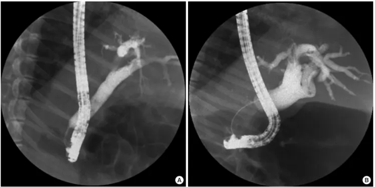
Endobiliary RFA of the CBD was successfully performed on all 12 swine. No technical difficulties or adverse events were en- countered during the procedures. No clinical signs of acute chol- angitis (e.g., fever, weight loss, decreased daily food intake) were noted in any animal two weeks after the procedure was com- pleted. However, a large amount of pus was gushing out of the major duodenal papilla during follow-up ERC in all animals, so that the occurrence of acute cholangitis was suspected. Labora- tory tests also indicated cholestasis. Median values of the liver profiles were as follows: total bilirubin, 4.4 mg/dL (range, 0.2- 7.9); alkaline phosphatase, 360 IU/L (188-1,078); gamma-glu- tamyl transpeptidase, 92 IU/L (56-150); asparatate aminotrans- ferase, 162 IU/L (20-840); alanin aminotransperase, 31 IU/L (20-70). On the follow-up ERC two weeks after the procedure, a segmental bile duct stricture was observed in all animals (Fig.
3A) (Table 1). Before the main experiment, pilot study using en- ergy dosages of 80 W and 100 W for each mini-pig was performed to evaluate the effect and adverse event of RFA. Bile duct perfo- ration was observed in pig that received the 100 W dosage of RFA, but those of 80 W did not occurred bile duct perforation on ERC 2 weeks after RFA. As the results of pilot study, further experimental procedures using 100 W were discontinued be- cause of an ethical problem. We decided to modify the study that the pigs initially allocated to 100 W group were reclassified Fig. 2. Endoscopic retrograde cholangiograms of animals obtained during endobiliary RFA. (A) Normal cholangiogram before endobiliary RFA. (B) RFA catheter positioned in dis- tal CBD.
A B
to 80 W group (Supplementary Fig. 1).
Finally, bile duct perforations were observed in two animals (1/1 [100%] for 100 W; 1/7 [14.3%] for 80 W) on ERC two weeks after the procedure; these animals were sacrificed immediately to investigate pathologic progression. Bile duct perforation was not observed in the 60 W group (n = 4, 0%).
Biliary irrigation with normal saline and pus aspiration were performed for treatment of acute cholangitis in the remaining pigs, and follow-up ERC was performed four weeks after the initial procedure. On ERC review, the silhouette of a stricture of proximal margin was more apparent and proximal duct dilata- tion had progressed (Fig. 3B). Bile duct perforation was no lon- ger apparent.
Macroscopic and microscopic assessment
On the macroscopic examination of extracted CBD, the CBD had filled with yellowish sludge. In addition, there was a fibrous band at the level of stricture (distal CBD) and diffuse reddish mucosal inflammation on the proximal part of the CBD. No
signs of other damage to the surrounding CBD were found. In the animals that experienced perforation, bile duct perforation with abscess formation was noted at the level of stricture.
Microscopic findings of CBD specimens in all animals, in- cluding perforation cases, demonstrated severe periductal fi- brosis and luminal obliteration with transmural inflammation.
These microscopic findings did not show significant difference according to the dose of thermal energy. The CBD, which was proximal to the stricture segment, maintained normal mucosa and muscular structure, but the lumen was markedly dilated (Fig. 4).
DISCUSSION
The current study demonstrated that the thermal energy in- duced by RFA was effective in producing bile duct stricture. A significant stricture was seen in the CBD of all animals, sup- porting the efficacy of endobiliary RFA for creating stricture.
The results of our experiments are in agreement with previously published data. Thermal energy generated by a heat probe or a multipolar electro-cautery probe was evaluated by Rumalla et al. (12) in an animal study with nine swine that was designed to develop a BBS model of the CBD. In their study, 6 of 7 heat probe treatments and 1 of 6 multipolar probe treatments resulted in stricture. This led the authors to report that the application of intraluminal thermal injury can result in a reproducible animal model of BBS; the application of a heat probe at 15 J produced bile duct stenosis in all animals. In addition, the investigators Table 1. Results of endobiliary radiofrequency ablation in a swine model*
Dose Time Stricture Perforation
14 d 28 d 14 d 28 d
100 W 60 sec 1/1 NA *1/1 NA
80 W 60 sec 7/7 6/6 *1/7 0/6
60 W 60 sec 4/4 4/4 0/4 0/4
NA, not applicable.
*The animals were euthanized early.
Fig. 3. Endoscopic retrograde cholangiograms after endobiliary RFA. (A) Cholangiogram at two weeks after endobiliary RFA showing stricture at site of procedure. (B) Cholan- giogram at four weeks after endobiliary RFA. The silhouette of the stricture proximal margin is more apparent and proximal duct dilatation has progressed.
A B
found that strictures tend to develop more effectively as the dose of thermal energy increases (> 10 J).
On the basis of these results, we planned to begin our endo- scopic experiment with a higher level of energy power to im- Fig. 4. Photomicrographs of a histologic section of bile duct with endobiliary RFA at a 60 W setting. (A) The distal part of the CBD, which is embedded in the duodenal muscle layer, reveals mild mucosal inflammation without ulcer or stricture in representative pathologic findings from the swine bile duct (H&E; original magnification 40 ×). (B) The muscle layer of distal CBD is well preserved (MT; original magnification 40 ×). (C) The RFA area of the CBD shows luminal obliteration with mucosal ulcer and transmural in- flammation (H&E; original magnification 40 ×). (D) The RFA area shows destruction of muscle layer with transmural inflammation and severe fibrosis (MT; original magnification 40 ×). (E) The lumen of the proximal part of the CBD is markedly dilated. Mucosa is intact without ulcer (H&E; original magnification 12.5 ×). (F) The proximal CBD shows at- tenuated muscle layer (MT; original magnification 12.5 ×).
A B
C D
E F
animal to adjust the risk of bile duct perforation and determine the threshold to create biliary stricture at the beginning of this study. The biliary stricture in this animal was formed properly, but perforation was detected two weeks after the procedure.
Subsequently, the doses of electrical power were decreased se- quentially down to 60 W in the other animals. All animals that received 60 W for 60 seconds survived at the end of study with- out bile duct perforation. In terms of the ability to create biliary stricture, this thermal energy option yielded satisfactory results.
On the serial cholangiograms two and four weeks after the pro- cedure, a tight biliary stricture with dilated proximal bile duct was confirmed in all four animals that treated with this thermal energy option. Moreover, severe fibrosis was observed at the site of injury and luminal obliteration was found on microscop- ic examination of CBD specimens. Therefore, we estimated that the optimal thermal energy option for creating a BBS model us- ing RFA would be 60 W for 60 seconds. This finding will be of great benefit for future studies in that it should reduce unneces- sary failure and improve the yield rate in making a BBS model.
The basic mechanism for creating BBS in this study was ther- mal injury. The RF waves passing through the electrode agitat- ed tissue ions around the electrode, thus increasing the temper- ature by frictional heat and resulting in destruction of the nor- mal bile duct wall located close to the electrode (14). Thereafter, biliary stricture gradually developed during the healing process.
Although the mechanism for induction of biliary stricture in the current study differs from those associated with other causes of BBS (15), the histologic features of this BBS model are similar to cases of BBS in patients with concentric periductal fibrosis and luminal obliteration. The histologic features we encountered are typical end-result findings detected in BBS (16). Therefore, we estimated that our stricture model is a proper animal model that can substitute for BBS in humans, and hopefully be helpful in the development of new device, technology, or treatment strategies. However, this BBS model would not be applicable to investigate adverse events resulting from biliary endoscopy pro- cedures. The biliary system of swine has relatively few differen- ces from that of humans. One difference is that the biliary sys-
these surgical approaches yielded satisfactory results in creat- ing strictures, they require complicated preparation and post- operative care and sometimes lead to mortality of the animal, whereas establishment of a model using endobiliary RFA is stra- ightforward and safe. All endoscopic procedures can be per- formed without technical difficulty within 20 minutes, and all animal subjects that underwent 60 W-RFA survived without se- rious adverse events until the end of the experiment. Further- more, strictures of the CBD in all animals were found and the degree of stricture was satisfactory upon pathological examina- tion. The second advantage of the current animal model is that the condition of the stricture in large animals is similar to the clinical manifestations seen in human patients with BBS, thus allowing surgeons to practice endoscopic therapeutic techni- ques or conduct preclinical experiments prior to working with humans.
The primary limitation of this study was the short observation time. Although RFA- induced bile duct strictures were maintained for four weeks in the current study, it is uncertain whether this model could be used for long-term observation experiments of therapeutic devices. These strictures have to be preserved with- out spontaneous improvement for a long time in order to inves- tigate the therapeutic effect of various devices. However, the aim of our study was to establish a simple and reproducible method for making an animal model of BBS. A further long-term follow- up study will be needed to determine the long-term durability of stricture.
In conclusion, we developed a swine model of bile duct stric- ture using endobiliary RFA. The endobiliary RFA with proper energy settings appears to be a feasible and safe method to cre- ate a BBS model.
ACKNOWLEDGMENT
We thank APRO Korea Inc. for providing the RFA catheters and generators used in this animal experiment.
DISCLOSURE
The authors have no potential conflicts of interest to disclose.
AUTHOR CONTRIBUTION
Conception and design of the study: Park JS, Jeong S, Lee DH.
Acquisition of data: Park JS, Jeong S, Park SS, Lee DH. Analysis and interpretation of data: Park JS, Jeong S. Pathologic evalua- tion of tissue specimens: Kim JM. Drafting of the article: Park JS, Jeong S. Critical revision: Lee DH. Final approval: all authors.
ORCID
Jin-Seok Park http://orcid.org/0000-0001-9911-8823 Seok Jeong http://orcid.org/0000-0001-6178-8338 Joon Mee Kim http://orcid.org/0000-0003-1355-4187 Sang Soon Park http://orcid.org/0000-0003-1609-1990 Don Haeng Lee http://orcid.org/0000-0003-0397-302X REFERENCES
1. Mahajan A, Ho H, Sauer B, Phillips MS, Shami VM, Ellen K, Rehan M, Sch
mitt TM, Kahaleh M. Temporary placement of fully covered selfexpand
able metal stents in benign biliary strictures: midterm evaluation (with video). Gastrointest Endosc 2009; 70: 3039.
2. Chan CH, Telford JJ. Endoscopic management of benign biliary strictures.
Gastrointest Endosc Clin N Am 2012; 22: 51137.
3. van Berkel AM, Cahen DL, van Westerloo DJ, Rauws EA, Huibregtse K, Bruno MJ. Selfexpanding metal stents in benign biliary strictures due to chronic pancreatitis. Endoscopy 2004; 36: 3814.
4. Devière J, Nageshwar Reddy D, Püspök A, Ponchon T, Bruno MJ, Bourke MJ, Neuhaus H, Roy A, GonzálezHuix Lladó F, Barkun AN, et al. Success
ful management of benign biliary strictures with fully covered selfexpand
ing metal stents. Gastroenterology 2014; 147: 38595.
5. Kountouras J, Billing BH, Scheuer PJ. Prolonged bile duct obstruction: a new experimental model for cirrhosis in the rat. Br J Exp Pathol 1984; 65:
30511.
6. Hirazawa K, Oka M, Ogura Y, Miyahara M, Hazama S, Suzuki T. New tech
nique for inducing reversible obstructive jaundice in the rat. Eur Surg Res 1997; 29: 195201.
7. Ker CG, Wu SC. A simple animal model for inducing and releasing surgi
cal jaundice in rats. Gaoxiong Yi Xue Ke Xue Za Zhi 1992; 8: 5204.
8. Cho YK, Kim JK, Kim MY, Rhim H, Han JK. Systematic review of random
ized trials for hepatocellular carcinoma treated with percutaneous abla
tion therapies. Hepatology 2009; 49: 4539.
9. Rustagi T, Jamidar PA. Intraductal radiofrequency ablation for manage
ment of malignant biliary obstruction. Dig Dis Sci 2014; 59: 263541.
10. Hu B, Gao DJ, Wu J, Wang TT, Yang XM, Ye X. Intraductal radiofrequency ablation for refractory benign biliary stricture: pilot feasibility study. Dig Endosc 2014; 26: 5815.
11. Steel AW, Postgate AJ, Khorsandi S, Nicholls J, Jiao L, Vlavianos P, Habib N, Westaby D. Endoscopically applied radiofrequency ablation appears to be safe in the treatment of malignant biliary obstruction. Gastrointest Endosc 2011; 73: 14953.
12. Rumalla A, Petersen BT, Baron TH, Burgart LJ, Herman LJ, Wiersema MJ, Gostout CJ. Development of a swine model for benign stenosis of the bile duct by endoscopic application of intraluminal thermal injury. Gastroin- test Endosc 2003; 57: 737.
13. Nishikawa H, Inuzuka T, Takeda H, Nakajima J, Sakamoto A, Henmi S, Matsuda F, Eso Y, Ishikawa T, Saito S, et al. Percutaneous radiofrequency ablation therapy for hepatocellular carcinoma: a proposed new grading system for the ablative margin and prediction of local tumor progression and its validation. J Gastroenterol 2011; 46: 141826.
14. Shin JH, Baek JH, Ha EJ, Lee JH. Radiofrequency ablation of thyroid nod
ules: basic principles and clinical application. Int J Endocrinol 2012; 2012:
919650.
15. Judah JR, Draganov PV. Endoscopic therapy of benign biliary strictures.
World J Gastroenterol 2007; 13: 35319.
16. Corvera CU, Blumgart LH, Darvishian F, Klimstra DS, DeMatteo R, Fong Y, D’Angelica M, Jarnagin WR. Clinical and pathologic features of proxi
mal biliary strictures masquerading as hilar cholangiocarcinoma. J Am Coll Surg 2005; 201: 8629.
17. Zhao DF, Chen DZ, Lv JS, Lang R, Jin ZK, Qing H. Establishment of an ani
mal model of biliary ischemic stenosis with clamping in mice. Transplant Proc 2008; 40: 13035.
18. Wang Y, Liang Y, Wang W, Jin R, Cai X. Management of electrothermal in
jury of common bile duct with a degradable biliary stent: an experimen
tal study in a porcine model. J Gastrointest Surg 2013; 17: 17605.
Supplementary Fig. 1. The flow chart of study and distribution of mini-pigs.
Outcome evaluation n = 12