ⓒ 2016 Korean Association of Physical Anthropologists
This is an Open Access article distributed under the terms of the Creative Commons Attribution Non-Commercial License(http://creativecommons.org/ licenses/by-nc/3.0) which permits unrestricted non-commercial use, distribution, and reproduction in any medium, provided the original work is properly cited.
ISSN 2287-626X (Online)·ISSN 1225-150X (Print) Korean J Phys Anthropol Vol. 29, No. 4(2016) pp.145~154
https://doi.org/10.11637/kjpa.2016.29.4.145 Original Article
서 론
일상으로의 빠른 복귀를 원하는 환자, 기저 질환들 때문 에 척추 수술의 대상이 되지 못하는 환자, 그리고 고령으 로 전통적인 수술 방법을 원하지 않는 허리통증 환자의 치 료에 새로운 치료법들이 사용되고 있는데 그중 하나가 경 막바깥신경성형술(epidural neuroplasty)이다. 경막바깥신 경성형술은 척수를 둘러싼 보호막인 경막과 척수관 사이
공간인 경막바깥공간(epidural space)에 정밀한 특수도구 인 카테터로 치료약물을 주입하여 척추사이구멍과 척추관 내 구조물의 자유로운 움직임을 저해하는 신경유착부위를 제거하고 또한, 병소에 치료 약물이 제대로 도달하게 하여 염증이나 부종을 감소시킴으로서 통증을 완화하는 시술이 다[1-4]. 경막바깥신경성형술은 엉치뼈틈새(sacral hiatus, SH)를 통해서 특수한 카테터를 경막바깥공간으로 삽입 목 적부위의 병소까지 도달하여 약물을 주입하는데 시술의 안전성과 부작용을 방지하기 위해서는 엉치뼈틈새에서부 터 경막주머니종말부(termination of dural sac, DS) 및 척 수원뿔(conus medullaris, CM)까지의 거리가 중요한 지표 가 된다.
엉치뼈틈새는 발생학적으로 다섯째엉치척추뼈(그리고
자기공명영상을 이용한 한국 성인의 엉치뼈틈새와 척수원뿔 사이 거리 연구
박태수
1, 황병욱
2, 박상준
2, 백선용
1, 윤 식
11부산대학교 의학전문대학원 해부학교실, 2부산우리들병원 신경외과
(2016년 10월 27일 접수, 2016년 11월 23일 수정접수, 2016년 12월 6일 게재승인)
간추림 : 이 연구에서는 경막바깥신경성형술 시행에 있어서 중요한 엉치뼈틈새에서 경막주머니종말부 및 척수원뿔
사이의 거리를 자기공명영상에서 계측하여 한국인의 생체 연구에 대한 기초 자료를 제공하고자 한다. 허리통증으로
자기공명영상을 촬영한 환자 중 척추압박골절이나 척추전방전위증 그리고 기형 등이 없는 환자 200명(남자 88명, 여 자 112명)을 대상으로 하였으며, 평균 연령은 54.3세(20~84세), 평균 신장은 161.3cm(135~187cm)이었다. T2강조 자기공명영상을 이용하여 엉치뼈틈새에서 경막주머니종말부 그리고 척수원뿔 사이 거리를 계측하여 성별 및 신장
에 따른 상관관계를 분석하였다. 엉치뼈틈새꼭지에서 경막주머니종말부까지 거리는 62.8±9.4mm, 엉치뼈틈새꼭지
에서 척수원뿔까지 거리는 232.2±21.8mm, 엉치뼈틈새꼭지에서 경막주머니종말부 사이 최소 거리는 34.8mm, 최대 거리는 93.9 mm, 엉치뼈틈새꼭지에서 척수원뿔 사이 최소 거리는 155.0mm, 최대 거리는 284.0mm이었다. 엉치뼈틈
새꼭지에서 경막주머니종말부까지, 그리고 엉치뼈틈새꼭지에서 척수원뿔까지 거리는 모두 여성이 남성보다 짧게 나
타났고(p<0.05), 엉치뼈틈새꼭지에서 경막 종말부 및 척수원뿔 사이 거리는 신장에 대해 유의한 상관관계를 보였다 (p<0.01). 이 연구의 결과는 한국 성인의 엉치뼈틈새에서 경막주머니종말부 및 척수원뿔 사이 거리에 대한 생체 계
측 자료를 제공하며, 경막바깥공간에 대한 임상적 시술의 안전성 확보와 한국인의 체형에 맞는 카테터 개발에 도움
이 될 것이라 생각한다.
찾아보기 낱말 : 엉치뼈틈새, 경막주머니종말부, 척수원뿔, 자기공명영상장치, 한국인
*이 논문은 부산대학교 자유과제 학술연구비(2년)에 의하여 연구되었음.
저자 (들)는 ‘의학논문 출판윤리 가이드라인’을 준수합니다.
저자 (들)는 이 연구와 관련하여 이해관계가 없음을 밝힙니다.
교신저자 : 윤 식(부산대학교 의과대학 해부학교실) 전자우편 : [email protected]
때로 넷째엉치척추뼈)의 척추고리판이 중앙선을 따라 융 합되지 않으면서 발생하는 구조물로서[5] 엉치뼈끝(apex of sacrum)과 양쪽의 엉치뼈뿔(sacral cornu)로 이루어져 있는 삼각형의 형태로 나타나며 엉치뼈틈새는 피부, 피하 지방, 엉치꼬리막(sacrococcygeal membrane)으로 덮여 있 어 경막바깥공간 진입시 중요한 지표가 된다[6]. 엉치뼈틈 새꼭지에 해당하는 엉치뼈끝은 넷째엉치척추뼈 또는 다섯 째엉치척추뼈에 80%가 위치한다고 보고되어 있다[7]. 일 반적으로 척수원뿔의 위치는 첫째허리뼈와 둘째허리뼈 사 이의 척추사이원반이지만, 열한째등뼈와 열두째등뼈 사이 의 척추사이원반 뿐만 아니라, 셋째허리뼈 아래 3분의 1 높이에 있기도 한다. 척수원뿔은 남성에 비해 여성에서 더 아래쪽에 위치한다고 알려져 있다[8]. 척수원뿔의 위치는 인종에 따라서도 차이가 있는 것으로 밝혀졌는데, 유럽인 에서는 첫째허리뼈 중간부위(44%)에 가장 많이 위치하고, 아프리카인에서는 첫째허리뼈와 둘째허리뼈 척추사이원 반 부위(52%)에 가장 많이 위치한다고 알려져 있으며[9], 최근 한국인을 대상으로 한 자기공명연구에서는 첫째허 리뼈 중간부위에 가장 많이 위치하고, 시체를 대상으로 한 연구에서는 둘째허리뼈 위 3분의 1 부위에 가장 많이 위치 한다고 보고되었다[10]. 또한 경막주머니종말부는 일반적 으로 둘째엉치척추뼈 아래부위에 위치한다고 알려져 있지 만 첫째엉치척추뼈 위모서리에서 넷째엉치척추뼈 위모서 리 사이에 다양하게 위치한다는 보고도 있다[11].
그러므로 엉치뼈틈새와 경막주머니종말부 및 척수원뿔 사이의 정확한 해부학적 지식은 경막바깥신경성형술과 같
은 임상적 술기 시 엉치뼈틈새를 통하여 경막바깥공간으 로 접근하는 카테터 유도 바늘로 인한 경막의 손상을 예방 하고, 경막주머니종말부의 위치에 따라 척수신경뿌리의 밀 도가 달라서 발생할 수 있는 척수신경뿌리 손상을 사전에 방지함을 기여할 수도 있다. 특히 척수원뿔부위 이상 높이 의 경막바깥공간에 카테터를 위치시켜 경막바깥신경성형 술시행 중 척수가 손상 받게 되면 운동마비, 감각이상, 대 소변 장애와 같은 심각한 문제가 발생할 가능성이 높다. 이에 이 연구에서는 임상 시술 중 고려되어야 할 엉치뼈틈 새에서 경막주머니종말부 및 척수원뿔까지 거리를 자기공 명에서 계측하여 한국인의 정상 성인에 대한 기초 자료를 제공하고자 한다.
Table 1. Basic information about research subjects
Group Frequency Percent
Male Female Total (%) Height group(cm)
≤149 0 33 33 16.5
150~159 6 55 61 30.5
160~169 44 24 68 34.0
≥170 38 0 38 19.0
Total 88 112 200 100
Age group(years)
≤39 25 17 42 21.0
40~49 16 20 36 18.0
50~59 17 20 37 18.5
60~69 17 23 40 20.0
≥70 13 32 45 22.5
Total 88 112 200 100
(A) (B)
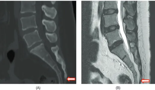
Fig. 1. Determination of the exact measurement of position in relation to the location and the type of variation of the sacral hiatus(SH): (A) Identification of the position of SH using bone setting sagittal images with CT, and (B) Determination of the exact location of measurement by using T2-weighted MR images(arrows; Apex of the SH).
재료 및 방법
허리통증으로 허리엉치부위의 자기공명영상(MRI, Ach- ieva 1.5T; Philips, Eindhoven, Netherlands)을 촬영한 환자 중 척수원뿔 및 경막주머니종말부의 위치나 형태의 변형 을 유발할 수 있는 척추압박골절이나 척추전방전위증 그 리고 기형 등의 질환이 없는 200명(남자 88명, 여자 112 명)의 영상을 사용하였으며, 연령 분포는 20세에서 84세
이었고 평균 나이는 54.3세이었으며, 신장은 135cm에서 187cm이었고, 평균 신장은 161.3cm이었다(Table 1). 자기 공명영상에서 뇌척수액은 고신호 강도로 관찰되고 척수, 척수신경, 그리고 뇌척수액의 구별이 분명한 터보스핀에코 (turbo spin echo) T2강조영상기법(TR 3670 msec, TE 120 msec)로 하고, 뇌척수액 같은 물 성분을 더욱 강조하는 필 립스사의 자기공명영상 획득 기능 중 하나인 DRIVE를 사 용하여 경막주머니종말부 경계부의 지방 신호를 줄여 시
(A) (B) (C)
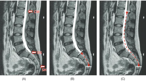
Fig. 2. Schematic illustration of the measurement methods: (A) Identification of the position of the sacral hiatus(SH). (B) Measurement of the distance between SH and the termination of dural sac(DS). (C) Measurement of the distance between SH and the conus medullaris(CM).
(A) (B) (C)
Fig. 3. MRI T2-weighted, midsagittal plane images using PACS(picture archiving communication system) were measured along the ver- tebral canal: (A) The position of the sacral hiatus(SH), termination of dural sac(DS) and conus medullaris(CM). (B) The distance between SH and DS. (C) The distance between SH and CM.
상면 영상을 획득하였다.
엉치뼈틈새의 형태 및 위치의 다양성에 대하여 알려진 바에 의하면, 엉치뼈틈새가 없는 경우가 4%, 엉치뼈뿔이
없는 경우가 54%이고, 그리고 엉치뼈틈새꼭지는 첫째엉치 척추뼈에서 다섯째엉치척추뼈 사이에 위치한다고 보고되 어 있다[7]. 그러므로 자기공명영상의 시상면에서 엉치뼈 틈새의 정확한 위치 설정이 중요하다. 이를 위해 먼저 계측 대상자의 엉치척추뼈가 잘 보이도록 재구성된 전산화단층 촬영장치의 시상면 영상을 가지고 엉치뼈틈새의 위치를 파악한 후, 자기공명영상의 시상면에서 최종 확인하는 방 법으로 정확한 위치를 설정하였다(Fig. 1). 엉치뼈틈새에서 경막주머니종말부 및 척수원뿔 사이 거리 계측은 영상의 확대 및 측정값의 수치 보정이 가능하고 재현성과 정확도 가 높고 이용하기 쉬운 PACS(picture archiving communi- cation system, G2, Infinitt health care, Seoul, Korea)를 사 용하였다[12](Figs. 2 and 3). 연구자의 계측 오류를 줄이기 위해서 한 명의 연구대상자당 각각 두 번씩 계측하여 평 균값을 이용하였고, 척도신뢰도분석(scale reliability anal- ysis)의 급내상관계수(intraclass correlation coefficient)를 이용한 신뢰도 분석 결과 엉치뼈틈새꼭지에서 경막주머니
Frequency
140 120 100 80 60 40 20
0 S3 S4 S5
Location
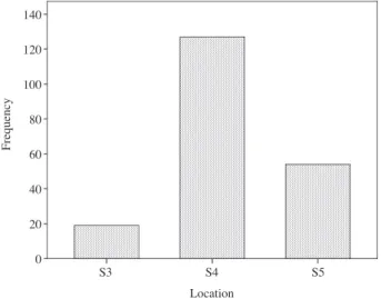
Fig. 4. Location of the Apex of the sacral hiatus(SH). The inci- dences of the apex of SH located at the level of S3 to S5 vertebrae were 9.5% at the level of S3, 63.5% at the level of S4, and 27% at the level of S5.
Frequency
80
60
40
20
0 S3 S4 S5
Location
Fig. 5. Histogram showing the frequency of the vertebral level of the apexes of the sacral hiatus in male and female subjects. 5.5%
of the male subjects, and 4% of the female subjects were located at the level of S3. 28% of the male subjects, and 35.5% of the female subjects were located at the level of S4. 10.5% of the male sub- jects, and 16.5% of the female subjects were located at the level of S5.
Sex Male Female
Frequency
50
40
30
20
10
0 S3 S4 S5
Location
Fig. 6. Histogram of the frequency of the vertebral level of the apexes of the sacral hiatus among the different height groups in both male and female subjects. 1.5% of the height group(≤149 cm), 2% of the height group(150~159cm), 3% of the height group(160~169cm), and 3% of the height group(≥170cm) were located at the level of S3. 11.5% of the height group(≤149cm), 22.5% of the height group(150~159cm), 18% of the height group (160~169cm), and 11.5% of the height group(≥170cm) were located at the level of S4. 3.5% of the height group(≤149cm), 6% of the height group(150~159cm), 13% of the height group (160~169cm), and 4.5% of the height group(≥170cm) were lo- cated at the level of S5.
Height group
≤149 150~159 160~169
≥170
종말부 사이 계측값이 99.5% 일치하여 통계적으로 유의하 였고(p<0.001), 엉치뼈틈새꼭지에서 척수원뿔 사이 계측 값도 99.9% 일치하여 통계적으로 유의하였다(p<0.001).
계측 자료 분석은 SPSS(Ver.21, IMB, USA), 성별 비교 는 t-test, 신장 그룹에 따른 성별은 one way ANOVA로, 그 리고 신장에 대한 계측치와의 상관성은 피어슨 상관계수 (correlation coefficient)를 이용하였다.
결 과
상기와 같은 방법으로 계측한 결과, 엉치뼈틈새꼭지는 셋째엉치척추뼈에 9.5%, 넷째엉치척추뼈에 63.5%, 그리고 다섯째엉치척추뼈에 27%가 위치하였다(Fig. 4). 그중 성 별에 따른 엉치뼈틈새꼭지의 위치는 셋째엉치척추뼈에 남 성 5.5%, 여성 4%가 위치하였고, 넷째엉치척추뼈에는 남 성 28%, 여성 35.5%가 위치하였으며, 다섯째엉치척추뼈에 는 남성 10.5%, 여성 16.5%로 나타났다(Fig. 5). 또한 신장 에 따른 엉치뼈틈새꼭지의 위치는 셋째엉치척추뼈에 149 cm 이하 그룹에서는 1.5%, 150~150cm 그룹은 2%, 160~
169cm 그룹에서는 3%, 그리고 170cm 이상 그룹에서는 3%가 위치하였고, 넷째엉치척추뼈에 149cm 이하 그룹은 11.5%, 150~159cm 그룹에서는 22.5%, 160~169cm 그 룹은 18%, 그리고 170cm 이상 그룹에서는 11.5%가 위치 하였으며, 다섯째엉치척추뼈에 149cm 이하 그룹에서는 3.5%, 150~159cm 그룹은 6%, 160~169cm 그룹에서는 13%, 그리고 170cm 이상 그룹에서 4.5%로 나타났다(Fig.
6).
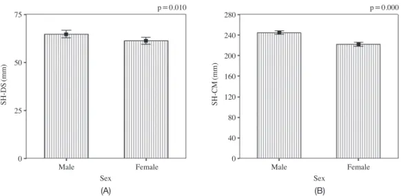
엉치뼈틈새꼭지에서 경막주머니종말부 사이 거리는 남 성의 경우 64.7±8.8mm, 여성은 61.3±9.6mm이었고, 평 균 62.8±9.4mm로서 여성이 남성보다 거리가 짧게 나타 났다(t-test, p<0.05). 엉치뼈틈새꼭지에서 척수원뿔까지 거리는 남성의 경우 244.5±16.0mm 그리고 여성의 경우 222.5±21.0mm이었고, 평균 232.2±21.8mm로서 여성이 남성보다 거리가 짧게 나타났다(t-test, p<0.05, Table 2, Fig. 7).
연구 대상자의 신장은 161.3±9.6cm이었고, 엉치뼈틈새 꼭지에서 경막주머니종말부 사이 최소 거리는 34.8mm로, 이에 해당하는 대상자는 신장 149cm 이하 그룹에 속하였 고, 엉치뼈틈새꼭지에서 경막주머니종말부 간의 최대 거리 는 93.9mm로, 이에 해당하는 대상자는 신장 160~169 cm 그룹에 속하였다(Table 3). 엉치뼈틈새꼭지에서 척수원뿔
Table 2. Comparison of the distance between SH and DS with that between SH and CM according to sex
Sex N Mean±SD
(mm) t p-value Total mean±SD
(mm) SH-DS* MaleFemale 112 61.3±9.688 64.7±8.8 2.612 0.010 62.8±9.4 SH-CM† Male 88 244.5±16.0
8.159 0.000 232.2±21.8 Female 112 222.5±21.0
The data were analysed by t-test and considered significant at p<0.05.
*SH-DS: The distance between the sacral hiatus and the termination of dural sac.
†SH-CM: The distance between the sacral hiatus and the conus medullaris.
SH-DS(mm)
p=0.010 p=0.000
SH-CM(mm)
75
50
25
0
280 240 200 160 120 80 40 0
(A) (B)
Male Female Male Female
Sex Sex
Fig. 7. Histogram of the distances between the sacral hiatus(SH) and termination of dural sac(DS), and between SH and conus medullaris (CM) according to the sex. (A) The distance between SH and DS. (B) The distance between SH and CM. The data were analyzed by t-test and were considered significant at p<0.05.
사이 최소 거리는 155.0mm로, 그 대상자는 신장 149cm 이하의 그룹에 속하였고, 엉치뼈틈새꼭지에서 척수원뿔 간 최대 거리는 284.0mm로서, 그 대상자는 신장 170cm 이 상 그룹에 속하였다(Table 3).
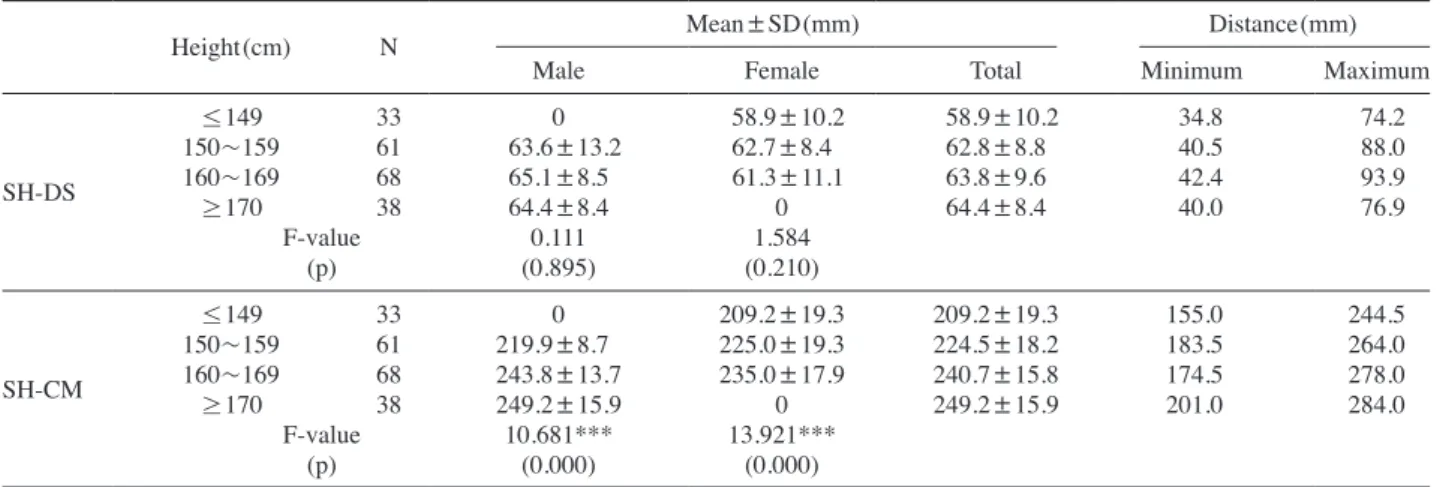
엉치뼈틈새꼭지에서 경막주머니종말부 사이의 성별에 따른 신장 그룹별 거리 비교에서는, 신장 150~159cm 그 룹에서 남성이 63.6±13.2mm 그리고 여성이 62.7±8.4 mm로 나타났고, 신장 160~169cm 그룹에서 남성이 65.1
±8.5mm 그리고 여성이 61.3±11.1mm로서 여성이 남성 보다 거리가 짧게 나타났다. 또한 엉치뼈틈새꼭지에서 척 수원뿔 사이의 성별에 따른 신장 그룹별 거리 비교에서 는, 신장 160~169cm 그룹에서 남성이 243.8±13.7mm
그리고 여성이 235.0±17.9mm로서 여성이 남성보다 거 리가 짧게 나타지만, 신장 150~159cm 그룹에서 남성은 219.9±8.7mm 로서 여성의 225.0±19.3mm보다 짧게 나 타났다(Table 3). 엉치뼈틈새꼭지에서 경막주머니종말부 사이의 신장 그룹에 따른 성별 거리 비교에서는 남성과 여 성 모두 신장에 따른 유의한 차이점을 보이지 않았지만, 엉치뼈틈새꼭지에서 척수원뿔 사이의 신장그룹에 따른 성 별 거리 비교에서는 남성과 여성 모두 신장에 따른 유의 한 차이점을 보였다(one way ANOVA, p<0.001, Table 3, Fig. 8). 하지만 성별의 구분 없이 엉치뼈틈새꼭지에서 경막주머니종말부 및 척수원뿔 사이 거리는 신장에 따라 서 각각 유의한 상관관계를 보였다(correlations, p<0.01, Table 3. Comparison of the distance between SH and DS with that between SH and CM according to height
Height(cm) N Mean±SD(mm) Distance(mm)
Male Female Total Minimum Maximum
SH-DS
≤149 33 0 58.9±10.2 58.9±10.2 34.8 74.2
150~159 61 63.6±13.2 62.7±8.4 62.8±8.8 40.5 88.0
160~169 68 65.1±8.5 61.3±11.1 63.8±9.6 42.4 93.9
≥170 38 64.4±8.4 0 64.4±8.4 40.0 76.9
F-value 0.111 1.584
(p) (0.895) (0.210)
SH-CM
≤149 33 0 209.2±19.3 209.2±19.3 155.0 244.5
150~159 61 219.9±8.7 225.0±19.3 224.5±18.2 183.5 264.0
160~169 68 243.8±13.7 235.0±17.9 240.7±15.8 174.5 278.0
≥170 38 249.2±15.9 0 249.2±15.9 201.0 284.0
F-value 10.681*** 13.921***
(p) (0.000) (0.000)
The data were analyzed by one way ANOVA. *p<0.05, **p<0.01, and ***p<0.001
SH-DS(mm)
p=0.000
MaleFemale Male
Female
SH-CM(mm)
90 80 70 60 50 40
280
240
200
160
(A) (B)
≤149 150~159 160~169 ≥170 ≤149 150~159 160~169 ≥170 Height group(cm) Height group(cm)
Fig. 8. Vertical box plots of the distances between the sacral hiatus(SH) and termination of dural sac(DS), and between SH and conus medullaris(CM) according to the height and sex. (A) The distance between SH and DS. (B) The distance between SH and CM. The data were analyzed by one way ANOVA.
Sex Sex
Fig. 9). 그러나 엉치뼈틈새꼭지에서 경막주머니종말부 및 척수원뿔 사이의 거리는 엉치뼈틈새꼭지의 위치와 유의한 관계를 보이지 않았다(Table 4).
고 찰
허리통증은 인류의 역사와 더불어 생겨난 질병으로 역 학적 조사에 의하면 인간은 직립보행 생활을 영위하는 동 안 약 80%가 허리통증을 겪으며, 연간 유병율은 15~40%
로 알려져 있다[13,14]. 허리통증의 가장 중요한 요인은 척 추사이원반의 퇴행성 변화에 의해 속질핵을 싸고 있는 섬 유테의 파열로 속질핵의 일부 또는 전부가 파열 부위로 돌 출되어 척수의 경막이나 척수신경뿌리를 압박하여 증상을 유발하는 질환인 척추사이원반탈출증이다. 젊은 연령에서 는 넷째허리뼈-다섯째허리뼈 보다는 다섯째허리뼈-첫째엉 치척추뼈에서 많이 발생하고, 고령에서는 보다 상위 구간
인 셋째허리뼈-넷째허리뼈나 둘째허리뼈-셋째허리뼈에서 많이 발생한다고 한다. 척추사이원반탈출증은 20세 이하 및 60세 이상에서는 드물고, 20대에서 40대 사이의 연령에 서 많이 발생하며, 남성의 척수사이원반이 여성에게서 보 다 더 초기에 퇴행성 변화가 생기며 40세 이후에서는 90%
이상 퇴행성 변화가 일어난다고 보고되어 있다[15].
허리뼈 척추사이원반탈출증이나 척추사이구멍협착증 치료에 새로운 치료법이 개발 사용되고 있는데, 요즘 많이 시술되고 있는 경막바깥신경성형술은 엉치뼈틈새에서 경 막주머니종말부 그리고 척수원뿔사이 거리가 시술의 안전 성에 있어서 중요한 정보를 제공한다. 척수원뿔 및 경막주 머니종말부의 위치에 대한 선행연구는 주로 시체해부를 통한 육안 해부학적인 방법으로 연구하였고, 오늘날에는 영상의학 기술이 발전함에 따라서 생체의 영상 해부학적 인 방법으로 연구가 이루어지고 있다. 하지만 많은 선행연 구 중 한국 성인을 대상으로 한 엉치뼈틈새에서 경막주머 니종말부 및 척수원뿔사이 거리 계측에 대한 연구가 없어,
SH-DS(mm)
p=0.007 p=0.000
SH-CM(mm)
90 80 70 60 50 40
280
240
200
160
(A) (B)
≤149 150~159 160~169 ≥170 ≤149 150~159 160~169 ≥170 Height group(cm) Height group(cm)
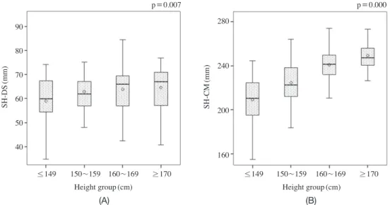
Fig. 9. Vertical box plots of the distances between the sacral hiatus(SH) and termination of dural sac(DS), and between SH and conus medullaris(CM) according to the height. (A) The distance between SH and DS. (B) The distance between SH and CM. The data were ana- lyzed by correlation coefficient and were considered significant at p<0.01.
Table 4. Analysis on Pearson’s correlation coefficients of height and location for the distances from SH to DS and CM
N Pearson
correlation p-value SH-DS Height
200
0.189* 0.007
Location† 0.808 0.260
SH-CM Height 0.636* 0.000
Location† -0.037 0.602
*Correlation was considerd significant at p<0.01.
†Location of the apex of the sacral hiatus.
Table 5. The frequency of different locations of the base of the sacral hiatus in relation to sacral vertebrae
Location of sacral hiatus apex
Total sacra
(n=200) Male sacra
(n=88) Female sacra (n=112)
No. % No. % No. %
S3 19 9.5 11 12.5 8 7.1
S4 127 63.5 56 63.6 71 63.4
S5 54 27.0 21 23.9 33 29.5
Total 200 100 88 100 112 100
이 연구에서는 시체의 육안 해부학적인 방법 이외에 생체 의 척수, 뇌척수액, 척수신경뿌리 등의 해부학적 구조를 잘 볼 수 있고, 비침습적이며, 척주관의 관상면, 시상면, 가로 면을 볼 수 있다는 점에서 많이 이용되고 있는 자기공명영 상[16]을 가지고 엉치뼈틈새꼭지에서 경막주머니종말부 및 척수원뿔사이의 거리를 계측하였다.
Barson[17]은 성인의 척수원뿔은 열두째등뼈와 셋째허 리뼈사이에 다양하게 위치하며, 여성과 흑인의 척수원뿔은 남성에 비해 더 낮게 위치한다고 보고하였다. Kim[18]은 한국인을 대상으로 한 육안 해부학적 연구에서 척수원뿔 의 위치는 남성은 열두째등뼈 척수사이원반에서부터 둘째 허리뼈 아래부위까지 분포하고 첫째허리뼈와 둘째허리뼈 위부위에 위치하는 경우가 많이 있었다고 하였고, 여성의 경우 첫째허리뼈 척수사이원반에서부터 둘째허리뼈 아래 부위까지 포함하여 남성보다 더 낮은 위치에 분포한다고 하였다. Joo 등[19]은 자기공명영상을 이용하여 정상 한국 인을 대상으로 연구한 결과로는 평균 척수원뿔의 위치는 첫째허리뼈 아래 3분의 1 지점이었고, 열두째등뼈 중간 3 분의 1에서 셋째허리뼈 중간 3분의 1 사이에 분포함을 보 고하였다. 또한, 여성이 남성보다 더 아래에 있다고 기술하 였다.
Kim[18]은 한국인의 경막주머니종말부 위치에 대한 육 안 해부학적 연구에서 남성의 경우 첫째엉치척추뼈 중간 부위에서 셋째엉치척추뼈 아래부위에 걸쳐 있고, 둘째엉 치척추뼈 위부위에 가장 많이 분포되어 있는 반면, 여성인 경우 첫째엉치척추뼈 중간부위에서 둘째엉치척추뼈 아래 부위에 걸쳐 있고, 둘째엉치척추뼈의 중간부위 및 아래부 위에 많은 분포를 보이며 여성이 남성보다 약간 아래에 위 치한다고 보고하였다. Park 등[20]은 자기공명영상을 이용 한 한국인 대상 연구에서 경막주머니종말부는 다섯째허리 뼈-첫째엉치척추뼈 척추사이원반에서 셋째엉치척추뼈 아 래 3분의 1 사이에 위치하며, 둘째엉치척추뼈 아래 3분의 1 부위가 가장 많고, 둘째엉치척추뼈 중간부위, 첫째엉치척 추뼈 아래 3분의 1 부위, 첫째엉치척추뼈-둘째엉치척추뼈 척수사이원반 부위의 순서라고 보고하였다.
이 연구에서는 엉치뼈틈새꼭지에서 척수원뿔사이 거리 가 여성에서 남성에 비해 유의하게 더 짧게 나타났다는 것 은, 척수원뿔의 위치가 여성이 남성보다 더 아래에 있다 는 것으로 의미 할 수 있으며, 이는 이 연구와는 연구방법 이 달라 직접적인 비교 대상이 될 수 없지만, 척수원뿔 위 치에 대한 한국인의 연구[18,19,21]에서 여성이 남성보 다 낮게 위치한다는 연구결과와 일치하는 것으로 해석할 수 있을 것이다. Ko 등[22]은 성별에 따른 경막주머니종말 부 위치의 유의성이 없는 것으로 보고하였으나, 이 연구에
서는 성별에 따라 엉치뼈틈새꼭지에서 경막주머니종말부 사이 거리가 유의한 차이(p<0.05)를 보였다는 것은, 성별 에 따른 경막주머니종말부의 위치에 유의성이 있는 것으 로 해석할 수 있을 것이다. 그리고 이 연구에서는 신장에 따른 엉치뼈틈새꼭지에서 경막주머니종말부 및 척수원뿔 사이 거리 또한 각각 유의한 상관관계를 보였다(p<0.01).
Nasr[23]는 자기공명영상을 이용하여 엉치뼈틈새꼭지에 서 경막주머니종말부 및 척수원뿔사이의 거리를 계측하였 다. 엉치뼈틈새꼭지에서 경막주머니종말부사이 거리는 남 성은 53.6±7.6mm이고 여성이 47.4±9.9mm로 기술하였 고, 엉치뼈틈새꼭지에서 척수원뿔사이 거리는 남성 210.±
81 mm 그리고 여성 201.1±8.2mm로 보고하여, 이 연구와 마찬가지로 엉치뼈틈새꼭지에서 경막주머니종말부 및 척 수원뿔사이 거리 모두 여성이 남성보다 더 짧게 나타났다. 그리고 엉치뼈틈새에서 경막주머니종말부 및 척수원뿔사 이의 거리 계측결과가 이 연구보다 짧게 나타난 이유는 측 정방법이 척주관을 따라서 곡선으로 계측한 것이 아니라, 각각의 계측 지점별 직선으로 계측한 결과의 차이도 있을 것으로 생각된다. Mustafa 등[24]은 이집트인을 대상으로 하였을 때 엉치뼈틈새꼭지의 위치는 셋째엉치척추뼈에서 다섯째엉치척추뼈 사이에 위치한다고 보고하였고, 그 중에 서도 넷째엉치척추뼈에 남성은 54% 그리고 여성은 83%
로 가장 많이 위치한다고 보고하였다. 이는 한국인을 대상 으로 한 이 연구에서도 남성은 63.6%, 여성은 63.4%로 넷 째엉치척추뼈에 가장 많은 위치를 보였다(Table 5).
이 연구에서 성별에 따른 신장 그룹별 표본 중, 149cm 이하의 남성 그룹과 170cm 이상의 여성 그룹이 없는 점이 연구의 단점으로 지적될 수 있다. 하지만 이 연구의 결과 는 한국인의 생체 계측 자료를 제공하고, 나아가 경막바깥 신경성형술과 같은 임상적 시술의 안전성 확보와 한국인 의 체형에 맞는 카테터 유도 바늘과 카테터의 개발에 도움 이 될 것이라 생각한다.
REFERENCES
1. Racz G, Heavner J, Trescot A. Percutaneous lysis of epi- dural adhesions: evidence for safety and efficacy. Pain Pract. 2008; 8:277-86.
2. Hsu E, Atanelov L, Plunkett A, Chai N, Chen Y, Cohen S. Epidural lysis of adhesions for failed back surgery and spinal stenosis: factors associated with treatment outcome.
Anesth Analg. 2014; 118:215-24.
3. Erdine S, Talu GK. Precautions during epidural neuroplas- ty. Pain Pract. 2002; 2:308-14.
4. Ho KY, Manghnani P. Acute monoplegia after lysis of epi- dural adhesions: A case Report. Pain Pract. 2008; 8:404-7.
5. Bogduk N. The sacrum. Clinical anatomy of the lumbar spine and sacrum. 4th ed. Philadelphia: Elsevier Churchill Livingstone; 2005. p. 59-61.
6. Senoglu N, Senoglu M, Oksuz H, Gumusalan Y, Yuksel KZ, Zencirci B, et al. Landmarks of the sacral hiatus for caudal epidural block: an anatomical study. Br J Anaesth.
2005; 95:692-5.
7. Sekiguchi M, Yabuki S, Satoh K, Kikuchi S. An anatomic study of the sacral hiatus: a basis for successful caudal epi- dural block. Clin J Pain. 2004; 20:51-4.
8. Demiryürek D, Aydingöz Ü, Akşit M, Yener N, Geyik P.
MR imaging determination of the normal level of conus medullaris. Clin Imaging. 2002; 26:375-7.
9. Louis R. Topographic relationship of the vertebral column, spinal-cord, and nerve roots. Anat Clin. 1978; 1:3-12.
10. Kwon SW, Kim TS, Kim HS, Rhyu IJ. The Tip Level of the Conus Medullaris by Magnetic Resonance Imaging and Cadaver Studies in Korean Adults. Korean J Phys An- thropol. 2016; 29:47-51.
11. Macdonald A, Chatrath P, Spector T, Ellis H. Level of ter- mination of the spinal cord and the dural sac: a magnetic resonance study. Clin Anat. 1999; 12:149-52.
12. Becker SH, Arenson RL. Cost benefits of picture archiving and communication system. J Am Med Assoc. 1994; 1:361- 71.
13. Deyo RA, Loeser JD, Bigos SJ. Herniated lumbar interver- tebral disk. Ann Intern Med. 1990; 112:598-603.
14. Shin SW. Low back pain: Review of anatomy and patho- physiology. J Korean Med Assoc. 2006; 49:656-64.
15. Suk SI. Lumbar disc disease. Spinal surgery. Revised edi-
tion. Seoul: Newest Medical Journal; 2004. p. 218-20.
16. Modic MT, Pavlicek W, Weinstein MA, Boumphrey F, Ngo F, Hardy R, et al. Magnetic rresonance imaging of interver- tebral disk disease: Clinical and pulse sequence consider- ations. Radiology. 1984; 152:103-11.
17. Barson AJ. The vertebral level of termination of spinal cord during normal and abnormal development. J Anat. 1970;
106:489-97.
18. Kim DC. Anatomical studies on the vertebral column and spinal cord of korean subjects: Part Ⅱ. Study on the adult.
Seoul J Med. 1961; 2:149-63.
19. Joo SP, Kim SH, Lee JK, Kim TS, Jung S, Kang SS, et al.
The variation of position of the conus medullaris in korean adults: A magnetic resonance imaging study. J Korean Neu- rosurg Soc. 2001; 30:451-5.
20. Park SJ, Choi WS, Kim EJ, Oh JH, Yoon Y. Normal vari- ation of lumbosacral thecal sac on MR. J Korean Radiol Soc. 1996; 34:565-69.
21. Ko HY, Kim K, Kim HJ. Normal variations of the spinal cord termination. J Korean Acad Rehabil Med. 1998;
22:1040-43.
22. Ko HY, Park JH, Kim HJ, Kim K. Variations of end level of the dural sac. J Korean Acad Rehabil Med. 1999; 23:805-8.
23. Nasr, AY. Vertebral level and measurements of conus medullaris and dural sac termination with special reference to the apex of the sacral hiatus: anatomical and MRI radio- logic study. Folia Morphol. 2016; 75:287-99. doi: 10.5603/
FM.a2016.0004
24. Mustafa MS, Mahmoud OM, El Raouf HH, Atef HM. Mor- phometric study of sacral hiatus in adult human Egyptian sacra: Their significance in caudal epidural anesthesia. Sau- di J Anaesth. 2012; 6:350-7.
Morphometric Analysis of Distances between Sacral Hiatus and
Conus Medullaris Using Magnetic Resonance Image in Korean Adult
Tai Soo Park
1, Byeong-Wook Hwang
2, Sang-Joon Park
2, Sun-Yong Baek
1, Sik Yoon
11
Department of Anatomy, College of Medicine, Pusan National University
2
Busan Wooridul Spine Hospital
Abstract : The purpose of this study was to provide basic biometric data on Korean adults through magnetic
resonance imaging(MRI)-based measurements of the distances between the apex of sacral hiatus(SH) and the termination of dural sac(DS), and between SH and conus medullaris(CM) because they are critical to the performance of epidural neuroplasty.A total of 200 patients(88 males and 112 females) with back pain, who had no spine fracture, significant spinal deformity, and spondyloisthesis were selected for this study. The subjects were of mean age 54.3(20~84) years and mean height 161.3cm(135~187). T2-weighted MRI images were used for correlation analysis to evaluate the relationships between the distances, and variables such as sex and height.
In all patients, the mean distance between SH and DS was 62.8±9.4mm and the mean distance between SH and CM was 232.2±21.8 mm. The minimum distance and the maximum distance between SH and DS were 34.8 mm and 93.9mm respectively, and the minimum distance and the maximum distance between SH and CM were 155.0mm and 284.0mm respectively.
In female patients, both the distances between the SH and DS, and between SH and CM were shorter when compared to those of the male patients(p<0.05). Both the distances between SH and DS and between SH and CM showed a significant correlation with height(p<0.01). The results of this study will provide a useful biometric data on the distances between SH and DS and between SH and CM in Korean in ensuring clinical safety and in the development of more effective catheterization techniques for epidural neuroplasty in Korean.
Keywords : Sacral hiatus, Termination of dural sac, Conus medullaris, Magnetic resonance imaging, Korean
Correspondence to : Sik Yoon(Department of Anatomy, College of Medicine, Pusan National University) E-mail : [email protected]