Brief Report
Vol. 30, No. 4, 2018 481
Received April 11, 2017, Revised August 11, 2017, Accepted for publication August 14, 2017
Corresponding author: Hei Sung Kim, Department of Dermatology, Incheon St. Mary’s Hospital, College of Medicine, The Catholic University of Korea, 56 Dongsu-ro, Bupyeong-gu, Incheon 21431, Korea. Tel: 82-32-280-5700, E-mail: [email protected]
ORCID: https://orcid.org/0000-0003-0576-0474
This is an Open Access article distributed under the terms of the Creative Commons Attribution Non-Commercial License (http://creativecommons.org/
licenses/by-nc/4.0) which permits unrestricted non-commercial use, distribution, and reproduction in any medium, provided the original work is properly cited.
Copyright © The Korean Dermatological Association and The Korean Society for Investigative Dermatology
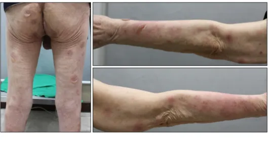
Fig. 1. Multiple erythematous tense bullae on the buttock and both extremities.
https://doi.org/10.5021/ad.2018.30.4.481
A Case of Stevens-Johnson Syndrome Probably Induced by Herbal Medicine
Ji Hong Lim, Sang Hyun Cho, Jeong Deuk Lee, Hei Sung Kim
Department of Dermatology, The Catholic University of Korea, Incheon St. Mary’s Hospital, Seoul, Korea
Dear Editor:
Stevens-Johnson syndrome (SJS) is a life-threatening skin reaction characterized by extensive epidermal detach- ment1. Drugs, especially sulfa drugs, anti-epileptics, and antibiotics, are the most common causes1, but recently, SJS associated with herbal medication has been reported2. Herein, we report a case of SJS probably induced by herb- al medicine. We received the patient’s consent form about publishing all photographic materials. The study protocol was approved by the Institutional Review Board of Incheon St. Mary’s Hospital, The Catholic University of Korea (IRB no. OC17ZESI0049).
A 77-year-old man presented with a sudden onset of bul- lous lesions on his trunk and extremities (Fig. 1). The his-
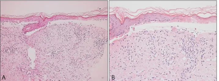
topathological findings were sub-epidermal split with ex- tensive epidermal necrosis (Fig. 2). The direct immuno- fluorescence findings were negative. Days after the skin biopsy, the vesicles and bullae began to fuse, rapidly pro- gressing into skin erosion and denudation. The mucous membranes of the mouth and conjunctiva were also affected. Epidermal detachment was seen in less than 10%
of the body surface area and the Nikolsky sign was present. The patient answered that there has been no change in his routine medication for the past 3 years, but mentioned that he started on herbal medication a month ago. The herbal medication was said to contain deer ant- lers, ginseng, camphor etc. Based on severity-of-illness score for toxic epidermal necrolysis, our patient’s ex-
Brief Report
482 Ann Dermatol
Fig. 2. (A) Biopsy specimens from the arm revealed a subepidermal split with bullae and epidermal necrosis (H&E, ×100). (B) (H&E,
×200).
pected mortality rate was 12.1%. He was asked to imme- diately stop the herbal medication. The patient made a full recovery after a course of intravenous steroid therapy, dai- ly dressings and supportive care.
According to prior reports, the patch test results varied among SJS patients with culprit drug. Lin et al.3 reported that while 62.5% of patients with carbamazepine-induced SJS/toxic epidermal necrolysis (TEN) show positive patch test results, patch tests for allopurinol-induced SJS/TEN are mostly negative. As for our case, we were not able to per- form a patch test on herbal medicine because the patient refused.
Differential diagnoses of SJS include exfoliative dermatitis, staphylococcal scalded skin syndrome (4S), bullous pem- phigoid (BP), paraneoplastic pemphigus and bullous eryth- ema multiforme (EM). Although exfoliative dermatitis re- sembles SJS clinically, it rarely affects the mucosa. There is an intra-epidermal separation in 4S unlike SJS, which shows a sub-epidermal split. BP usually shows a gradual onset and spares the mucosal area. Paraneoplastic pemphi- gus is usually associated with an underlying cancer. As for our patient, the cancer screening tests were negative.
Bullous EM is commonly triggered by herpes simplex virus infection and presents with characteristic targetoid lesions.
More than 100 drugs have been reported as potential cause of SJS4, but herbal medicine induced SJS has not yet been reported in the Korean literature. Recently, several studies have reported the relationship between the human leukocyte antigen (HLA) genotype and drug hypersen- sitivity. Although the HLA genotype of the herbal induced SJS remained uncertain. Herbal medications carry a mix- ture of ingredients that originate from plants and animals.
Because of this characteristic, identifying the culprit in-
gredient within the herbal medication is extremely difficult. Also, since there is no obligation to notify the in- gredient within the medication packet, scientific evalua- tion in case of adverse events are nearly impossible.
Herbal medication induced drug eruption can be caused by herbal medicine itself, but also by added impurities and the combination of ingredients5. With the lack of patch testing and being unable to identify the full in- gredient of the herbal medicine, the authors were not able to confirm that our SJS is caused by herbal medicine.
However with the clinical circumstances, we believe that herbal medicine is most likely the culprit drug in our case.
We report this case because we think it is important that dermatologists consider the possibility of herbal medi- cine-induced drug eruption and notify the public about the potential serious side effects of herbal medicine.
CONFLICT OF INTEREST
The authors have nothing to disclose.
REFERENCES
1. Gerull R, Nelle M, Schaible T. Toxic epidermal necrolysis and Stevens-Johnson syndrome: a review. Crit Care Med 2011;39:1521-1532.
2. Bonhomme A, Poreaux C, Jouen F, Schmutz JL, Gillet P, Barbaud A. Bullous drug eruption to Nigella sativa oil:
Consideration of the use of a herbal medicine - clinical report and review of the literature. J Eur Acad Dermatol Venereol 2017;31:e217-e219.
3. Lin YT, Chang YC, Hui RC, Yang CH, Ho HC, Hung SI, et al. A patch testing and cross-sensitivity study of carbama-
Brief Report
Vol. 30, No. 4, 2018 483
Received December 12, 2016, Revised July 25, 2017, Accepted for publication August 16, 2017
Corresponding author: Jun Young Lee, Department of Dermatology, Seoul St.
Mary’s Hospital, College of Medicine, The Catholic University of Korea, 222 Banpo-daero, Seocho-gu, Seoul 06591, Korea. Tel: 82-2-2258-6222, Fax: 82-2-599-9950, E-mail: [email protected]
ORCID: https://orcid.org/0000-0002-8650-1759
This is an Open Access article distributed under the terms of the Creative Commons Attribution Non-Commercial License (http://creativecommons.
org/licenses/by-nc/4.0) which permits unrestricted non-commercial use, distribution, and reproduction in any medium, provided the original work is properly cited.
Copyright © The Korean Dermatological Association and The Korean
Society for Investigative Dermatology Fig. 1. On physical examination, wheals and erythematous
patches were found on the trunk and both extremities.
zepine-induced severe cutaneous adverse drug reactions. J Eur Acad Dermatol Venereol 2013;27:356-364.
4. Yoon J, Oh CW, Kim CY. Stevens-johnson syndrome induced by vandetanib. Ann Dermatol 2011;23(Suppl 3):
S343-S345.
5. Lim YL, Thirumoorthy T. Serious cutaneous adverse reactions to traditional Chinese medicines. Singapore Med J 2005;46:714-717.
https://doi.org/10.5021/ad.2018.30.4.483
Schnitzler Syndrome: A Case Report and Review of Literature
Yoon Seob Kim, Yu Mee Song, Chul Hwan Bang, Hyun-Min Seo, Ji Hyun Lee, Young Min Park, Jun Young Lee
Department of Dermatology, Seoul St. Mary’s Hospital, College of Medicine, The Catholic University of Korea, Seoul, Korea
Dear Editor:
Schnitzler syndrome (SchS) is a rare autoinflammatory dis- ease characterized by a recurrent urticaria and mono- clonal gammopathy1. Herein, to our knowledge, we re- port the first case of SchS in Korea. The study protocol was approved by the Institutional Review Board of Seoul St.
Mary’s Hospital, The Catholic University of Korea (KC16ZISE0262).
A 64-year-old man presented with two year history of dai- ly urticaria. On physical examination, wheals and eryth- ematous patches were found on the trunk and both ex- tremities (Fig. 1). In contrast to most patients with urti- caria, there was no pruritus, and antihistamine therapies did not have any effect. Only systemic steroid treatment yielded transient symptom improvement. The individual lesions lasted about 24 hours and resolved completely.
Associated symptoms were musculoskeletal pain, and bouts of fever. Laboratory investigations showed leukocy- tosis (10.57×10⁹/L), an elevated erythrocyte sedimen- tation rate (77 mm/hr; 0∼20 mm/hr) and an increased C-re- active protein (CRP) level (11.95 mg/L; 0.01∼0.47 mg/L).
Increased immunoglobulin (Ig)M levels (852 mg/dL; 46∼
260 mg/dL), decreased IgG (831 mg/dL; 870∼1,700 mg/dL) and IgA (99 mg/dL; 110∼410 mg/dL) were detected. Elevated levels of free kappa light chain (32.95