Introduction
Trunk stability is essential to maintain an anti- gravity posture, such as when sitting and standing, and also to move the limbs smoothly (Hodes and Richardson, 1997). After stroke, problems in trunk con-
trol are common and have an effect on balance, gait and function (Ryeson et al, 2008; Verheyden et al, 2006). Trunk control is important to walk stably and reduce the falling risk in stroke patients (Neckel et al, 2008). Trunk control is an early predictor that may be used to anticipate activities of daily living in stroke Corresponding author: Hye-seon Jeon [email protected]
The Effects of a Bridging Exercise With Hip Adductor Contraction on the EMG Activities of Abdominal Muscles in Patients With Sub-Acute Stroke
Chan-bum Park1,2, MSc, PT, Jin-young Ahn3, MSc, OT, Ho-young Kim4, PhD, PT, Jong-ha Lee5, PhD, MD, Hye-seon Jeon6, PhD, PT
1Dept. of Physical Therapy, The Graduate School, Yonsei University
2Dept. of Physical Therapy, Kyunghee University Medical Center
3Dept. of Occupational Therapy, Kyungbok University
4Dept. of Physical Therapy, The Graduate School of Physical Therapy, Sahmyook University
5Dept. of Rehabilitation Medicine, Kyunghee University Medical Center
6Dept. of Ergonomic Therapy, The Graduate School of Health and Environment, Yonsei University
Abstract
1)Background: Muscle weakness and impaired trunk muscle control are common in stroke patients. The bridging exercise (BE) is generally used for trunk stabilization and improving the overall function of stroke patients. The effectiveness of the BE with hip adductor contraction (BEHA) in facilitating trunk muscle activation has been well studied in healthy adults. However, the impact of BEHA in sub-acute stroke patients has not yet been investigated.
Objects: The purpose of this study was to determine the effects of BEHA on the electromyography (EMG) activities and the asymmetry of the rectus abdominis (RA), external oblique (EO) and internal oblique (IO) abdominal muscles.
Methods: Twenty participants with sub-acute stroke (11 males and 9 females) were recruited. Each participant was asked to perform bridging exercises for five seconds under three different conditions: BE in a neutral position (BEN), BEHA with a large ball (BEHAL) and BEHA with a small ball (BEHAS).
The EMG amplitudes of the bilateral RA, EO and IO and the asymmetry of the EMG activity between the sound and affected sides were compared among the conditions. The significance level was set at α=.05.
Results: The EMG activities of RA, EO and IO were significantly greater during BEHAL and BEHAS than during BEN (p<.05); the asymmetry of the RA, EO and IO decreased significantly during BEHAL and BEHAS compared to BEN (p<.05). However, no measured variables showed any significant differences between BEHAL and BEHAS (p>.05).
Conclusion: This study compared the EMG activities of the RA, EO and IO on both sides and the asymmetry of the RA, EO and IO during BEN, BEHAL and BEHAS. Our findings suggest that BEHA was more effective for individuals with hemiplegic stroke at facilitating and normalizing abdominal muscle control than BEN.
Key Words: Asymmetry; Bridging exercise; Hip adduction; Stroke; Trunk.
patients six months after the onset of stroke (Hsieh et al, 2002); trunk strength is related with the Berg bal- ance score at hospital discharge (Karata et al, 2004).
Contrary to common perception that only the affected side of the trunk is impaired following a stroke, stroke patients experience effects on both the sound side and the affected side of the trunk (Tsuji et al, 2003).
The bridging exercise (BE) is commonly used therapeutically for trunk stabilization and has a pos- itive effect on coordinated global and local muscle development (Stevens et al, 2006). Performing the BE on the floor lowers the center of gravity, reduces fear, increases the stability of weight-bearing during gait, and helps to stabilize posture (Schunk, 1982). A modified BE reportedly improved balance and weight bearing in a standing position in stroke patients (Song and Heo, 2015). Similarly, a core stability ex- ercise involving BE improved stroke patients’ dy- namic balance and gait (Chung et al, 2013), while BE with abdominal drawing-in increased the balance ability in patients with stroke (Song and Heo, 2016).
Previous studies has reported the positive effects of various bridging exercises with hip adductor con- traction (BEHA) in healthy adults. BEHA using a resistive Pilates device increased the electro- myography (EMG) activity of the internal obliques (IO), adductor longus (AL) and gluteus maximus (GM) (Kim et al, 2007); BEHA with biofeedback us- ing a stabilizer increased the EMG activity of the rectus abominis (RA), external obliques (EO), IO and GM (Jang et al, 2013), and BEHA with an exercise ball increased the EMG activity of the transversus abominis, adductor magnus (AM) and gluteus medius (Gmed) (Lee and Lee, 2012). Hip adductors could play a role in increasing contraction of abdominal muscles (Kibler et al, 1996) and different hip abduc- tion angles during BE contributed to different muscle activities (Kang et al, 2016). Hip abduction or adduc- tion angles could be controlled by using different sized balls. However, no previous study has exam- ined the effects of a BEHA with using different sized balls in sub-acute stroke patients.
Therefore, the purpose of this study was to de- termine the effects of the BEHA on the EMG activ- ities and asymmetry of the abdominal muscles (RA, EO and IO) in sub-acute stroke patients and to in- vestigate whether there were any differences related to ball sizes for adduction. We hypothesized that BEHA would generate increased abdominal muscle EMG activity and produce more symmetrical EMG activities on both sides of the abdominal muscles to BE. Additionally, BEHA using a small ball (BEHAS) should be more effective than BEHA using a large ball (BEHAL).
Methods
Participants
Twenty sub-acute stroke inpatients who visited Kyunghee University Medical Center were included in this study. The selection criteria were: 1) after two weeks and within six months from stoke onset; 2) a Mini-Mental State Examination-Korean version score greater than 24/30 and the ability to comprehend sim- ple commands; 3) modified Ashworth scale 0 or 1; 4) affected hip extensor strength F- or above on the manual muscle test; 5) affected lower extremity 3 or above on the Brunnstrum stage; 6) ability to perform the BEN, BEHAL and BEHAS independently; 7) abil- ity to lift both shoulders from the mat in curling mo- tion; and 8) above 20 years of age. Participants with any neurological disorder other than stroke were excluded. All of the protocols used in this study were approved by the Institutional Review Board at Kyunghee University Medical Center (approval num-
Variable Mean (SDa)
Age (year) 57.7 (4.88)
Time post stroke (day) 31.8 (10.01)
MMES-Kb 26.4 (1.27)
astandard deviation, ba mini-mental state examination-Korean version.
Table 1. Characteristics of the subjects (N=20)
ber: KMC IRB 1621-01). Before their participation, the procedures, risks and benefits were explained to all the participants, who provided their informed consent.
The characteristics of the participants are summarized in Table 1.
Instruments
Surface EMG data were collected using a DataLog MWX8 (DataLog MWX8, Biometrics Ltd., Newport, UK). The signal was full-wave rectified, and the root mean square value was calculated over 100 millisecond intervals. The EMG data were sampled at a frequency of 2,000 ㎐ and filtered using standard band-pass fil- tering techniques with cutoffs of 20 and 450 ㎐.
Procedures
The surface electrodes were placed over both sides (Pereira et al, 2011) of three muscle groups (Jang et al, 2013): 1) the RA centered on the muscle belly midway between the pubis and the umbilicus; 2) the EO located 5 ㎝ above the anterior superior iliac spine (ASIS); and 3) the IO located 2 ㎝ medial to the ASIS.
Before performing the BEs, all participants did three curl-ups for the reference voluntary contraction (RVC) (Pereira et al, 2011). Participants lied on the mat and maintained their hip joints at 45˚ and their knee joints at 90˚ of flexion with their foot and knees kept apart at shoulder width (Joo et al, 2012). For both sides of the RA, participants curled up, lifting both scapulae from the mat. For the right side of the EO and the left side of the IO, participants curled up with left rotation of the trunk, lifting the right side of the scapula from the mat. For the left side of the EO and the right side of the IO, participants curled up with right rotation of the trunk while lifting the left side of the scapula from the mat. All participants performed three sets of each curl-up. One set con- sisted of holding for five seconds and resting one minute. The RVC data of the first and last second of each contraction were discarded, and the middle three seconds were used instead (Jang et al, 2013).
The participants performed the BE in a neutral
position (BEN) and the BEHA in the same supine position to allow for RVC. For the BEN, the partic- ipants lifted their buttocks as high as they could from the mat. The participants performed two BEHAs using different sized balls for controlling hip angles during BE. One was a BEHAL of a size sim- ilar to the width between both medial femoral epi- condyles, and the other was a BEHAS of lesser width than that between medial femoral epicondyles in the same supine position to allow for RVC. When performing the BEHAL and BEHAS, the participants lifted their buttocks from the mat and then adducted their knees, pressing each ball as hard as they could.
All participants performed three sets of the BEN, BEHAL and BEHAS. One set consisted of holding the position for five seconds and then resting for one minute. All data of the first and last second of each contraction were discarded, and the middle three seconds were used. The order of the BEN, BEHAL and BEHAS was performed randomly to reduce any order effects. Data for each muscle activation were normalized as percentages of the RVC.
Raw data were used to calculate the asymmetry of the RA, EO and IO using the following formula: (sound side-affected side)/(sound side+affected side)×100. When the score is close to zero, the asymmetry is decreased.
Statistical analysis
Statistical analysis was performed using the Statistical Package for the Social Sciences (SPSS) version 12.0 (SPSS Inc., Chicago, IL, USA). A re- peated-measure analysis of variance with Bonferroni corrections as post hoc was used to evaluate the differences in EMG activities and asymmetry among the BEN, BEHAL and BEHAS. All statistical tests were performed at the 5% level of significance.
Results
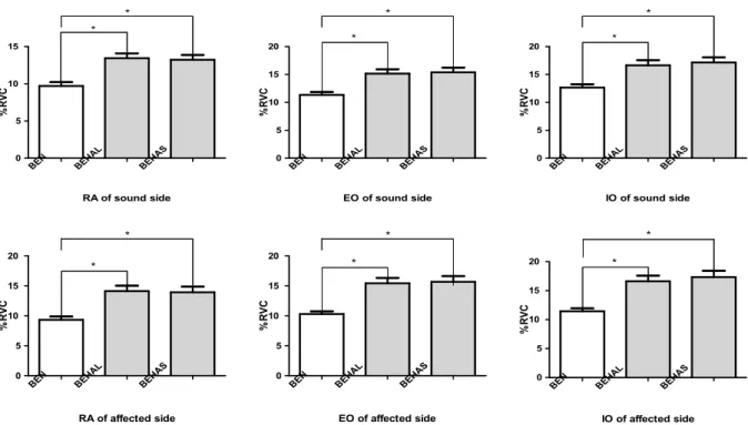
The activation values of each abdominal muscle during BEN, BEHAL and BEHAS before Bonferroni
corrections are summarized in Table 2, while Figure 1 shows those after the Bonferroni corrections.
Significant differences can be seen among the BEN, BEHAL and BEHAS (Table 2): RA (F=8.047, p=.003), EO (F=6.484, p=.008) and IO (F=6.963, p=.006) of the sound side and RA (F=10.042, p=.001), EO (F=9.251, p=.002) and IO (F=9.401, p=.002), respectively. After
the Bonferroni corrections (Figure 1), there were sig- nificant increases of muscle activity from BEN on both the sound side and the affected side during BEHAL. The increases on the sound side of the RA, EO and IO were 13.45 (p=.002), 15.16 (p=.006) and 16.65 (p=.006), respectively, while the increases on the affected side of the RA, EO and IO were 14.12
BEN BEHAL
BEHAS 0
5 10 15
RA of sound side
%RVC
*
*
BEN BEHAL
BEHAS 0
5 10 15
20 *
*
EO of sound side
%RVC
BEN BEHAL
BEHAS 0
5 10 15
20 *
*
IO of sound side
%RVC
BEN BEHAL
BEHAS 0
5 10 15 20
*
*
RA of affected side
%RVC
BEN BEHAL
BEHAS 0
5 10 15
20 *
*
EO of affected side
%RVC
BEN BEHAL
BEHAS 0
5 10 15
20 *
*
IO of affected side
%RVC
Figure 1. Comparison of the muscle activities among the different bridging exercise types. (RVC:
reference voluntary contraction, BEN: bridging exercise in a neutral position, BEHAL: bridging exercise with hip adductor contraction with a large ball, BEHAS: bridging exercise with hip adductor contraction with a small ball, RA: rectus abdominis, EO: external oblique, IO: internal oblique, *p<.05).
Muscle Mean %RVCa (SDb)
F-value p-value
BENc BEHALd BEHASe
Sound side
RAf 9.70 (2.32) 13.45 (2.90) 13.23 (2.93) 8.047 .003*
EOg 11.34 (2.36) 15.16 (3.50) 15.39 (3.81) 6.484 .008*
IOh 12.67 (2.59) 16.65 (4.11) 17.15 (4.13) 6.963 .006*
Affected side
RA 9.33 (2.46) 14.12 (4.03) 13.93 (4.18) 10.042 .001*
EO 10.30 (2.07) 15.42 (4.06) 15.67 (4.29) 9.251 .002*
IO 11.45 (2.20) 16.63 (4.31) 17.34 (4.86) 9.401 .002*
areference voluntary contraction, bstandard deviation, cbridging exercise in a neutral position, dbridging exercise with hip adductor contraction with a large ball, ebridging exercise with hip adductor contraction with a small ball, frectus abdominis, gexternal olique, hinternal oblique, *p<.05.
Table 2. Comparison of the muscle activities among the different bridging exercise types
(p=.001), 15.42 (p=.001) and 16.63 (p=.001), respectively.
In addition, during BEHAS, there were significant increases of muscle activity during BEN on both the sound side and the affected side. The increases on the sound side of the RA, EO and IO were 13.23 (p=.003), 15.39 (p=.005) and 17.15 (p=.004), respectively, and those on the affected side for the RA, EO and IO were 13.93 (p=.002), 15.67 (p=.001) and 17.34 (p=.001), respectively. However, there were no significant differ- ences in the activation of the sound side RA (p=.496), EO (p=1.000) and IO (p=.08) and the affected side RA (p=.621), EO (p=.821) and IO (p=.05) between the BEHAL and BEHAS.
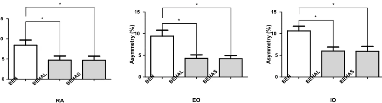
The asymmetry of each abdominal muscle during the BEN, BEHAL and BEHAS before the Bonferroni corrections is summarized in Table 3; Figure 2 shows the same values after Bonferroni corrections were made. There were significant differences among the BEN, BEHAL and BEHAS (Table 3): RA (F=6.701, p=.007), EO (F=7.442, p=.004) and IO (F=9.960, p=.001).
After the Bonferroni corrections, there were significant decreases of muscle asymmetry from the BEN during BEHAL (Figure 2). The asymmetry values of RA, EO and IO were 4.74 (p=.04), 4.29 (p=.003) and 5.98 (p=.001), respectively, during the BEHAL, while those during BEHAS were 4.70 (p=.008) for RA, 4.20 (p=.004) for EO and 5.91 (p=.013) for IO. However, there were no significant differences in the asymmetry of RA (p=1.000), EO (p=1.000) and IO (p=1.000) between the BEHAL and BEHAS.
Discussion
This study evaluated the abdominal muscle activ- ities of sub-acute stroke patients during BEN, BEHAL and BEHAS. Trunk muscles are responsible for stabilizing proximal body segments during volun- tary limb movements (Verheyden et al, 2006); there- fore, the proper activation of core muscles is essential
BEN BEHAL
BEHAS 0
5 10
15 *
*
RA
Asymmetry (%)
BEN BEHAL
BEHAS 0
5 10 15
*
*
EO
Asymmetry (%)
BEN BEHAL
BEHAS 0
5 10 15
*
*
IO
Asymmetry (%)
Figure 2. Comparison of the asymmetry among the different types of bridging exercises. (BEN:
bridging exercise in a neutral position, BEHAL: bridging exercise with hip adductor contraction with a large ball, BEHAS: bridging exercise with hip adductor contraction with a small ball, RA:
rectus abdominis, EO: external oblique, IO: internal oblique, *p<.05).
Muscle Mean %unita (SDb)
F-value p-value
BENc BEHALd BEHASe
RAf 8.43 (5.78) 4.75 (4.58) 4.70 (4.55) 6.701 0.007*
EOg 9.43 (6.12) 4.29 (3.54) 4.20 (3.40) 7.442 0.004*
IOh 10.63 (4.94) 5.97 (4.16) 5.91 (5.14) 9.960 0.001*
a(sound side-affected side)/(sound side+affected side)×100, bstandard deviation, cbridging exercise in a neutral position, dbridging exercise with hip adductor contraction with a large ball, ebridging exercise with hip adductor contraction with a small ball, frectus abdominis, gexternal oblique, hinternal oblique, *p<.05.
Table 3. Comparison of the asymmetry among the different types of bridging exercises
for optimal functioning of the lumbopelvic-hip complex (McGill and Cholewichi, 2001). Trunk muscle strength is often impaired after stroke (Dickstein et al, 2004);
contrary to common consideration, stroke patients are typically affected on both sides of the trunk instead of only on the affected side (Tsuji et al, 2003). In addi- tion, stroke patients have trunk asymmetry, damaged trunk performance and problems in balance and gait (Ryerson et al, 2008; Verheyden et al, 2006). Although it has been acknowledged that impaired selective trunk muscle control is related to balance, gait and hand function following stroke (Davies, 1990), a ma- jority of the post-stroke rehabilitation studies have focused only on the recovery of upper and lower ex- tremity control (Moreland and Thomason, 1994;
Moreland et al, 1998; van der Lee et al, 2001).
The BE is commonly used therapeutically for core muscle stabilization (Stevens et al, 2006). The BE increases abdominal pressure and induces muscle co-contraction to reduce hyperlordosis or anterior pelvic tilt (Kisner and Colby, 2007). Previous studies have reported positive effects of the BEHA on the EMG activities of trunk muscles in healthy adults (Jang et al, 2013; Joo et al, 2012; Kim et al, 2007;
Lee and Lee, 2012). Our results showed that the EMG activities in both the sound and affected sides of the RA, EO and IO were increased during both BEHAL and BEHAS compared to during BEN and are similar the results of BEHA seen in healthy adults. Because both the sound and affected sides of the trunk are impaired in stroke patients (Tsuji et al, 2003), it is important to increase the muscle strength of the sound side as well as the affected side.
Our results could be explained as follows. First, increased EMG activities of the RA, EO and IO dur- ing the BEHA could be explained by the BEHA in- creased the activities of the abdominal muscles (Jang et al, 2013). Second, decreased asymmetry of the trunk during the BEHA could be explained by the hip adductor contraction increased the abdominal sta- bility (Kibler et al, 1996) that may contribute to in- creasing contraction of the affected side of the trunk.
Third, no significant differences in the activation and asymmetry of the trunk between the BEHAL and BEHAS could be explained by the torque-angle rela- tionship on post-stroke was divergent from normal (Hedlund et al, 2012).
Our study is somewhat different than previous studies. First, we studied the effects of BEHA on the EMG activities of trunk muscles in sub-acute stroke patients. Previous studies have only reported the impact of BE without hip adduction in stroke patients or the effects of BEHA in healthy adults.
Second, we assessed both the sound and the affected sides of the trunk muscles. Our findings that BEHA increased the EMG activity of both the sound and affected sides are meaningful because both sides of the trunk are impaired in stroke patients. Third, we measured the asymmetry of trunk muscles; it is im- portant to decrease the asymmetry of muscle activity in stroke patients because this asymmetry produces asymmetrical movements. Fourth, although no meas- ured variables showed any significant differences be- tween the BEHAL and BEHAS, we attempted to compare the different conditions of the BEHA.
Our study had some limitations. First, we included a relatively small sample of participants. Second, be- cause we only measured the EMG activity of the RA, EO and IO muscles, future studies will need to assess the EMG of the GM and trunk extensor muscles that contribute to bridging. Third, we only assessed adduction conditions during the BE.
Therefore, longitudinal, randomized clinical studies to evaluate the long-term effects of the BEHA on more functional activity, such as balance and walking, will be needed in the future.
Conclusion
This study compared the EMG activities and asymmetry of both the affected and unaffected sides of the RA, EO and IO during BEN, BEHAL and BEHAS. We found that the BEHAL and BEHAS
were more effective at increasing the activity of both sides of the RA, EO and IO and at decreasing the asymmetry of RA, EO and IO than the BEN.
However, there were no significant differences in the activation of both sides of the RA, EO and IO or in the asymmetry of the RA, EO and IO between the BEHAL and BEHAS.
Our findings suggest that the BEHAL and BEHAS increase activities of RA, EO and IO and decrease the asymmetry of RA, EO and IO; there- fore, they could be used clinically in sub-acute stroke patients. Further studies with a large sample size that involve other muscles and investigate the long-term effects of BEHA are recommended.
References
Chung EJ, Kim JH, Lee BH. The effects of core stabili- zation exercise on dynamic balance and gait func- tion in stroke patients. J Phys Ther Sci.
2013;25(7):803-806. https://doi.org/10.1589/jpts.25.803 Davies PM. Right in the middle: Selective trunk ac-
tivity in the treatment of adult hemiplegia.
Springer-Verlag, Berlin Heidelberg, 1990:31-65.
Dickstein R, Shefi S, Marcovitz E, et al.
Electromyographic activity of voluntarily activated trunk flexor and extensor muscles in post-stroke hemiparetic subjects. Clin Neurophysiol. 2004;115(4):
790-796.
Hedlund N, Sojka P, Lundström R, Lindstroöm B.
Torque-angle relationship are better preserved during eccentric compared to concentric con- traction in patients with stroke. Isokinet Exerc Sci. 2012;20:129-140.
Hsieh CL, Sheu CF, Hsueh IP, et al. Trunk control as an early predictor of comprehensive activities of daily living function in stroke patients.
Stroke. 2002;33(11):2626-2630.
Jang EM, Kim MH, Oh JS. Effects of a bridging ex- ercise with hip adduction on the emg activities of the abdominal and hip extensor muscles in
females. J Phys Ther Sci. 2013;25(9):1147-1149.
https://doi.org/10.1589/jpts.25.1147
Joo S, Park Y, Lee H, et al. Comparison of trunk muscles activity during bridging exercise with hip adduction and hip abduction. J Phys Ther Sci. 2012;24(10):1077-1078.
Kang SY, Choung SD, Jeon HS. Modifying the hip abduction angle during brdging exercise can fa- cilitate gluteus maximus activity. Man Ther.
2016;22:211-215.
Kibler WB, Chandler TJ, Livingston BP, Roetert EP.
Shoulder range of motion in elite tennis players.
Effect of age and years of tournament paly. AM J Sports Med. 1996;24(3).279-285.
Kim SJ, Yoo WG, Kim MH. EMG activities of core muscles during bridging exercises with and without a pilates resistive device. Phys Ther Korea. 2007;14(4):21-27.
Kisner C, Colby LA. Therapeutic exercise: Foundations and techniques. 5th ed. Philadelphia, PA, F.A.
Davis Co., 2007:300-320.
Lee SY, Lee SK. The impact of abductor and adduc- tor contraction in a bridging exercise on muscle activities in of the abdominal region and the lower extremities. J Phys Ther Sci. 2012;24(11):
1095-1097.
McGill SM, Cholewichi J. Biomechanical basis for stability: An explanation to enhance clinical utility. J Orthop Sports Phys Ther. 2001;31(2):
96-100.
Moreland J, Thomason MA. Efficacy of electromyo- graphic biofeedback compared with conventional physical therapy for upper-extremity of function in patients following stroke: A research over- view and meta-analysis. Phys Ther. 1994;74(6):
534-547.
Moreland JD, Thomason MA. Fuoco AR.
Electromyographic biofeedback to improve lower extremity function after stroke: A meta-analysis.
Arch Phys Med Rehabil. 1998;79(2):134-140.
Neckel ND, Blonien N, Nichols D, et al. Abnormal joint torque patterns exhibited by chronic stroke
subjects while walking with a prescribed phys- iological gait pattern. J Neuroeng Rehabil.
2008;5:19. https://doi.org/10.1186/1743-0003-5-19 Pereira LM, Marcucci FCI, de Oliveira Menacho M,
Garanhani MR, Lavado EL, Cardoso JR.
Electromyographic activity of selected trunk muscles in subjects with and without hemiparesis during therapeutic exercise. J Electromyogr Kinesiol. 2011;32(2):327-332. https://doi.org/10.1016/
j.jelekin.2010.10.003
Ryerson S, Byl NN, Brown DA, et al. Altered trunk position sense and its relation to balance func- tions in people post-stroke. J Neurol Phys Ther.
2008;32(1):14-20.
Schunk MC. Electromyographic study of the per- oneus longus muscle during bridging activities.
Phys Ther. 1982;62(7):970-975.
Song GB, Heo JY. The effects of modified bridge ex- ercise on balance ability of stroke patients. J Phys Ther Sci. 2015;27(12):3807-3810. https://do- i.org/10.1589/jpts.27.3807
Song GB, Heo JY. The effects of bridge exercise with abdominal drawing-in on balance in patients with stroke. J Kor Phys Ther. 2016;28(1):1-7.
Stevens VK, Bouche KG, Mahieu NN, et al. Trunk muscle activity in healthy subjects during bridg- ing stabilization exercises. BMC Musculoskelet Disord. 2006;7:75.
Tsuji T, Liu M, Hase K, et al. Trunk muscles in persons with hemiparetic stroke evaluated with computed tomography. J Rehabil Med. 2003;
35(4):184-188.
van der Lee JH, Snels IA, Beckerman H, et al.
Exercise therapy for arm function in stroke pa- tients: A systematic review of randomized con- trolled trials. Clin Rehabil. 2001;15(1):20-31.
Verheyden G, Vereeck L, Truijen S, et al. Trunk performance after stroke and the relationship with balance, gait and functional ability. Clin Rehabil. 2006;20(5):451-458.
This article was received December 29, 2016, was reviewed December 29, 2016, and was accepted January 31, 2017.