Biomedical Science Letters 2014, 20(3): 139~146 http://dx.doi.org/10.15616/BSL.2014.20.3.139 eISSN : 2288-7415
Evaluation of a PCR-Reverse Blot Hybridization Assay to Identify Six Dermatophytes Predominant in the Republic of Korea
Hyunwoo Jin 1,§ , Hyunjung Kim 2,§ , Sunghyun Kim 2,3 , Yeonim Choi 4 , Hyeeun Bang 2 , Sangjung Park 5 , Hyeyoung Wang 6 , Jang-Ho Lee 7 , In Ho Jang 8 ,
Young-Kwon Kim 9,10 and Hyeyoung Lee 2,†
1
Department of Clinical Laboratory Science, College of Health Sciences, Catholic University of Pusan, Busan 609-757, Korea,
2Department of Biomedical Laboratory Science, College of Health Sciences, Yonsei University, Wonju 220-710, Korea,
3Institute for Life Science and Biotechnology, Yonsei University, Seoul 120-749, Korea,
4Department of Biomedical Laboratory Science, Songho College, Hoengseong 225-704,
Korea,
5Department of Clinical Laboratory Science, College of Medical Science, Daegu Haany University, Daegu 712-715, Korea,
6M&D, Inc., Wonju Eco Environmental Technology Center, Wonju 220-710, Korea,
7
Department of Laboratory Medicine, Samsung Medical Center, Seoul 135-710, Korea,
8Department of Biomedical Laboratory Science, College of Health Sciences, Sangji University, Wonju 220-702, Korea,
9
Department of Biomedical Laboratory Science, College of Medical Sciences, Konyang University, Daejeon 302-718, Korea,
10Korean Collection of Medical Fungi (KCMF), Daejeon 302-718, Korea
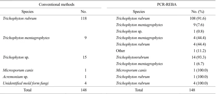
Accurate and rapid diagnosis of dermatophytosis, a disease whose prevalence has been steadily increased, is important for successful treatment. Current laboratory methods for diagnosing dermatophytosis rely on KOH mount and fungal culture method. However, these methods have low sensitivity and are time-consuming (2~4 weeks to diagnosis). In our previous study, a rapid molecular diagnostic assay (PCR-reverse blot hybridization assay, REBA) was developed to identify the following 6 main species of dermatophytes: Trichophyton rubrum, T. mentagrophytes, T. tonsurans, Microsporum canis, M. gypseum, and Epidermophyton floccosum. However, the REBA required more evaluation to validate its use in clinical examinations. The aim of the present study was to evaluate and validate the ability of the PCR-REBA to successfully identify dermatophytes in clinical isolates from dermatophytosis patients. Both conventional identification methods and the PCR-REBA were used to assess the presence of species of dermatophytes in 148 clinical isolates. The results of the two approaches were compared, and discrepancies between the two approaches were resolved by fungal ITS1 sequence analysis. T. rubrum was the most prevalent dermatophyte identified by conventional identification methods (118/148, 79.7%) and the PCR-REBA (131/148, 88.4%). The overall rate of consistency between conventional identification methods and the PCR-REBA was 79.0% (117/148 samples). Fungal ITS1 sequence analysis showed that PCR-REBA results were correct for 93.5% (29/31) of the discrepant samples. The PCR-REBA is rapid, sensitive, and highly specific compared with conventional identification methods. Thus, the PCR-REBA is a potentially useful tool for identifying dermatophytes in clinical settings.
Key Words: Dermatophytes, Identification, Molecular diagnosis, PCR-REBA
INTRODUCTION
Dermatophytosis is caused by fungal infection, especially by superficial fungi on the skin, hair, and nails.
These dermatophytes generally contain the genera Tricho- Original Article
*
Received: July 15, 2014 / Revised: August 7, 2014 Accepted: August 7, 2014
§
Equal contributors
†